

Lumbar transpedicular implant failure: a clinical and surgical challenge and its radiological assessment
- PMID: 24967042
- PMCID: PMC4068848
- DOI: 10.4184/asj.2014.8.3.281
Lumbar transpedicular implant failure: a clinical and surgical challenge and its radiological assessment
Abstract
Study design: It is a multicenter, controlled case study review of a big scale of pedicle-screw procedures from January 2000 to June 2010. The outcomes were compared to those with no implant failure.
Purpose: The purpose of this study was to review retrospectively the outcome of 100 patients with implant failure in comparison to 100 control-patients, and to study the causes of failure and its prevention.
Overview of literature: Transpedicular fixation is associated with risks of hardware failure, such as screw/rod breakage and/or loosening at the screw-rod interface and difficulties in the system assembly, which remain a significant clinical problem. Removal or revision of the spinal hardware is often required.
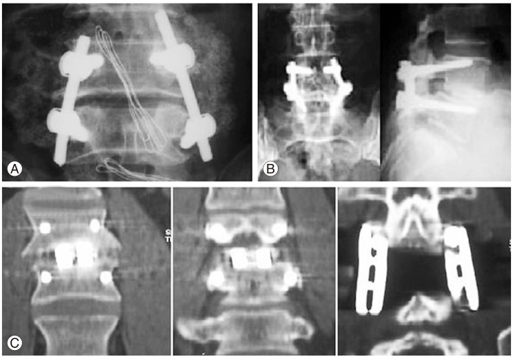
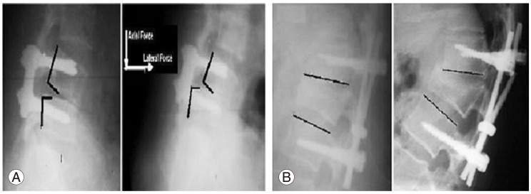
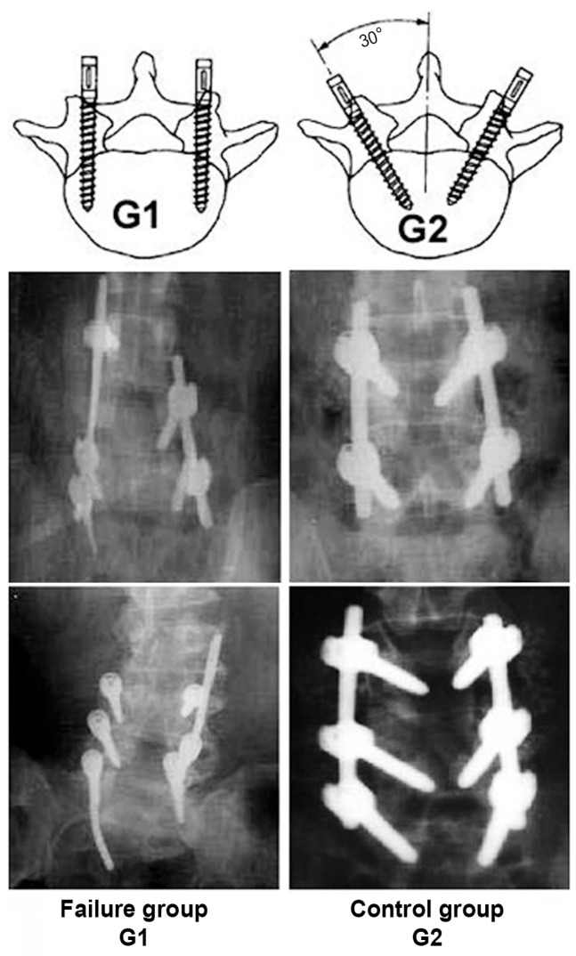
Methods: Two hundred patients (88 women, 112 men) were divided into 2 major groups, with 100 patients in group I (implant failure group G1) and 100 patients in group II (successful fusion, control group G2). We subdivided the study groups into two subgroups: subgroup a (single-level instrumented group) and subgroup b (multilevel instrumented group). The implant status was assessed based on intraoperative and follow-up radiographs.
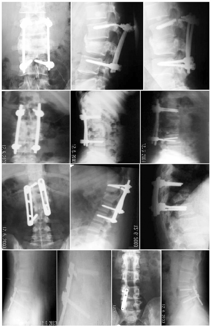
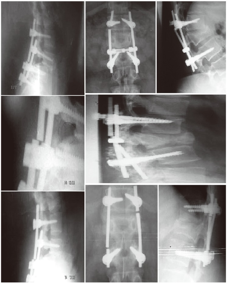
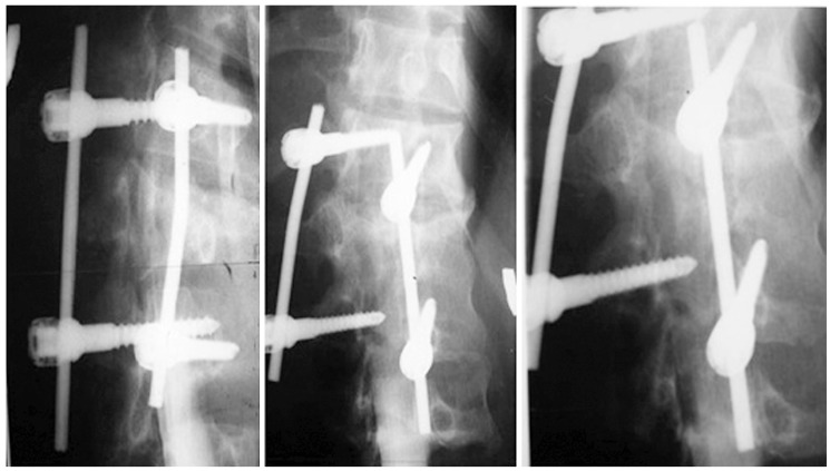
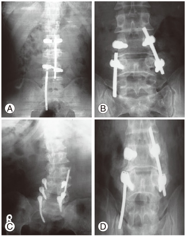
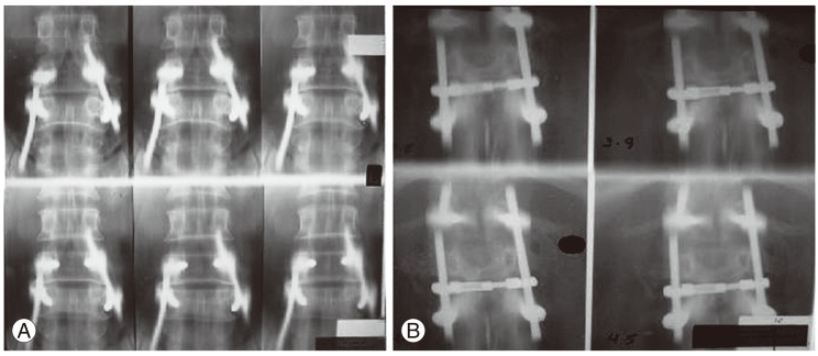
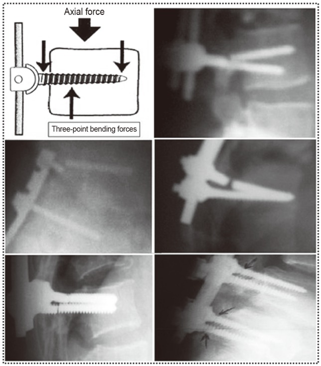
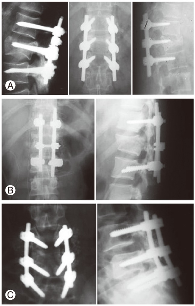
Results: Implant failure in general was present in 36% in G1a, and in 64% in G1b, and types of implant failure included screw fracture (34%), rod fracture (24%), rod loosening (22%), screw loosening (16%), and failure of both rod and screw (4%). Most of the failures (90%) occurred within 6 months after surgery, with no reported cases 1 year postoperatively.
Conclusions: We tried to address the problem and study the causes of failure, and proposed solutions for its prevention.
Keywords: Fracture fixations, prosthesis; Fusion; Loosening; Lumbar, fixation; Screw, failure.
Conflict of interest statement
No potential conflict of interest relevant to this article was reported.
Figures
Similar articles
-
The treatment of unstable thoracic spine fractures with transpedicular screw instrumentation: a 3-year consecutive series.Spine (Phila Pa 1976). 2002 Dec 15;27(24):2782-7. doi: 10.1097/00007632-200212150-00008. Spine (Phila Pa 1976). 2002. PMID: 12486347
-
Finite element analysis of the effects of pedicle screw fixation nut loosening on lumbar interbody fusion based on the elasto-plateau plasticity of bone characteristics.Spine (Phila Pa 1976). 2010 Mar 15;35(6):599-606. doi: 10.1097/BRS.0b013e3181b6258a. Spine (Phila Pa 1976). 2010. PMID: 20139810
-
[Two different fixation methods combined with lumbar interbody fusion for the treatment of two-level lumbar vertebra diseases: a clinical comparison study].Zhongguo Gu Shang. 2015 Oct;28(10):903-9. Zhongguo Gu Shang. 2015. PMID: 26727781 Chinese.
-
Pedicle Screw Fixation in Single-Level, Double-Level, or Multilevel Posterior Lumbar Fusion for Osteoporotic Spine: A Retrospective Study with a Minimum 2-Year Follow-Up.World Neurosurg. 2020 Aug;140:e121-e128. doi: 10.1016/j.wneu.2020.04.198. Epub 2020 May 4. World Neurosurg. 2020. PMID: 32376379
-
[Unilateral pedicle screw fixation versus its combination with contralateral translaminar facet screw fixation for the treatment of single segmental lower lumbar vertebra diseases].Zhongguo Gu Shang. 2015 Apr;28(4):306-12. Zhongguo Gu Shang. 2015. PMID: 26072610 Chinese.
Cited by
-
An Unusual Cause of Buttock Pain after Posterior Thoracolumbar Fixation: Rod Migration into the Pelvis.J Orthop Case Rep. 2019;9(5):31-34. doi: 10.13107/jocr.2019.v09.i05.1520. J Orthop Case Rep. 2019. PMID: 32547999 Free PMC article.
-
Continuous Rod Load Monitoring to Assess Spinal Fusion Status-Pilot In Vivo Data in Sheep.Medicina (Kaunas). 2022 Jul 6;58(7):899. doi: 10.3390/medicina58070899. Medicina (Kaunas). 2022. PMID: 35888618 Free PMC article.
-
Tulip-Screw Head Disjunction from Posterior C2 Fracture Fixation Instrumentation.Case Rep Orthop. 2020 Feb 24;2020:5824383. doi: 10.1155/2020/5824383. eCollection 2020. Case Rep Orthop. 2020. PMID: 32158578 Free PMC article.
-
Wearable bio-adhesive metal detector array (BioMDA) for spinal implants.Nat Commun. 2024 Sep 6;15(1):7800. doi: 10.1038/s41467-024-51987-2. Nat Commun. 2024. PMID: 39242511 Free PMC article.
-
Basic concepts in metal work failure after metastatic spine tumour surgery.Eur Spine J. 2018 Apr;27(4):806-814. doi: 10.1007/s00586-017-5405-z. Epub 2017 Dec 4. Eur Spine J. 2018. PMID: 29204734 Review.
References
-
- Masferrer R, Gomez CH, Karahalios DG, Sonntag VK. Efficacy of pedicle screw fixation in the treatment of spinal instability and failed back surgery: a 5-year review. J Neurosurg. 1998;89:371–377. - PubMed
-
- Arnold PM, Strang RD, Roussel D. Efficacy of variable-angle screws in transpedicular fixation. Neurosurg Focus. 1999;7:e1. - PubMed
-
- Bennett GJ, Serhan HA, Sorini PM, Willis BH. An experimental study of lumbar destabilization. Restabilization and bone density. Spine (Phila Pa 1976) 1997;22:1448–1453. - PubMed
-
- Esses SI, Sachs BL, Dreyzin V. Complications associated with the technique of pedicle screw fixation. A selected survey of ABS members. Spine (Phila Pa 1976) 1993;18:2231–2238. - PubMed
-
- Schnee CL, Freese A, Ansell LV. Outcome analysis for adults with spondylolisthesis treated with posterolateral fusion and transpedicular screw fixation. J Neurosurg. 1997;86:56–63. - PubMed
LinkOut - more resources
Full Text Sources
Other Literature Sources
Research Materials
