

Arteriovenous fistula of the filum terminale misdiagnosed and previously operated as lower lumbar degenerative disease
- PMID: 24967053
- PMCID: PMC4068859
- DOI: 10.4184/asj.2014.8.3.365
Arteriovenous fistula of the filum terminale misdiagnosed and previously operated as lower lumbar degenerative disease
Abstract
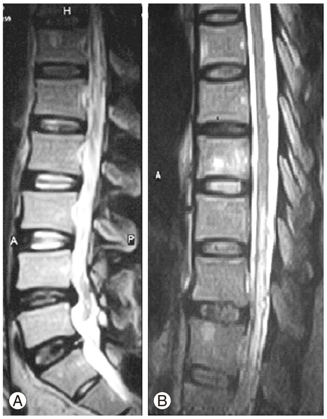
Filum terminale arteriovenous fistula (FTAVF) presenting as a cause of failed back surgery syndrome is a rare entity. We report a 48-year-old male patient who presented with clinical features of a conus medullaris/cauda equina lesion. He had upper and lower motor neuron signs in both the lower limbs with autonomic dysfunction. The patient was misdiagnosed and was operated twice earlier for lumbar canal stenosis and disc prolapse. After reviewing his clinical and radiological findings a diagnosis of FTAVF was made. He underwent surgery and there was a significant improvement in his neurological functions. We discuss the case and review the literature on FTAVF's.
Keywords: Failed back surgery syndrome; Filum terminale arteriovenous fistula.
Conflict of interest statement
No potential conflict of interest relevant to this article was reported.
Figures


Similar articles
-
Filum Terminale Arteriovenous Fistula Coexisting with a Large L2-L3 Disc Sequestration and Associated Diffuse Lumbar Arachnoiditis.Asian J Neurosurg. 2021 May 28;16(2):412-417. doi: 10.4103/ajns.AJNS_489_20. eCollection 2021 Apr-Jun. Asian J Neurosurg. 2021. PMID: 34268177 Free PMC article.
-
Spinal Sparganosis Coexisting with Acquired Arteriovenous Fistula of the Filum Terminale.World Neurosurg. 2020 Apr;136:341-347. doi: 10.1016/j.wneu.2020.01.132. Epub 2020 Jan 26. World Neurosurg. 2020. PMID: 31996338
-
Arteriovenous fistula of the filum terminale masqueraded as a failed back surgery syndrome - A case report and review of literature.Surg Neurol Int. 2021 Feb 10;12:53. doi: 10.25259/SNI_651_2020. eCollection 2021. Surg Neurol Int. 2021. PMID: 33654556 Free PMC article.
-
Spinal arteriovenous fistula located in the filum terminale externa: A case report and review of the literature.Interv Neuroradiol. 2021 Jun;27(3):451-455. doi: 10.1177/1591019920968363. Epub 2020 Oct 22. Interv Neuroradiol. 2021. PMID: 33092430 Free PMC article. Review.
-
Arteriovenous Fistula of the Filum Terminale: A Case Report and Review of the Literature.World Neurosurg. 2019 Oct;130:42-49. doi: 10.1016/j.wneu.2019.06.136. Epub 2019 Jun 26. World Neurosurg. 2019. PMID: 31254689 Review.
Cited by
-
Coexisting filum terminale arteriovenous fistula and filum terminale lipoma treated with single-stage surgery: illustrative case.J Neurosurg Case Lessons. 2023 Jan 16;5(3):CASE22474. doi: 10.3171/CASE22474. Print 2023 Jan 16. J Neurosurg Case Lessons. 2023. PMID: 36647255 Free PMC article.
-
CircRNA GRB10 is a Novel Biomarker for the Accurate Diagnosis of Lumbar Degenerative Disc Disease.Mol Biotechnol. 2023 May;65(5):816-821. doi: 10.1007/s12033-022-00574-1. Epub 2022 Oct 17. Mol Biotechnol. 2023. PMID: 36251122
-
Spinal Arteriovenous Fistulas of the Filum Terminale: Case Report and Literature Review.Asian J Neurosurg. 2019 Nov 25;14(4):1277-1282. doi: 10.4103/ajns.AJNS_100_19. eCollection 2019 Oct-Dec. Asian J Neurosurg. 2019. PMID: 31903378 Free PMC article.
-
Cauda Equina and Filum Terminale Arteriovenous Fistulas: Anatomic and Radiographic Features.AJNR Am J Neuroradiol. 2020 Nov;41(11):2166-2170. doi: 10.3174/ajnr.A6813. Epub 2020 Oct 8. AJNR Am J Neuroradiol. 2020. PMID: 33033040 Free PMC article.
-
Surgical Obliteration of a Filum Terminale Arteriovenous Fistula Following Aborted Endovascular Embolization: Illustrative Case Report and Literature Review.Cureus. 2024 Dec 4;16(12):e75083. doi: 10.7759/cureus.75083. eCollection 2024 Dec. Cureus. 2024. PMID: 39759643 Free PMC article.
References
-
- Kim LJ, Spetzler RF. Classification and surgical management of spinal arteriovenous lesions: arteriovenous fistulae and arteriovenous malformations. Neurosurgery. 2006;59(5 Suppl 3):S195–S201. - PubMed
-
- Mourier KL, Gobin YP, George B, Lot G, Merland JJ. Intradural perimedullary arteriovenous fistulae: results of surgical and endovascular treatment in a series of 35 cases. Neurosurgery. 1993;32:885–891. - PubMed
-
- Tender GC, Vortmeyer AO, Oldfield EH. Spinal intradural arteriovenous fistulas acquired in late adulthood: absent spinal venous drainage in pathogenesis and pathophysiology. Report of two cases. J Neurosurg Spine. 2005;3:488–494. - PubMed
-
- Mitha AP, Murphy EE, Ogilvy CS. Type A intradural spinal arteriovenous fistula. Case report. J Neurosurg Spine. 2006;5:447–450. - PubMed
-
- Jin YJ, Kim KJ, Kwon OK, Chung SK. Perimedullary arteriovenous fistula of the filum terminale: case report. Neurosurgery. 2010;66:E219–E220. - PubMed
LinkOut - more resources
Full Text Sources
Other Literature Sources
