

Hippocampal neurogenesis and antidepressive therapy: shocking relations
- PMID: 24967107
- PMCID: PMC4055571
- DOI: 10.1155/2014/723915
Hippocampal neurogenesis and antidepressive therapy: shocking relations
Abstract
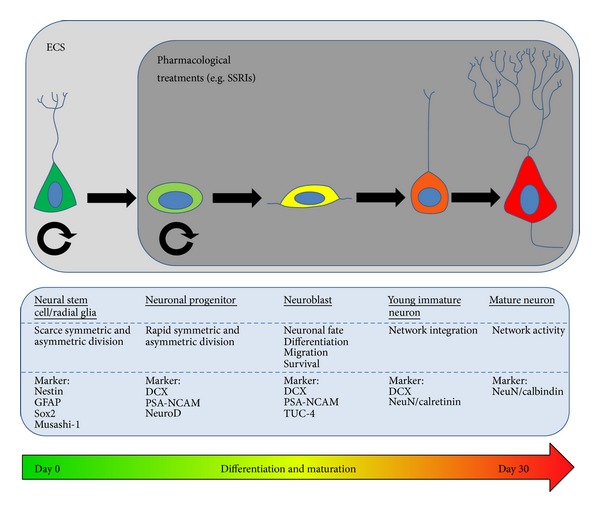
Speculations on the involvement of hippocampal neurogenesis, a form of neuronal plasticity, in the aetiology of depression and the mode of action of antidepressive therapies, started to arise more than a decade ago. But still, conclusive evidence that adult neurogenesis contributes to antidepressive effects of pharmacological and physical therapies has not been generated yet. This review revisits recent findings on the close relation between the mode(s) of action of electroconvulsive therapy (ECT), a powerful intervention used as second-line treatment of major depression disorders, and the neurogenic response to ECT. Following application of electroconvulsive shocks, intricate interactions between neurogenesis, angiogenesis, and microglia activation, the hypothalamic-pituitary-adrenal axis and the secretion of neurotrophic factors have been documented. Furthermore, considering the fact that neurogenesis strongly diminishes along aging, we investigated the response to electroconvulsive shocks in young as well as in aged cohorts of mice.
Figures


Similar articles
-
How does electroconvulsive therapy work? Theories on its mechanism.Can J Psychiatry. 2011 Jan;56(1):13-8. doi: 10.1177/070674371105600104. Can J Psychiatry. 2011. PMID: 21324238 Review.
-
The hippocampus, neurotrophic factors and depression: possible implications for the pharmacotherapy of depression.CNS Drugs. 2011 Nov 1;25(11):913-31. doi: 10.2165/11595900-000000000-00000. CNS Drugs. 2011. PMID: 22054117 Review.
-
Functional role of adult hippocampal neurogenesis as a therapeutic strategy for mental disorders.Neural Plast. 2012;2012:854285. doi: 10.1155/2012/854285. Epub 2012 Dec 31. Neural Plast. 2012. PMID: 23346419 Free PMC article. Review.
-
Electroconvulsive therapy in melancholia: the role of hippocampal neurogenesis.Acta Psychiatr Scand Suppl. 2007;(433):130-5. doi: 10.1111/j.1600-0447.2007.00971.x. Acta Psychiatr Scand Suppl. 2007. PMID: 17280579
-
Increase in hippocampal volume after electroconvulsive therapy in patients with depression: a volumetric magnetic resonance imaging study.J ECT. 2010 Mar;26(1):62-7. doi: 10.1097/YCT.0b013e3181a95da8. J ECT. 2010. PMID: 20190603
Cited by
-
Tamoxifen Activation of Cre-Recombinase Has No Persisting Effects on Adult Neurogenesis or Learning and Anxiety.Front Neurosci. 2017 Feb 1;11:27. doi: 10.3389/fnins.2017.00027. eCollection 2017. Front Neurosci. 2017. PMID: 28203140 Free PMC article.
-
Trajectories of Efficacy and Cognitive Function During Electroconvulsive Therapy Course in Young Adults with Treatment-Resistant Depression.Neuropsychiatr Dis Treat. 2023 Jan 29;19:267-281. doi: 10.2147/NDT.S394155. eCollection 2023. Neuropsychiatr Dis Treat. 2023. PMID: 36744206 Free PMC article.
-
FOXG1 Dose in Brain Development.Front Pediatr. 2019 Nov 22;7:482. doi: 10.3389/fped.2019.00482. eCollection 2019. Front Pediatr. 2019. PMID: 31824897 Free PMC article. Review.
-
Role of inflammatory cytokines in depression: Focus on interleukin-1β.Biomed Rep. 2017 Jan;6(1):15-20. doi: 10.3892/br.2016.807. Epub 2016 Nov 10. Biomed Rep. 2017. PMID: 28123701 Free PMC article.
-
Pathophysiological basis and promise of experimental therapies for Gulf War Illness, a chronic neuropsychiatric syndrome in veterans.Psychopharmacology (Berl). 2023 Apr;240(4):673-697. doi: 10.1007/s00213-023-06319-5. Epub 2023 Feb 15. Psychopharmacology (Berl). 2023. PMID: 36790443 Review.
References
-
- Ingram A, Saling MM, Schweitzer I. Cognitive side effects of brief pulse electroconvulsive therapy: a review. Journal of ECT. 2008;24(1):3–9. - PubMed
-
- Group TUE. Efficacy and safety of electroconvulsive therapy in depressive disorders: a systematic review and meta-analysis. The Lancet. 2003;361(9360):799–808. - PubMed
-
- Baghai T, Frey R, Kasper S, Möller H-J. Elektrokonvulsionstherapie: Klinische und Wissenschaftliche Aspekte. Vol. 483. Vienna, Austria: Springer; 2004.
-
- Couillard-Despres S, Wuertinger C, Kandasamy M, et al. Ageing abolishes the effects of fluoxetine on neurogenesis. Molecular Psychiatry. 2009;14(9):856–864. - PubMed
Publication types
MeSH terms
Substances
LinkOut - more resources
Full Text Sources
Other Literature Sources
