Emergent treatment of aortic rupture in acute type B dissection
- PMID: 24967173
- PMCID: PMC4052411
- DOI: 10.3978/j.issn.2225-319X.2014.05.05
Emergent treatment of aortic rupture in acute type B dissection
Abstract
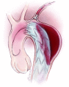
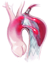
Massive left hemothorax is a rare and dramatic complication of acute type B aortic dissection. The primary endpoint is to treat the aortic rupture, stop the bleeding and stabilize the hemodynamic status, with the aim to prevent mortality and major cardiac, cerebral, visceral and renal complications. Thoracic endovascular repair (TEVAR) is the most frequent management, although its planning, in these emergent patients, may be very difficult and sub-optimal imaging may result at post-operative examination (CT and MRI). In case of TEVAR is not the definitive treatment of the aortic disease, a second stage surgical management can be performed in elective status, in a patient with a total clinical recover. In acute and dramatic circumstances, like ruptured type B dissection, TEVAR is a valid and suitable bridge procedure to open surgery, reducing the overall risk for mortality and major complications.
Keywords: Acute type B dissection; aortic rupture; frozen elephant trunk; thoracic endovascular repair (TEVAR).
Figures


Publication types
LinkOut - more resources
Full Text Sources
