

Ultrasonographic features of kidney transplants and their complications: an imaging review
- PMID: 24967275
- PMCID: PMC4045518
- DOI: 10.5402/2013/480862
Ultrasonographic features of kidney transplants and their complications: an imaging review
Abstract
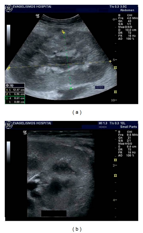
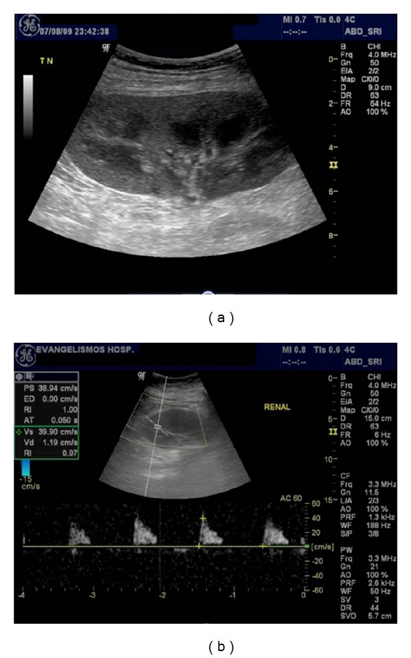
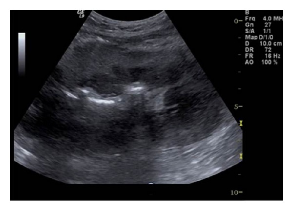
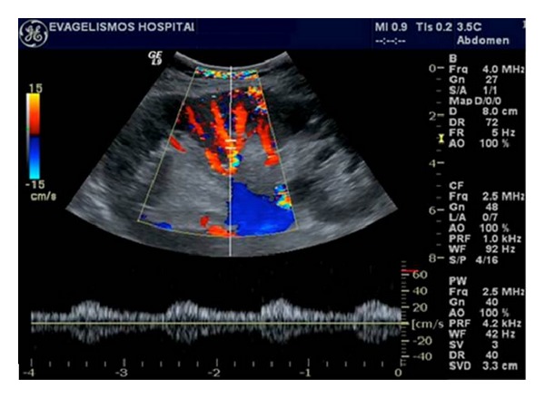
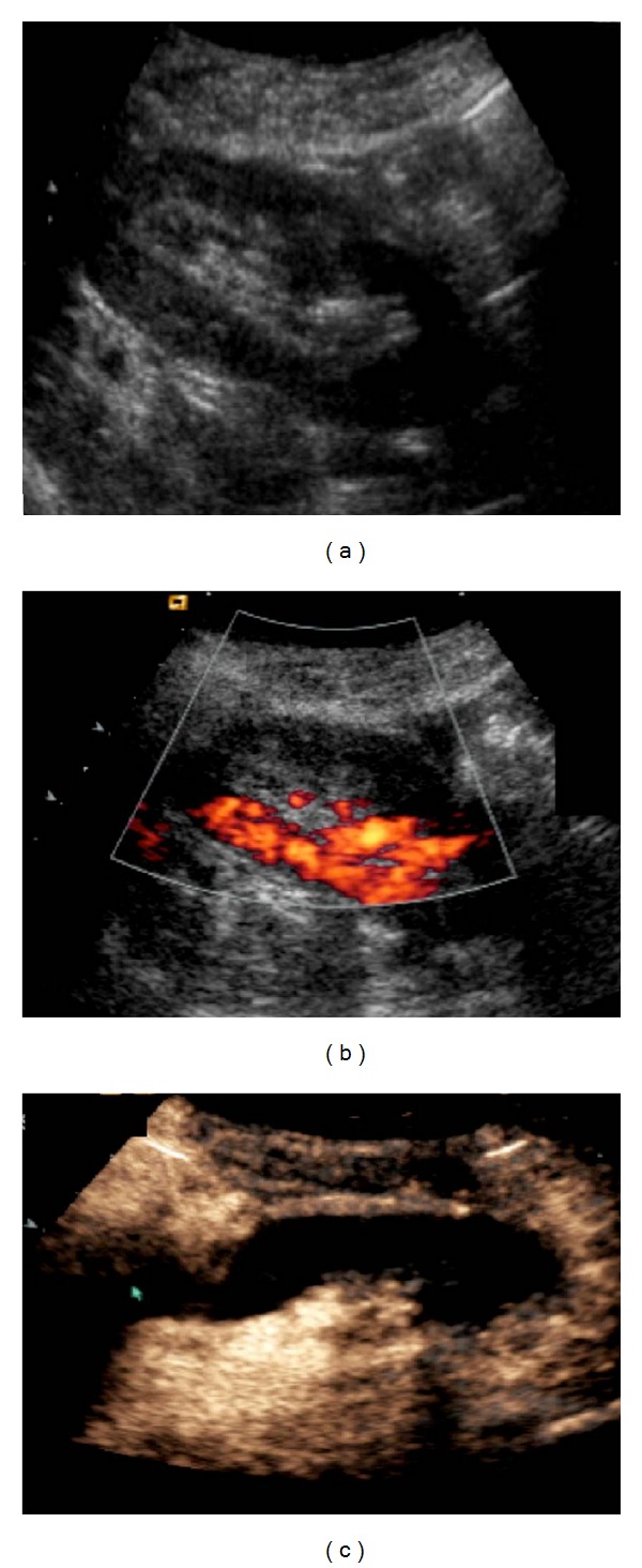
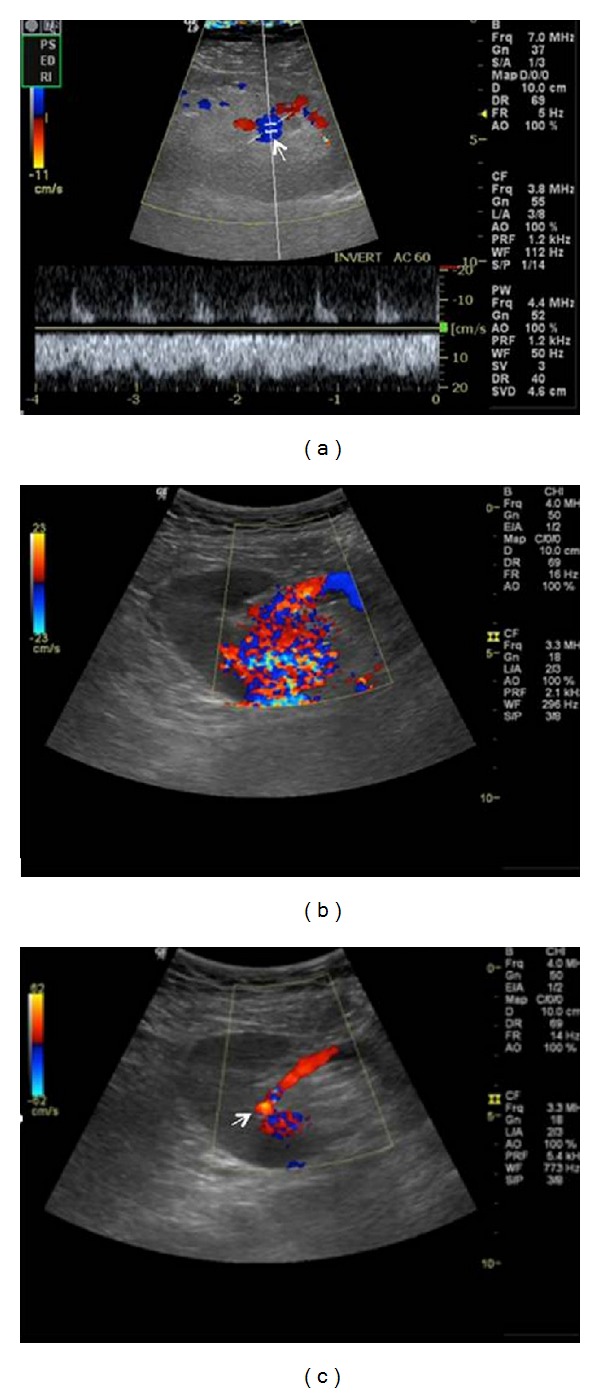
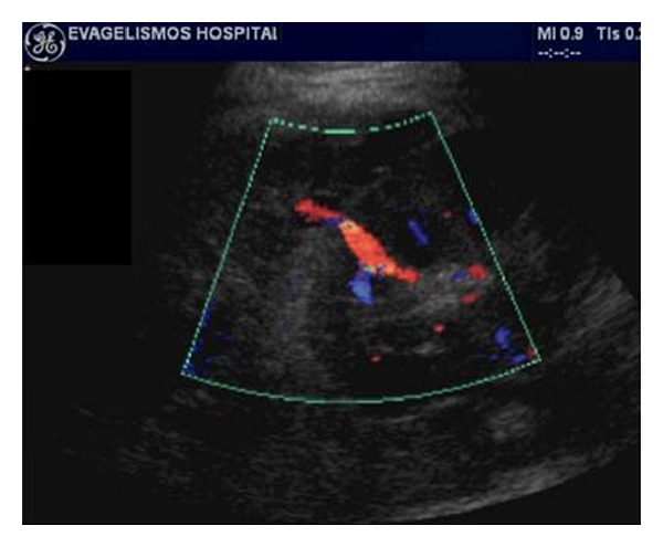
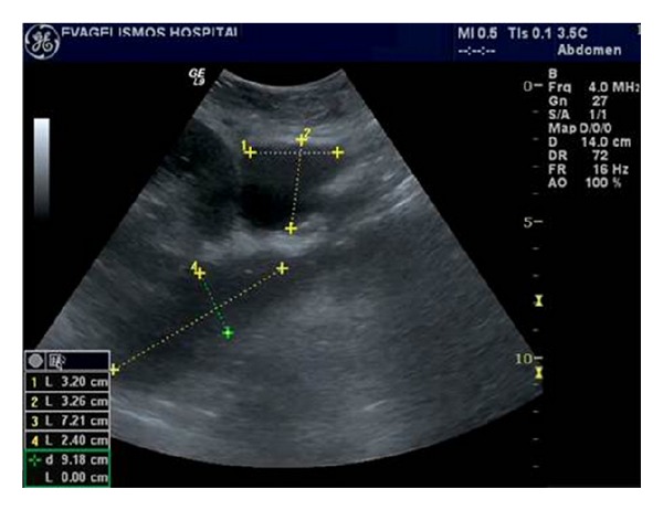
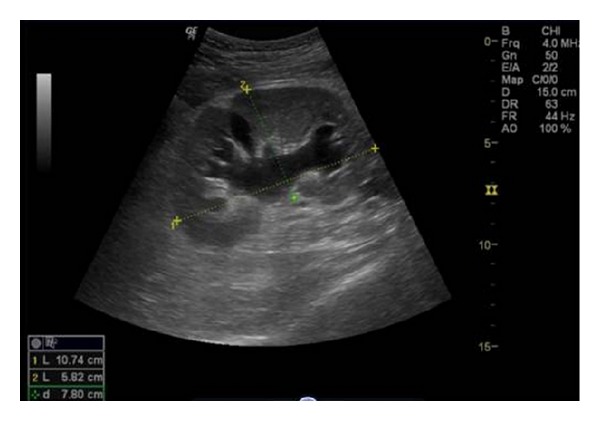
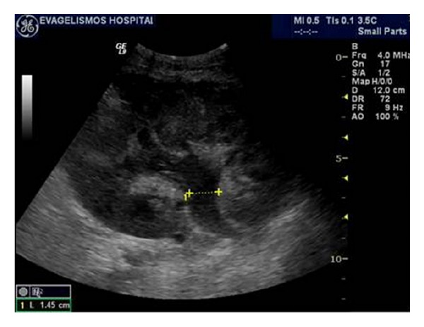
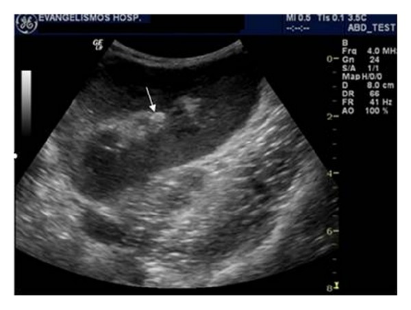
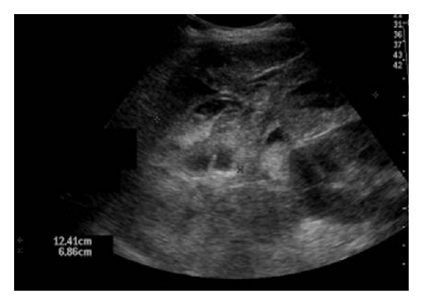
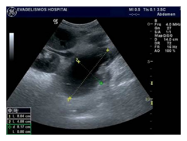
Renal transplantation is the treatment of choice for managing patients with end-stage kidney disease. Being submitted to a very serious surgical procedure, renal transplant recipients can only benefit from follow-up imaging and monitoring strategies. Ultrasound is considered as the principal imaging test in the evaluation of renal transplants. It is an easily applied bedside examination that can detect possible complications and guide further imaging or intervention. In this imaging review, we present essential information regarding the sonographic features of healthy renal transplants, detailing the surgical technique and how it affects the sonoanatomy. We focus on various complications that occur following renal transplantation and their sonographic features by reviewing pertinent literature sources and our own extensive imaging archives.
Figures


References
-
- Vollmer WM, Wahl PW, Blagg CR. Survival with dialysis and transplantation in patients with end-stage renal disease. The New England Journal of Medicine. 1983;308(26):1553–1558. - PubMed
-
- Cecka JM, Terasaki PI. The UNOS scientific renal transplant registry. Clinical Transplants. 1992:1–16. - PubMed
-
- Brown ED, Chen MYM, Wolfman NT, Ott DJ, Watson NE. Complications of renal transplantation: evaluation with US and radionuclide imaging. Radiographics. 2000;20(3):607–622. - PubMed
-
- Cosgrove DO, Chan KE. Renal transplants: what ultrasound can and cannot do. Ultrasound Quarterly. 2008;24(2):77–87. - PubMed
-
- Park SB, Kim JK, Cho KS. Complications of renal transplantation: ultrasonographic evaluation. Journal of Ultrasound in Medicine. 2007;26(5):615–633. - PubMed
Publication types
LinkOut - more resources
Full Text Sources
Other Literature Sources