Review
. 2013 Sep 2:2013:483069.
doi: 10.5402/2013/483069. eCollection 2013.

Abdominal manifestations of lymphoma: spectrum of imaging features

Affiliations
Review

Abdominal manifestations of lymphoma: spectrum of imaging features

Adonis Manzella et al. ISRN Radiol. .

Abstract

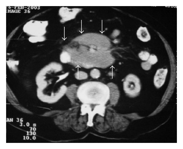
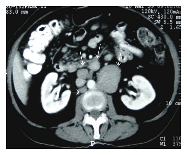
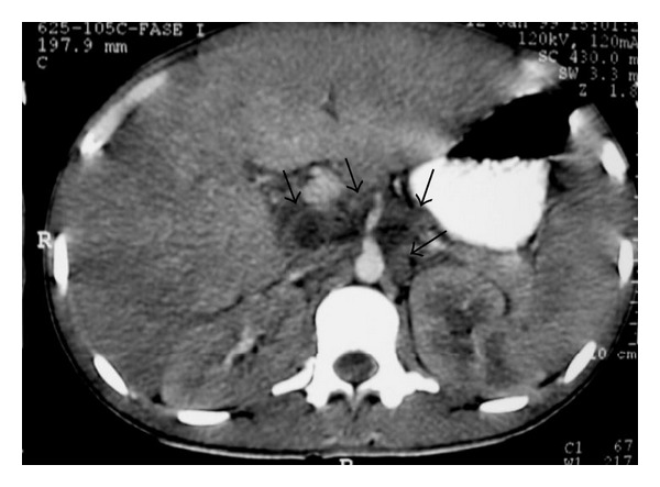
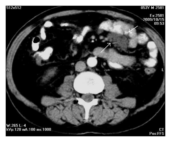
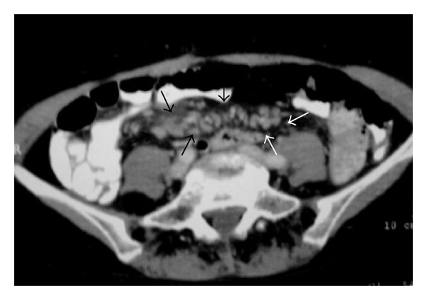
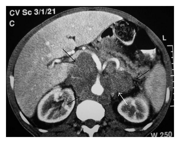
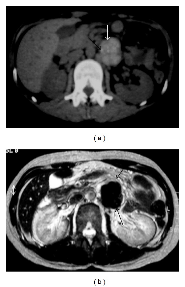
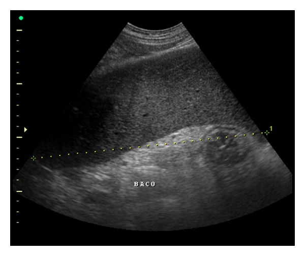
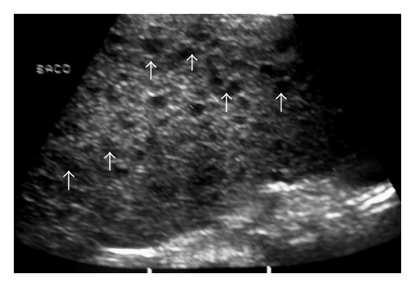
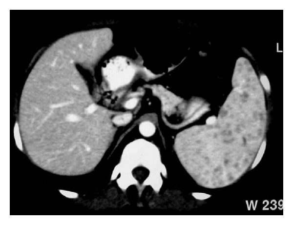
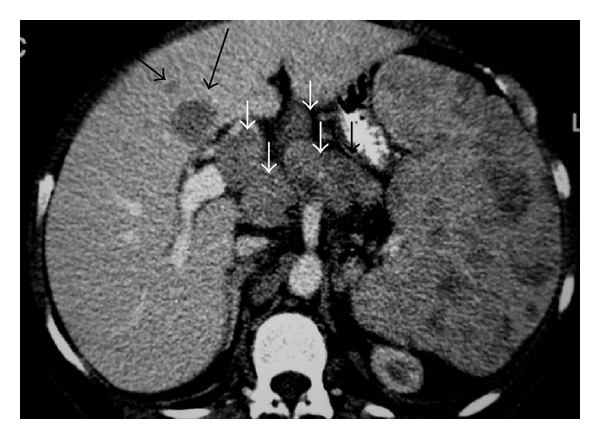
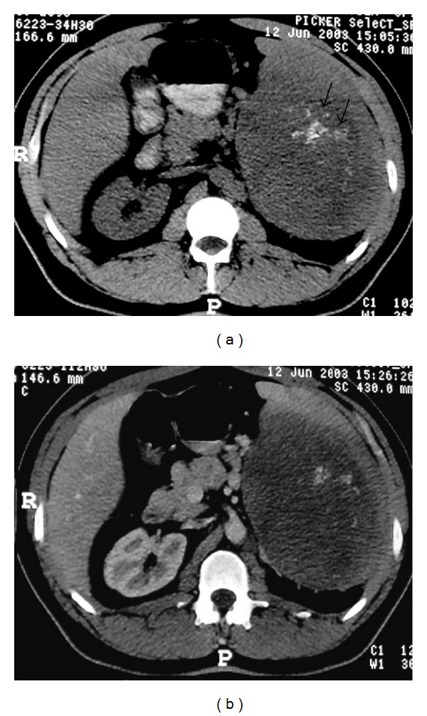
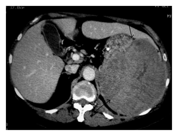
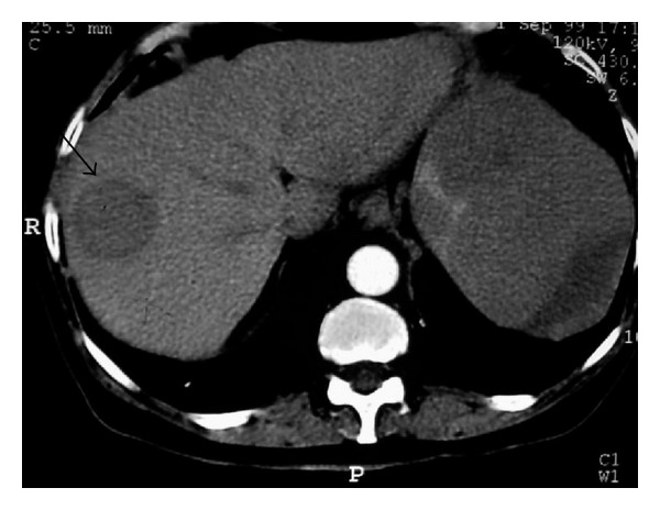
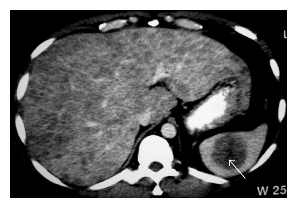
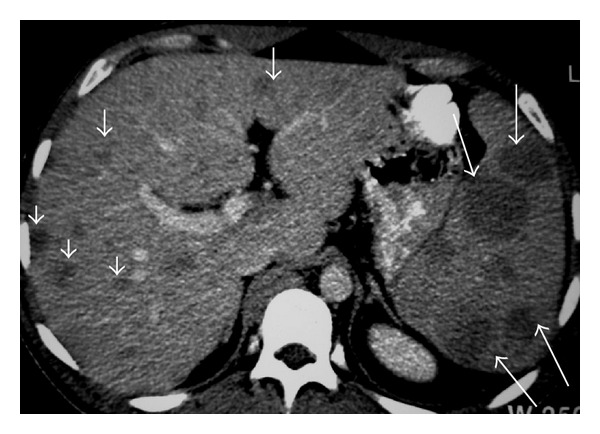
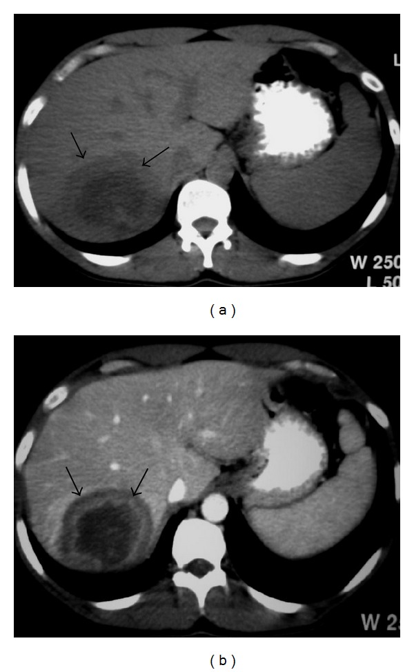
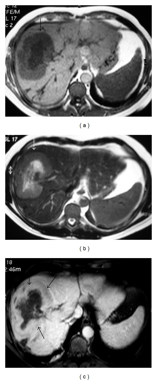
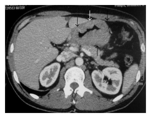
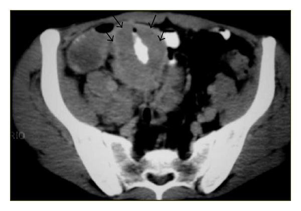
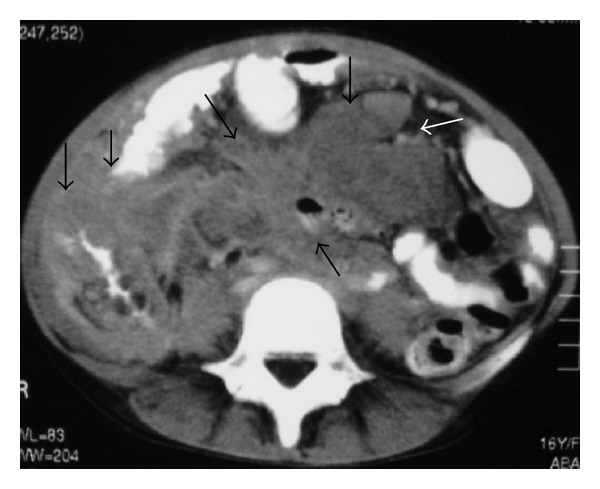
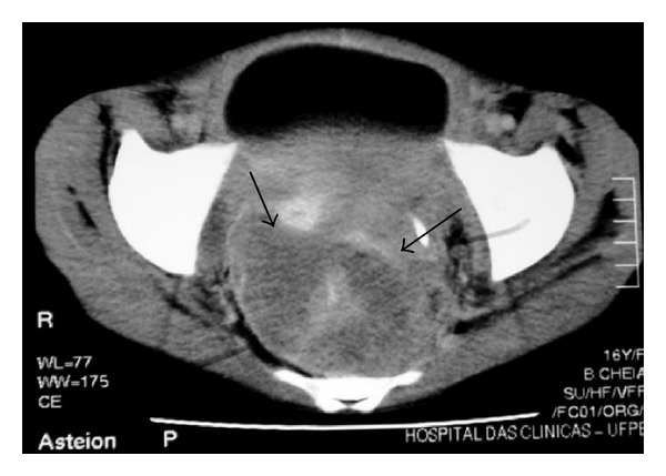
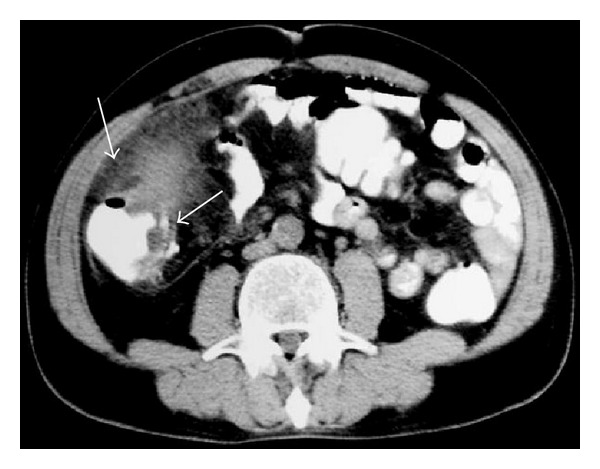
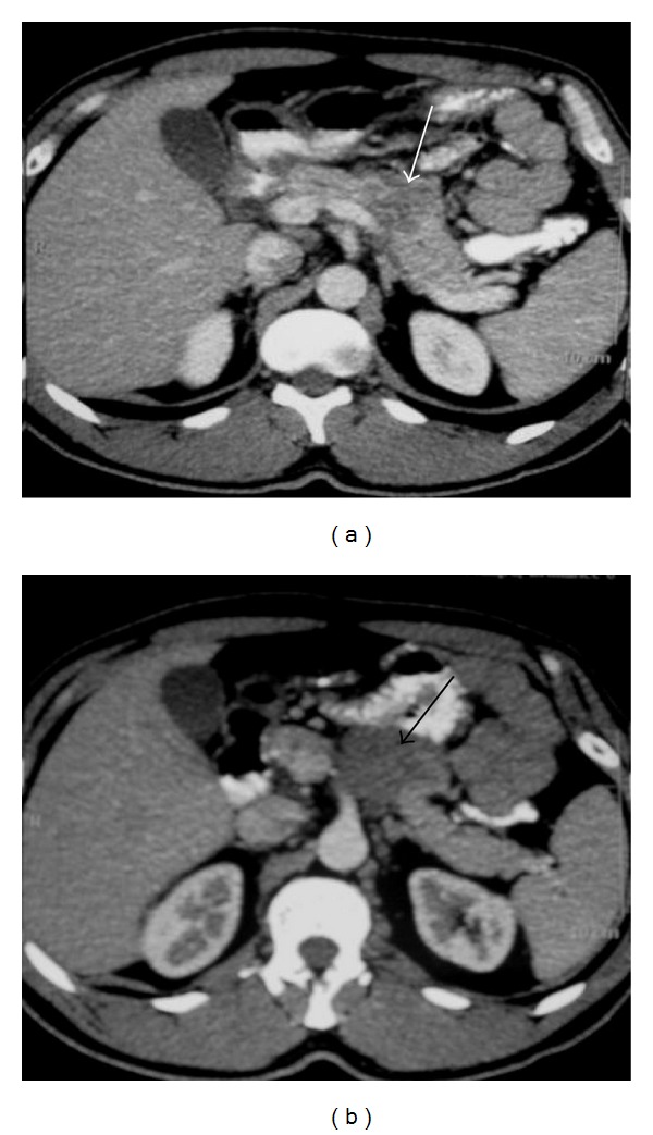
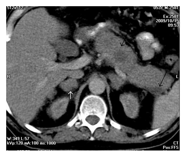
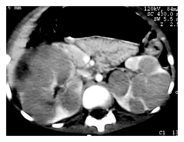
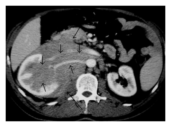
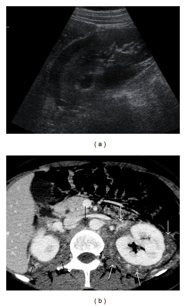
Non-Hodgkin and Hodgkin lymphomas frequently involve many structures in the abdomen and pelvis. Extranodal disease is more common with Non-Hodgkin's lymphoma than with Hodgkin's lymphoma. Though it may be part of a systemic lymphoma, single onset of nodal lymphoma is not rare. Extranodal lymphoma has been described in virtually every organ and tissue. In decreasing order of frequency, the spleen, liver, gastrointestinal tract, pancreas, abdominal wall, genitourinary tract, adrenal, peritoneal cavity, and biliary tract are involved. The purpose of this review is to discuss and illustrate the spectrum of appearances of nodal and extranodal lymphomas, including AIDS-related lymphomas, in the abdominopelvic region using a multimodality approach, especially cross-sectional imaging techniques. The most common radiologic patterns of involvement are illustrated. Familiarity with the imaging manifestations that are diagnostically specific for lymphoma is important because imaging plays an important role in the noninvasive management of disease.


Figures

Figure 1
Figure 1
CT of the abdomen demonstrates lymph nodes involving mesenteric vessels (sandwich sign) anterior to aorta and inferior to vena cava in a patient with lymphoma (arrows).
Figure 2
Figure 2
NHL. Axial contrast-enhanced abdominal CT shows retroperitoneal lymph nodes (arrows).
Figure 3
Figure 3
Nodal disease. Axial contrast-enhanced CT shows retroperitoneal lymph nodes (arrows).
Figure 4
Figure 4
A 53-year-old man with lymphoma. Axial contrast-enhanced CT demonstrates mesenteric lymph nodes.
Figure 5
Figure 5
NHL in a 53-year-old woman. Axial pelvic contrast-enhanced CT shows mesenteric lymph nodes (arrows).
Figure 6
Figure 6
NHL. Axial contrast-enhanced abdominal CT shows retroperitoneal lymph nodes (arrows).
Figure 7
Figure 7
NHL. (a) Nonenhanced axial abdominal CT shows left paraaortic node (arrows) with small calcifications. (b) Axial T2-weighted image demonstrates left paraaortic hypointense mass (arrows).
Figure 8
Figure 8
US of the spleen reveals splenomegaly.
Figure 9
Figure 9
Young man with NHL. Sonogram of the spleen shows multiple hypoechoic nodules (arrows).
Figure 10
Figure 10
NHL. Axial contrast-enhanced CT image demonstrates multiple low-attenuation nodules in the spleen.
Figure 11
Figure 11
NHL. Axial contrast-enhanced CT image shows multiple low-attenuation splenic nodules. Note multiple enlarged LN (small arrows) and hepatic lesions (long arrows).
Figure 12
Figure 12
A 40-year-old woman with lymphoblastic lymphoma. Axial contrast-enhanced abdominal CT shows splenomegaly with multiple low-attenuation nodules (arrows).
Figure 13
Figure 13
A 29-year-old man with NHL involving the spleen. Axial abdominal CT before (a) and after administration of intravenous CM (b) showing a large mass in the spleen (long arrows) with calcifications (small arrows).
Figure 14
Figure 14
NHL involving the spleen. Axial abdominal CT after administration of intravenous CM reveals a large mass in spleen (arrows).
Figure 15
Figure 15
A 40-year-old woman with lymphoblastic lymphoma. Axial contrast-enhanced abdominal CT demonstrates circumscribed low-attenuation mass in the liver (arrow). Hypodense lesions are also seen in spleen.
Figure 16
Figure 16
NHL. Axial contrast-enhanced CT image reveals multiple small low-attenuation nodules in the liver. Note also hypodense mass involving the spleen (arrow).
Figure 17
Figure 17
NHL. Axial contrast-enhanced CT image shows low-attenuation nodules in the liver (short arrows) and spleen (long arrows).
Figure 18
Figure 18
A 44-year-old man with NHL involving liver. (a) Nonenhanced axial CT of the liver reveals heterogeneous mass with central hypoattenuation (arrows). (b) Axial contrast-enhanced CT shows peripheral enhancement of the lesion (arrows).
Figure 19
Figure 19
NHL. Axial liver MRI. (a) T1WI and (b) T2WI show heterogeneous mass (arrows) with central area of low signal on T1WI and high signal on T2WI (arrows). (c) The lesion shows enhancement.
Figure 20
Figure 20
A 33-year-old man with gastric lymphoma. Axial contrast-enhanced abdominal CT demonstrates irregular thickening of the wall of the stomach (arrows).
Figure 21
Figure 21
NHL. CT of the pelvis shows concentric thickening of small bowel loop on the right reducing its lumen (arrows).
Figure 22
Figure 22
NHL. Axial CT shows masses involving small bowel loops and ascending colon (arrows).
Figure 23
Figure 23
NHL in a 16-year-old girl. Axial contrast-enhanced CT of the pelvis reveals soft-tissue mass involving the rectal region (arrows).
Figure 24
Figure 24
NHL. CT shows soft-tissue mass involving the ascending colon (arrows).
Figure 25
Figure 25
CT images of a 44-year-old man with pancreatic lymphoma. (a) Axial contrast-enhanced abdominal CT shows hypodense lesion involving the body of the pancreas (arrow). (b) Axial contrast-enhanced CT at a lower level demonstrates the lobulated hypodense mass (arrow).
Figure 26
Figure 26
Lymphoma involving pancreas and adrenals. Axial contrast-enhanced CT demonstrates enlarged pancreas with low-attenuation nodules (arrows) and enlarged adrenal glands (small arrows).
Figure 27
Figure 27
Burkitt lymphoma. Axial contrast-enhanced abdominal CT demonstrates multiple hypodense lesions involving the kidneys.
Figure 28
Figure 28
NHL. Axial contrast-enhanced CT reveals soft-tissue mass involving the right kidney (long arrows) with lymphomatous infiltration of the renal sinus and encasement of vessels (short arrows).
Figure 29
Figure 29
NHL. (a) Right kidney sonogram shows hypoechoic perinephric mass (arrows). (b) Axial contrast-enhanced CT scan shows a large soft-tissue mass (arrows) infiltrating the retroperitoneum and extending into the perinephric space bilaterally.
Figure 30
Figure 30
Lymphoma involving both kidneys. Axial gadolinium-enhanced T1-WI demonstrates lymphomatous infiltration of the retroperitoneum with extension to perinephric space bilaterally.
Figure 31
Figure 31
Adrenal lymphoma. Axial unenhanced CT (a) shows irregular large bilateral adrenal glands. MR axial images (b) in phase and (c) out of phase demonstrate enlarged adrenal glands.
Figure 32
Figure 32
Patient with peritoneal lymphomatosis. Axial contrast-enhanced CT image shows ascites and infiltration of mesenteric fat.

References

    1. Kwee TC, Kwee RM, Nievelstein RAJ. Imaging in staging of malignant lymphoma: a systematic review. Blood. 2008;111(2):504–516. - PubMed
    1. Lee W-K, Lau EWF, Duddalwar VA, Stanley AJ, Ho YY. Abdominal manifestations of extranodal lymphoma: spectrum of imaging findings. American Journal of Roentgenology. 2008;191(1):198–206. - PubMed
    1. Yu R-S, Zhang W-M, Liu Y-Q. CT diagnosis of 52 patients with lymphoma in abdominal lymph nodes. World Journal of Gastroenterology. 2006;12(48):7869–7873. - PMC - PubMed
    1. Metser U, Goor O, Lerman H, Naparstek E, Even-Sapir E. PET-CT of extranodal lymphoma. American Journal of Roentgenology. 2004;182(6):1579–1586. - PubMed
    1. Guermazi A, Brice P, De Kerviler E, et al. Extranodal Hodgkin disease: spectrum of disease. Radiographics. 2001;21(1):161–179. - PubMed

LinkOut - more resources