

Detection of Early Ischemic Changes in Noncontrast CT Head Improved with "Stroke Windows"
- PMID: 24967315
- PMCID: PMC4045559
- DOI: 10.1155/2014/654980
Detection of Early Ischemic Changes in Noncontrast CT Head Improved with "Stroke Windows"
Abstract
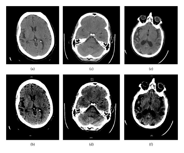
Introduction. Noncontrast head CT (NCCT) is the standard radiologic test for patients presenting with acute stroke. Early ischemic changes (EIC) are often overlooked on initial NCCT. We determine the sensitivity and specificity of improved EIC detection by a standardized method of image evaluation (Stroke Windows). Methods. We performed a retrospective chart review to identify patients with acute ischemic stroke who had NCCT at presentation. EIC was defined by the presence of hyperdense MCA/basilar artery sign; sulcal effacement; basal ganglia/subcortical hypodensity; and loss of cortical gray-white differentiation. NCCT was reviewed with standard window settings and with specialized Stroke Windows. Results. Fifty patients (42% females, 58% males) with a mean NIHSS of 13.4 were identified. EIC was detected in 9 patients with standard windows, while EIC was detected using Stroke Windows in 35 patients (18% versus 70%; P < 0.0001). Hyperdense MCA sign was the most commonly reported EIC; it was better detected with Stroke Windows (14% and 36%; P < 0.0198). Detection of the remaining EIC also improved with Stroke Windows (6% and 46%; P < 0.0001). Conclusions. Detection of EIC has important implications in diagnosis and treatment of acute ischemic stroke. Utilization of Stroke Windows significantly improved detection of EIC.
Figures
Similar articles
-
Preliminary study of time maximum intensity projection computed tomography imaging for the detection of early ischemic change in patient with acute ischemic stroke.Medicine (Baltimore). 2018 Mar;97(9):e9906. doi: 10.1097/MD.0000000000009906. Medicine (Baltimore). 2018. PMID: 29489691 Free PMC article.
-
Time dependence of reliability of noncontrast computed tomography in comparison to computed tomography angiography source image in acute ischemic stroke.Int J Stroke. 2015 Jan;10(1):55-60. doi: 10.1111/j.1747-4949.2012.00859.x. Epub 2012 Sep 13. Int J Stroke. 2015. PMID: 22974504
-
The CT-Defined Hyperdense Arterial Sign as a Marker for Acute Intracerebral Large Vessel Occlusion.J Neuroimaging. 2018 Mar;28(2):212-216. doi: 10.1111/jon.12484. Epub 2017 Nov 14. J Neuroimaging. 2018. PMID: 29134723
-
Associations between early ischemic signs on non-contrast CT and time since acute ischemic stroke onset: A scoping review.Eur J Radiol. 2022 Oct;155:110455. doi: 10.1016/j.ejrad.2022.110455. Epub 2022 Jul 26. Eur J Radiol. 2022. PMID: 35973304
-
ASPECTS CT in Acute Ischemia: Review of Current Data.Top Magn Reson Imaging. 2017 Jun;26(3):103-112. doi: 10.1097/RMR.0000000000000122. Top Magn Reson Imaging. 2017. PMID: 28277460 Review.
Cited by
-
Accuracy and time efficiency of a novel deep learning algorithm for Intracranial Hemorrhage detection in CT Scans.Radiol Med. 2024 Oct;129(10):1499-1506. doi: 10.1007/s11547-024-01867-y. Epub 2024 Aug 9. Radiol Med. 2024. PMID: 39123064 Free PMC article.
-
Ischemic infarct detection, localization, and segmentation in noncontrast CT human brain scans: review of automated methods.PeerJ. 2020 Dec 18;8:e10444. doi: 10.7717/peerj.10444. eCollection 2020. PeerJ. 2020. PMID: 33391867 Free PMC article.
-
A Review on Computer Aided Diagnosis of Acute Brain Stroke.Sensors (Basel). 2021 Dec 20;21(24):8507. doi: 10.3390/s21248507. Sensors (Basel). 2021. PMID: 34960599 Free PMC article. Review.
-
Endovascular thrombectomy with or without intravenous alteplase in large-core ischemic stroke: a systematic review and meta-analysis.Neurol Sci. 2024 Nov;45(11):5129-5140. doi: 10.1007/s10072-024-07653-y. Epub 2024 Jun 19. Neurol Sci. 2024. PMID: 38896187
-
Computed tomography patterns of intracranial infarcts in a Ghanaian tertiary facility.Ghana Med J. 2022 Mar;56(1):28-37. doi: 10.4314/gmj.v56i1.5. Ghana Med J. 2022. PMID: 35919779 Free PMC article.
References
-
- Lev MH, Farkas J, Gemmete JJ, et al. Acute stroke: improved nonenhanced CT detection—benefits of soft-copy interpretation by using variable window width and center level settings. Radiology. 1999;213(1):150–155. - PubMed
-
- Tanne D, Kasner SE, Demchuk AM, et al. Markers of increased risk of intracerebral hemorrhage after intravenous recombinant tissue plasminogen activator therapy for acute ischemic stroke in clinical practice: the multicenter rt-PA acute stroke survey. Circulation. 2002;105(14):1679–1685. - PubMed
-
- Derex L, Nighoghossian N. Intracerebral haemorrhage after thrombolysis for acute ischaemic stroke: an update. Journal of Neurology, Neurosurgery and Psychiatry. 2008;79(10):1093–1099. - PubMed
-
- Barber PA, Demchuk AM, Zhang J, Buchan AM. Validity and reliability of a quantitative computed tomography score in predicting outcome of hyperacute stroke before thrombolytic therapy. The Lancet. 2000;355(9216):1670–1674. - PubMed
-
- Truwit CL, Barkovich AJ, Gean-Marton A, Hibri N, Norman D. Loss of the insular ribbon: another early CT sign of acute middle cerebral artery infarction. Radiology. 1990;176(3):801–806. - PubMed
LinkOut - more resources
Full Text Sources
Other Literature Sources