. 2014 Jun 26;9(6):e101071.
doi: 10.1371/journal.pone.0101071. eCollection 2014.

Vimentin mediates uptake of C3 exoenzyme

Affiliations

Vimentin mediates uptake of C3 exoenzyme

Astrid Rohrbeck et al. PLoS One. .

Abstract

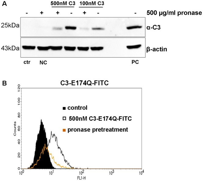
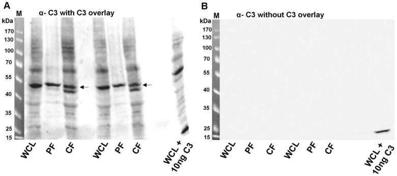
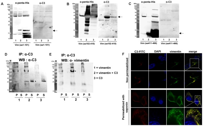
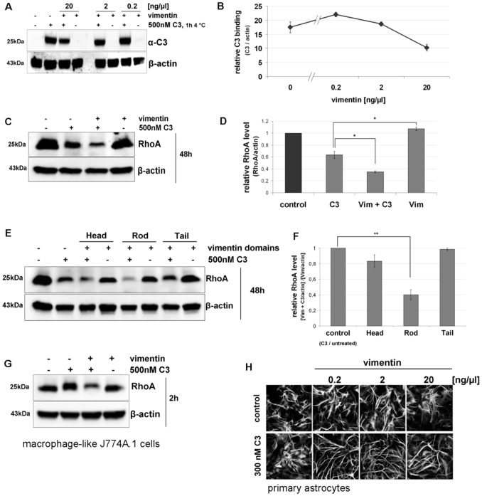
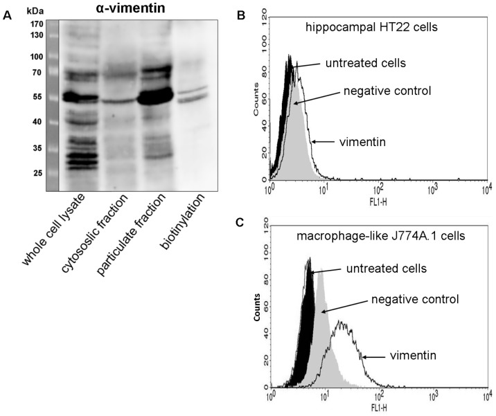
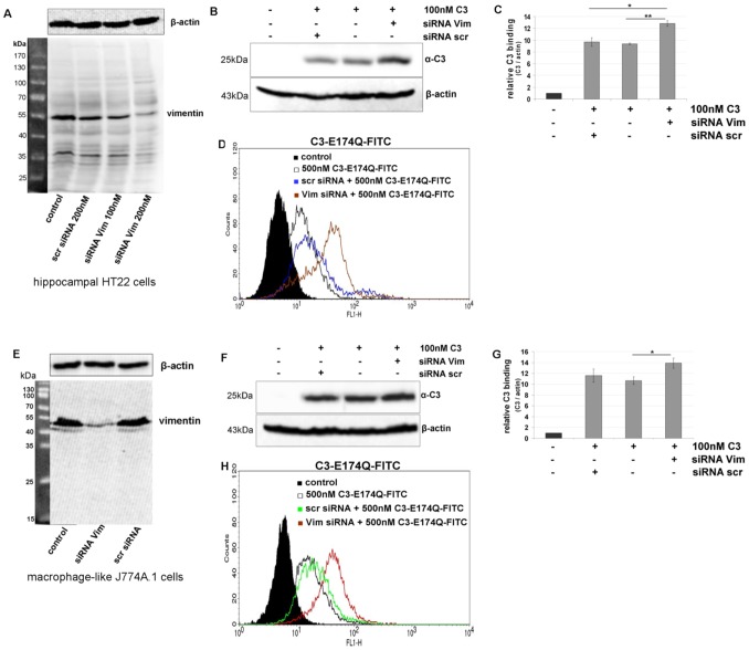
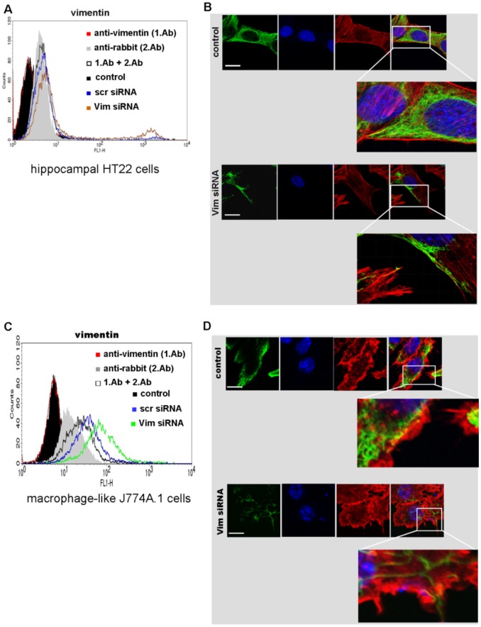
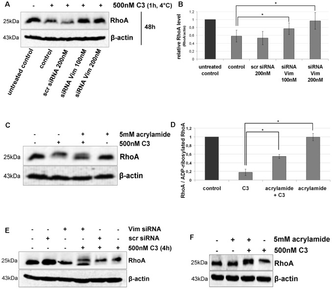
Clostridium botulinum C3 exoenzyme (C3) selectively inactivates RhoA/B/C GTPases by ADP-ribosylation. Based on this substrate specificity C3 is a well-established tool in cell biology. C3 is taken up by eukaryotic cells although lacking an uptake and translocation domain. Based on different approaches vimentin was identified as membranous C3-interaction partner by mass spectrometry. Vimentin in fact was partly localized at the outer surface of hippocampal HT22 cells and J744A.1 macrophages. Domain analysis identified the rod domain as binding partner of C3. Vimentin was also involved in uptake of C3 as shown by knock down of vimentin in HT22 and J774A.1 cells. The involvement of vimentin in uptake of C3 was further supported by the findings that the vimentin disruptor acrylamide blocked uptake of C3. Vimentin is not only a major organizing element of the intermediate filament network but is also involved in both binding and uptake of C3 exoenzyme.


Conflict of interest statement

Competing Interests: The authors have declared that no competing interests exist.

Figures

Figure 1
Figure 1. Binding of C3 to HT22 cells after pronase treatment.
A) Pronase pre-incubated HT22 cells were exposed to 100 or 500 nM of C3 for 1 h at 4°C. Subsequently, β-actin and bound C3 were detected by Western blot. NC = negative control without C3, PC = positive control lysate with 10 ng C3. One representative experiment is shown (n = 3 independent experiments). B) Pronase-treated HT22 cells were exposed to 500 nM of C3-E174Q-FITC for 1 h at 4°C and bound C3- E174Q-FITC was analyzed by FACS.
Figure 2
Figure 2. C3-overlay (binding of C3 to HT22 proteins).
A) Whole cell lysate, cytosolic fraction or particulate fraction from HT22 cells were generated as described in material and methods followed by separation through SDS-PAGE and transfer onto nitrocellulose. Nitrocellulose was incubated with 10 µg/ml of C3 for 60 min at 4°C. After washing bound C3 was detected by anti-C3. Arrows indicate the protein of interest (55 kDa). B) The right panel shows the anti-C3 Western blot without C3-overlay. M = molecular mass marker, WCL = whole cell lysate, PF = particulate fraction, CF = cytosolic fraction, WCL +10 ng C3 = C3 was added to whole cells lysate prior to SDS-PAGE and blotting to generate a positive C3 signal.
Figure 3
Figure 3. Binding of C3 to the rod domain of vimentin.
His-tagged vimentin domains (A) head: amino acids 1 to 101; (B) rod: amino acids 102 to 410; (C) tail: amino acids 411 to 466 were expressed in E. coli. Crude extracts from non-induced E. coli (lane 1), crude extracts from induced E. coli (lane 2) and His-tagged vimentin domains purified through Ni-IDA column (lane 3) were separated by 15% SDS-PAGE and electroblotted. Left panels of A, B and C show probing with penta-His polyclonal antibody to detect vimentin domains. Right panels of A, B and C show probing with anti-C3 to detect bound C3. Predicted apparent mol masses of each vimentin domain are indicated by arrow. Arrows indicate the protein of interest. D) Immunoprecipitation of C3 with C3 antibody. Full length recombinant vimentin was incubated with C3 in solution. The vimentin-C3 complex was immunoprecipitated with C3 antibody. P = pellet of agarose beads, S = supernatant. E) Immunoprecipitation of vimentin with C3 antibody and detection of vimentin. P = pellet of agarose beads, S = supernatant. F) For imaging vimentin and C3 at the cell surface (upper panel), cells were incubated with C3-FITC for 1 h at 4°C. After washing with PBS cells were fixed (without permeabilization) and incubated with vimentin antibody following by an Alexa-555 conjugated secondary antibody. For imaging intracellular vimentin (lower panel), cells were fixed, permeabilized and incubated as described above. For imaging intracellular C3, living cells were incubated with C3-FITC for 1 h at 37°C. After washing with PBS cells were fixed, permeabilized with 0.02% saponin for 30 min and imaged by confocal microscopy.
Figure 4
Figure 4. Binding and uptake of C3 in cultivated cells dependent on vimentin.
A) The Western blot shows the binding of C3 in presence and absence of extracellular added vimentin (n = 3 independent experiments). B) Densitometric evaluation of C3 (from A) and adjustment to the corresponding actin band are shown. C) The Western blot shows the degradation of RhoA as marker for C3 uptake and Rho-ADP-ribosylation. HT22 cells were treated with C3 (500 nM) alone or C3 (500 nM) plus vimentin (1 ng/µl) for 48 h. Cell lysates were submitted to Western blot analysis probing RhoA and β-actin. One representative Western blot experiment is shown (n = 3 independent experiments). D) Densitometric evaluation of RhoA (from C) and adjustment to the corresponding actin band are shown; the bars give the relative RhoA level. E) HT22 cells were incubated with C3 (500 nM) or C3 (500 nM) plus 1 ng/µl of either vimentin head-, rod- or tail-domain for 48 h. Cell lysates were submitted to Western blot analysis probing RhoA and β-actin. Decreased signal of RhoA reflects degradation of RhoA after ADP-ribosylation and thus, enhanced C3 uptake. One representative Western blot experiment is shown (n = 3 independent experiments). F) Densitometric evaluation of RhoA (from E) and adjustment to the corresponding actin band are shown; the bars give the relative RhoA level. G) J774A.1 macrophages were treated with C3 (500 nM) alone or C3 (500 nM) plus vimentin (1 ng/µl) for 2 h. Cell lysates were submitted to Western blot analysis probing RhoA and β-actin. One representative Western blot experiment is shown (n = 3 independent experiments). H) Primary astrocytes were exposed to C3 (300 nM) alone or a combination of C3 (300 nM) with different concentrations of vimentin (0.2, 2 and 20 ng/µl) for 6 h at 37°C. After incubation time the astrocytes were stained for the intermediate filament protein GFAP to visualize morphological changes.
Figure 5
Figure 5. Vimentin is present at the cell surface of HT22 cells and J774A.1 macrophages.
A) Intact HT22 cells were biotinylated for 1 h at 4°C. Whole cell lysates, cytosolic and particulate fractions were prepared. In addition, cell surface biotinylated proteins were enriched by precipitation with NeutrAvidin beads. The fractions and precipitation, respectively, were immunoblotted and probed with anti-vimentin. Biotinylation fraction represents the extracellular proteins exclusively. One representative Western blot experiment is shown (n = 3 independent experiments). Presence of vimentin at the cell surface of HT22 cells (B) and J774A.1 cells (C) was analyzed by FACS cytometry using anti-vimentin. Oregon green-488 conjugated goat anti-rabbit antibody alone served as negative control. Untreated cells were used as control.
Figure 6
Figure 6. Knock down of vimentin in hippocampal HT22 cells and J774A.1 macrophages.
A) HT22 cells were transfected with siRNA for 48 h (scr = scrambled, Vim = vimentin). Vimentin and β-actin were detected by Western blot analysis of cell lysates. B) After siRNA transfection for 48 h, HT22 cells were exposed to C3 (100 nM) for 1 h at 4°C. Bound C3 was detected in Western blot with anti-C3. β-actin was used as internal control. C) Densitometric evaluation of bound C3 (from B) and adjustment to the corresponding actin band are shown; the bars give the relative C3 binding. D) HT22 cells transfected with siRNA for 48 h were incubated with C3-E174Q-FITC (500 nM) for 1 h at 4°C and bound C3-E174Q-FITC was analyzed by FACS cytometry. E – G) Same experiments for J774A.1 macrophages. E) Knock down of vimentin. F) Binding of C3 to cells with vimentin knock down. G) Densitometric evaluation of F. H) Binding of C3-E174Q-FITC to cells with vimentin knock down and FACS analysis.
Figure 7
Figure 7. Analysis of vimentin distribution in analyzed cells.
A) Vimentin was detected by anti-vimentin at the cell surface of HT22 cells transfected with siRNA for 48 h at 37°C by FACS analysis. B) Confocal microscopy of vimentin in HT22 cells transfected for 48 h at 37°C. The green (oregon green 488) anti-vimentin, DNA staining in blue (Dapi), rhodamine red-staining for actin and a merge image are shown for each panel. In the enlarged images the cell boundaries are shown. Significant difference between the vimentin distribution was detected for the transfected cells (lower panel) in comparison to the control (upper panel). Scale bar = 20 µM. C) Detection of vimentin at the cell surface of J774A.1 cells transfected with siRNA. D) Confocal microscopy of vimentin distribution in J774A.1 cells transfected for 48 h.
Figure 8
Figure 8. Uptake of C3 in HT22 and J744A.1 cells is dependent on vimentin distribution and integrity.
A) Influence of Vim-siRNA knock down (for 48 h) on the uptake of C3 into HT22 cells detected as RhoA degradation (induced by C3-catalysed ADP-ribosylation). In a pulse-chase experiment, HT22 cells were incubated with C3 (500 nM) at 4°C for 60 min. Afterwards unbound C3 was removed by washing the cells three times with PBS and fresh medium was added. Cells were then cultivated for further 48 h. Cell lysates were generated and separated by SDS-PAGE followed by Western blot analysis probing RhoA and β-actin. One representative experiment is shown (n = 3 independent experiments). B) Cellular levels of RhoA proteins were quantified by densitometric evaluation of RhoA (from A) and adjusted to the corresponding actin band. C) HT22 cells were pre-treated with acrylamide (5 mM) for 30 min followed by incubation with C3 (500 nM) for 24 h. Cells were lysed and submitted to Western blot analysis probing RhoA and β-actin. C3 alone causes a complete mol weight shift of RhoA in SDS-PAGE. Western blot analysis of one representative experiment is shown (n = 3 independent experiments). D) RhoA shift (indicative of Rho-ADP-ribosylation) by quantified by densitometric evaluation of RhoA (from C) and adjusted to the corresponding β-actin signal. E) Influence of Vim-siRNA knock down (for 48 h) on the uptake of C3 into J774A.1 cells detected as incomplete RhoA ADP-ribosylation. J774A.1 macrophages were incubated with C3 (500 nM) at 37°C for 4 h. Cell lysates were generated and separated by SDS-PAGE followed by Western blot analysis probing RhoA and β-actin. One representative experiment is shown (n = 3 independent experiments). F) J774A.1 cells were pre-treated with acrylamide (5 mM) for 30 min followed by incubation with C3 (500 nM) for 4 h. Cells were lysed and submitted to Western blot analysis probing RhoA and β-actin.

Similar articles

Cited by

References

    1. Aktories K, Frevert J (1987) ADP-ribosylation of a 21–24 kDa eukaryotic protein(s) by C3, a novel botulinum ADP-ribosyltransferase, is regulated by guanine nucleotide. Biochem J 247: 363–368. - PMC - PubMed
    1. Just I, Selzer J, Jung M, van Damme J, Vandekerckhove J, et al. (1995) Rho-ADP-ribosylating exoenzyme from Bacillus cereus. Purification, characterization, and identification of the NAD-binding site. Biochemistry 34: 334–340 10.1021/bi00001a041 - DOI - PubMed
    1. Wilde C, Chhatwal GS, Schmalzing G, Aktories K, Just I (2001) A novel C3-like ADP-ribosyltransferase from Staphylococcus aureus modifying RhoE and Rnd3. J Biol Chem 276: 9537–42 10.1074/jbc.M011035200 - DOI - PubMed
    1. Sehr P, Joseph G, Genth H, Just I, Pick E, et al. (1998) Glucosylation and ADP-ribosylation of Rho proteins: Effects on nucleotide binding, GTPase activity, and effector coupling. Biochemistry 37: 5296–5304 10.1021/bi972592c - DOI - PubMed
    1. Genth H, Gerhard R, Maeda A, Amano M, Kaibuchi K, et al. (2003) Entrapment of rho ADP-ribosylated by Clostridium botulinum C3 exoenzyme in the Rho-guanine nucleotide dissociation inhibitor-1 complex. J Biol Chem 278: 28523–28527 10.1074/jbc.M301915200 - DOI - PubMed

Publication types

MeSH terms

LinkOut - more resources