

Fractal dimension of EEG activity senses neuronal impairment in acute stroke
- PMID: 24967904
- PMCID: PMC4072666
- DOI: 10.1371/journal.pone.0100199
Fractal dimension of EEG activity senses neuronal impairment in acute stroke
Abstract
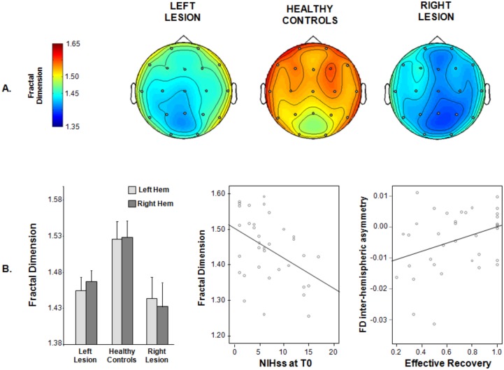
The brain is a self-organizing system which displays self-similarities at different spatial and temporal scales. Thus, the complexity of its dynamics, associated to efficient processing and functional advantages, is expected to be captured by a measure of its scale-free (fractal) properties. Under the hypothesis that the fractal dimension (FD) of the electroencephalographic signal (EEG) is optimally sensitive to the neuronal dysfunction secondary to a brain lesion, we tested the FD's ability in assessing two key processes in acute stroke: the clinical impairment and the recovery prognosis. Resting EEG was collected in 36 patients 4-10 days after a unilateral ischemic stroke in the middle cerebral artery territory and 19 healthy controls. National Health Institute Stroke Scale (NIHss) was collected at T0 and 6 months later. Highuchi FD, its inter-hemispheric asymmetry (FDasy) and spectral band powers were calculated for EEG signals. FD was smaller in patients than in controls (1.447±0.092 vs 1.525±0.105) and its reduction was paired to a worse acute clinical status. FD decrease was associated to alpha increase and beta decrease of oscillatory activity power. Larger FDasy in acute phase was paired to a worse clinical recovery at six months. FD in our patients captured the loss of complexity reflecting the global system dysfunction resulting from the structural damage. This decrease seems to reveal the intimate nature of structure-function unity, where the regional neural multi-scale self-similar activity is impaired by the anatomical lesion. This picture is coherent with neuronal activity complexity decrease paired to a reduced repertoire of functional abilities. FDasy result highlights the functional relevance of the balance between homologous brain structures' activities in stroke recovery.
Conflict of interest statement
Figures
Similar articles
-
Prognostic Value of EEG Microstates in Acute Stroke.Brain Topogr. 2017 Sep;30(5):698-710. doi: 10.1007/s10548-017-0572-0. Epub 2017 May 25. Brain Topogr. 2017. PMID: 28547185
-
Longitudinal quantitative electroencephalographic study in mono-hemispheric stroke patients.Neural Regen Res. 2019 Jul;14(7):1237-1246. doi: 10.4103/1673-5374.251331. Neural Regen Res. 2019. PMID: 30804255 Free PMC article.
-
Elevated EEG fractal dimension in Parkinson's during timing tasks.Chaos. 2025 Jun 1;35(6):063107. doi: 10.1063/5.0274411. Chaos. 2025. PMID: 40455198
-
A contralesional EEG power increase mediated by interhemispheric disconnection provides negative prognosis in acute stroke.Restor Neurol Neurosci. 2013;31(2):177-88. doi: 10.3233/RNN-120244. Restor Neurol Neurosci. 2013. PMID: 23254689
-
Fractal Dimension Analysis in Neurological Disorders: An Overview.Adv Neurobiol. 2024;36:313-328. doi: 10.1007/978-3-031-47606-8_16. Adv Neurobiol. 2024. PMID: 38468040 Review.
Cited by
-
Magnetoencephalography in the study of brain dynamics.Funct Neurol. 2014 Oct-Dec;29(4):241-53. Funct Neurol. 2014. PMID: 25764254 Free PMC article. Review.
-
Comprehensive Analysis of Feature Extraction Methods for Emotion Recognition from Multichannel EEG Recordings.Sensors (Basel). 2023 Jan 12;23(2):915. doi: 10.3390/s23020915. Sensors (Basel). 2023. PMID: 36679710 Free PMC article.
-
Electroencephalographic Fractal Dimension in Healthy Ageing and Alzheimer's Disease.PLoS One. 2016 Feb 12;11(2):e0149587. doi: 10.1371/journal.pone.0149587. eCollection 2016. PLoS One. 2016. PMID: 26872349 Free PMC article.
-
The EEG Split Alpha Peak: Phenomenological Origins and Methodological Aspects of Detection and Evaluation.Front Neurosci. 2017 Sep 12;11:506. doi: 10.3389/fnins.2017.00506. eCollection 2017. Front Neurosci. 2017. PMID: 28955192 Free PMC article.
-
Cortical gamma-synchrony measured with magnetoencephalography is a marker of clinical status and predicts clinical outcome in stroke survivors.Neuroimage Clin. 2019;24:102092. doi: 10.1016/j.nicl.2019.102092. Epub 2019 Nov 12. Neuroimage Clin. 2019. PMID: 31795062 Free PMC article.
References
-
- Cannon WB (1929) Organization For Physiological Homeostasis. Physiol Rev 9: 399–431.
-
- Meyer M, Stiedl O (2003) Self-affine fractal variability of human heartbeat interval dynamics in health and disease. Eur J Appl Physiol 90: 305–316. - PubMed
-
- Fadel PJ, Barman SM, Phillips SW, Gebber GL (2004) Fractal fluctuations in human respiration. J Appl Phys 97: 2056–2064. - PubMed
-
- Hausdorff JM, Ashkenazy Y, Peng CK, Ivanov PC, Stanley HE, et al. (2001) When human walking becomes random walking: fractal analysis and modeling of gait rhythm fluctuations. Physica A 302: 138–147. - PubMed
Publication types
MeSH terms
LinkOut - more resources
Full Text Sources
Other Literature Sources
Medical
