. 2014 Jun 26;5(6):e1306.
doi: 10.1038/cddis.2014.272.

Dysferlin regulates cell membrane repair by facilitating injury-triggered acid sphingomyelinase secretion

Affiliations

Dysferlin regulates cell membrane repair by facilitating injury-triggered acid sphingomyelinase secretion

A Defour et al. Cell Death Dis. .

Abstract

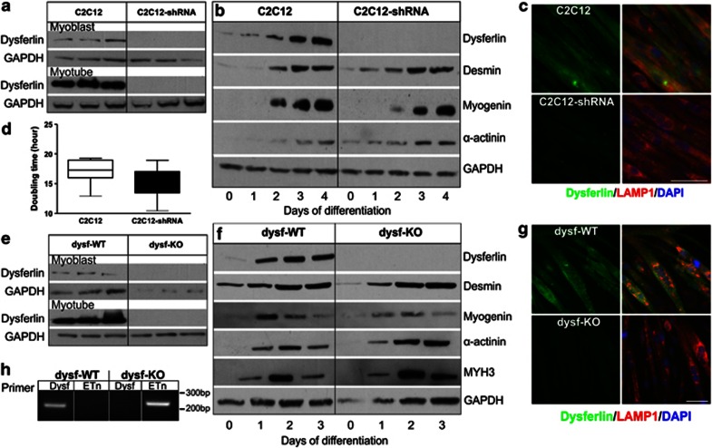
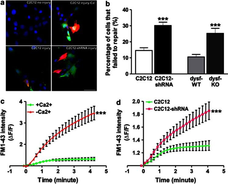
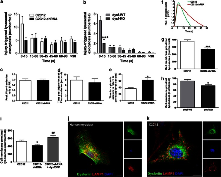
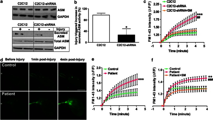
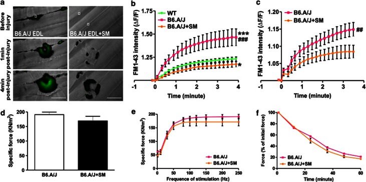
Dysferlin deficiency compromises the repair of injured muscle, but the underlying cellular mechanism remains elusive. To study this phenomenon, we have developed mouse and human myoblast models for dysferlinopathy. These dysferlinopathic myoblasts undergo normal differentiation but have a deficit in their ability to repair focal injury to their cell membrane. Imaging cells undergoing repair showed that dysferlin-deficit decreased the number of lysosomes present at the cell membrane, resulting in a delay and reduction in injury-triggered lysosomal exocytosis. We find repair of injured cells does not involve formation of intracellular membrane patch through lysosome-lysosome fusion; instead, individual lysosomes fuse with the injured cell membrane, releasing acid sphingomyelinase (ASM). ASM secretion was reduced in injured dysferlinopathic cells, and acute treatment with sphingomyelinase restored the repair ability of dysferlinopathic myoblasts and myofibers. Our results provide the mechanism for dysferlin-mediated repair of skeletal muscle sarcolemma and identify ASM as a potential therapy for dysferlinopathy.


Figures

Figure 1
Figure 1
Dysferlin deficiency does not alter myoblast proliferation or differentiation. (a and e) Western blot analysis of dysferlin expression in three independent cultures each of myoblasts and myotubes (following 4 days of differentiation): (a) C2C12 and C2C12-shRNA and (e) dysf-WT and dysf-KO myoblasts. (b and f) Western blot analysis of expression of various muscle differentiation markers tested following the indicated days of differentiation in (b) C2C12 and C2C12-shRNA and (f) dysf-WT and dysf-KO myoblasts. (c and g) Immunostaining of dysferlin (green) and LAMP1 (red) in (c) C2C12 and C2C12-shRNA and (g) dysf-WT and dysf-KO myotubes following 4 days of differentiation. Scale bar: 50 μm. (d) Plot showing doubling time for C2C12 and C2C12-shRNA myoblasts in culture during 4 days of proliferation (n=8 cell cultures; error bars show the extreme values). (h) PCR analysis of genomic DNA from dysf-WT and dysf-KO myoblasts using primers to identify the presence of the full-length dysferlin allele (Dysf) and the dysferlin allele with the insertion of a retrotransposon (Etn) in the dysferlin gene
Figure 2
Figure 2
Dysferlinopathic myoblasts show poor cell membrane repair. (a) C2C12 cells injured in the presence of FITC-dextran (green) were allowed to undergo repair in TRITC-dextran (red) and then stained with Hoechst 33342 (blue). Uninjured cells are not labeled with dextran (upper left panel); injured cells are labeled green, and cells that failed to repair themselves are labeled red and green. Upper panels show uninjured myoblasts and myoblasts that failed to repair themselves because of a lack of Ca2+. Lower panel shows C2C12 and C2C12-shRNA myoblasts injured and allowed to repair themselves in the presence of Ca2+; Scale bar: 50 μm. (b) Images of glass bead-injured cells were quantified (>100 cells each) and are presented as the fraction of cells that failed to show repair in the presence of Ca2+. (c) FM1-43 influx following focal laser injury in >10 myoblasts injured in the presence of Ca2+ shows efficient repair from laser injury; cells injured in the absence of Ca2+ do not. (d) Quantification of FM1-43 influx by time-lapse imaging of >15 myoblasts each following injury in the presence of Ca2+, showing poor healing of C2C12-shRNA myoblasts. Data in b, c and d show means±S.E.M.; ***P<0.001 by unpaired Student's t-test
Figure 3
Figure 3
Dysferlin deficiency reduces injury-triggered lysosomal exocytosis. (a) dysf-WT and dysf-KO myoblasts wounded in the presence of Ca2+ by glass beads in medium containing TRITC-dextran were stained for cell-surface LAMP1 (left panel); White arrows indicate the injured cells labeled (red) by TRITC dextran. Scale bar: 50 μm. (b and c) Reduced cell-surface LAMP1 staining in dysferlinopathic myoblasts following injury in the presence of Ca2+: cell-surface LAMP1 staining was quantified for >400 dysf-WT, dysf-KO, C2C12, and C2C12-shRNA myoblasts each. (d and f) TIRF images showing dysf-WT myoblasts with FITC-dextran-labeled lysosomes. ‘X' marks the sites of lysosome fusion following (d) laser injury or (f) ionomycin treatment. The orange line denotes the cell boundary. The inset in d shows a zoomed view of one lysosome as it underwent fusion, releasing FITC-dextran. Scale bar: 10 μm. (e and g) Quantification of lysosome fusions in >13 dysf-WT and dysf-KO myoblasts, showing a reduced number of fusion events in dysferlinopathic myoblasts following (e) laser injury (168 lysosome fusions for dys-WT and 88 for dys-KO) and (g) ionophore treatment (264 fusions for dys-WT and 112 for dys-KO). (h and i) Localization of lysosome exocytosis sites (168 for dys-WT, 88 for dys-KO, 350 for C2C12, and 385 for C2C12-shRNA) with respect to the site of injury in (h) C2C12 or (i) primary myoblasts (n≥10 cells each). Data are mean±S.E.M. * P<0.05 and ***P<0.001 by unpaired Student's t-test
Figure 4
Figure 4
Dysferlin regulates tethering of lysosomes to the cell membrane and kinetics of injury-triggered lysosome exocytosis. (a and b) Histogram showing averaged kinetics of lysosome exocytosis following laser injury (n≥10 cells) in (a) C2C12 and (b) primary myoblasts. Note that it is only the earliest exocytic events that are reduced in dysferlinopathic myoblasts. (c) Quantification of the peak increase in Fluo-4 emission following laser injury in C2C12 and C2C12-shRNA myoblasts (n>7 cells). (d) Time taken following laser injury for the Fluo-4 intensity to reach the peak value (n>7 cells) in C2C12 cells. (e) Time following laser injury for the increase in Fluo-4 intensity to return to pre-injury level (n>7 cells). (f) Plot showing injury-triggered change in Fluo-4 intensity for a representative C2C12 and C2C12-shRNA cell. (g and h) Quantification of cell membrane-proximal lysosomes in (g) C2C12 and C2C12-shRNA and (h) dysf-WT and dysf-KO myoblasts (n≥20 cells each), with fewer cell membrane-proximal lysosomes in dysferlinopathic cells. (i) Quantification of cell membrane-proximal lysosomes, showing that transient expression of dysferlin-GFP in C2C12-shRNA myoblasts (n=10) increases the number of cell membrane-proximal lysosomes to a level similar to C2C12. (j) Representative confocal image of a human myoblast immunolabeled for endogenous dysferlin (green) and LAMP1 (red), showing little lysosomal localization of dysferlin. (k) Confocal image of a C2C12 myoblast expressing dysferlin-GFP also shows lack of lysosomal localization of dysferlin. Scale bar: 10 μm. All data are means±S.E.M. *P<0.05 and ***P<0.001 for the comparison of mutant and corresponding WT samples; ##P<0.01 for the comparison to untransfected C2C12-shRNA; all compared by unpaired Student's t-test
Figure 5
Figure 5
Reduced ASM secretion by dysferlinopathic cells and rescue of their repair ability by sphingomyelinase (SM) treatment. (a) Western blot for cellular ASM expression in three independent C2C12 cultures (upper panel). Lower panel shows the same for a representative experiment in which the scrape injury-triggered secretion of ASM was compared in C2C12 and C2C12-shRNA cells. (b) Activity levels of secreted ASM after scrape injury of C2C12 and C2C12-shRNA myoblast cultures (n=4). (c) Quantification of FM1-43 influx following laser injury of C2C12 myoblasts and SM-treated and untreated C2C12-shRNA myoblasts (n=17 cells each). Note the rescue of the repair deficit in C2C12-shRNA cells by SM treatment. (d) Time-lapse images and (e) quantification (n>16 cells each) showing FM1-43 influx in control or LGMD2B patient myoblasts following focal laser injury, with poor repair of patient myoblasts. Scale bar: 100 μm. White box marks the injured region. (f) Quantification of FM1-43 influx following laser injury in control and SM-treated (2 U/ml) or untreated LGMD2B patient myoblasts (n>19 myoblasts each), showing full rescue of the repair defect in patient myoblasts. Data are means±S.E.M. *P<0.05, **P<0.01, and ***P<0.001 (comparison of mutant and corresponding WT samples); ##P<0.01 and ###P<0.001 (treated versus untreated dysferlinopathic samples) compared by unpaired Student's t-test
Figure 6
Figure 6
Sphingomyelinase (SM) treatment rescues the repair ability of dysferlinopathic myofibers without affecting their contractile force. (a) Time-lapse images and (b) quantification of FM1-43 influx, following laser injury, into SM (0.5 U/ml) -treated and untreated EDL myofibers (n=15) isolated from healthy (C57BL/6) and dysferlinopathic (B6.A/J) mice. Treatment of dysferlinopathic myofibers with SM (0.5 U/ml) fully rescued the sarcolemmal repair deficit. Scale bar: 50 μm. White box marks the region injured by the laser. (c) Quantification of FM1-43 influx following laser injury into SM-treated and untreated biceps myofibers (n>14) isolated from dysferlinopathic (B6.A/J) mice. (d) Specific force of SM-treated and untreated dysferlinopathic EDL (n=5) muscle from B6.A/J. (e) Frequency–force curve of SM-treated and untreated dysferlinopathic EDL (n=5) muscle from B6.A/J. (f) Fatigue characteristics (n>5) of SM-treated or untreated dysferlinopathic EDL muscle from B6.A/J. Data are means±S.E.M. *P<0.05 and ***P<0.001 (comparison of mutant and corresponding WT samples); ##P<0.01 and ###P<0.001 (treated versus untreated dysferlinopathic samples); all compared by unpaired Student's t-test

References

    1. Bashir R, Britton S, Strachan T, Keers S, Vafiadaki E, Lako M et al. A gene related to Caenorhabditis elegans spermatogenesis factor fer-1 is mutated in limb-girdle muscular dystrophy type 2B. Nat Genet 1998; 20: 37–42. - PubMed
    1. Liu J, Aoki M, Illa I, Wu C, Fardeau M, Angelini C et al. Dysferlin, a novel skeletal muscle gene, is mutated in Miyoshi myopathy and limb girdle muscular dystrophy. Nat Genet 1998; 20: 31–36. - PubMed
    1. Cenacchi G, Fanin M, De Giorgi LB, Angelini C. Ultrastructural changes in dysferlinopathy support defective membrane repair mechanism. J Clin Pathol 2005; 58: 190–195. - PMC - PubMed
    1. Demonbreun AR, Fahrenbach JP, Deveaux K, Earley JU, Pytel P, McNally EM. Impaired muscle growth and response to insulin-like growth factor 1 in dysferlin-mediated muscular dystrophy. Hum Mol Genet 2011; 20: 779–789. - PMC - PubMed
    1. Bansal D, Miyake K, Vogel SS, Groh S, Chen CC, Williamson R et al. Defective membrane repair in dysferlin-deficient muscular dystrophy. Nature 2003; 423: 168–172. - PubMed

Publication types

MeSH terms

Supplementary concepts