. 2014 Jun 26;9(6):e101133.
doi: 10.1371/journal.pone.0101133. eCollection 2014.

Identification of novel elements of the Drosophila blisterome sheds light on potential pathological mechanisms of several human diseases

Affiliations

Identification of novel elements of the Drosophila blisterome sheds light on potential pathological mechanisms of several human diseases

Oleksii Bilousov et al. PLoS One. .

Abstract

Main developmental programs are highly conserved among species of the animal kingdom. Improper execution of these programs often leads to progression of various diseases and disorders. Here we focused on Drosophila wing tissue morphogenesis, a fairly complex developmental program, one of the steps of which--apposition of the dorsal and ventral wing sheets during metamorphosis--is mediated by integrins. Disruption of this apposition leads to wing blistering which serves as an easily screenable phenotype for components regulating this process. By means of RNAi-silencing technique and the blister phenotype as readout, we identify numerous novel proteins potentially involved in wing sheet adhesion. Remarkably, our results reveal not only participants of the integrin-mediated machinery, but also components of other cellular processes, e.g. cell cycle, RNA splicing, and vesicular trafficking. With the use of bioinformatics tools, these data are assembled into a large blisterome network. Analysis of human orthologues of the Drosophila blisterome components shows that many disease-related genes may contribute to cell adhesion implementation, providing hints on possible mechanisms of these human pathologies.


Conflict of interest statement

Competing Interests: The authors have declared that no competing interests exist.

Figures

Figure 1
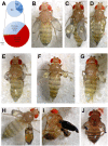
Figure 1. Results of wing-specific RNAi expression and the most typical manifestations of resulting blistered and “burned” wings.
(A) General statistics of the RNAi screening. Blistered and “burned” wings were the most frequent phenotypes. (B–G) Examples of blistered wings with the blister occupying the whole wing (B, RNAi-Fas1) or the central position in the wing (C, RNAi-rok), or blisters accompanied with other wing defects such as narrow wings (D, RNAi-CG10754), “horned” wings (E, RNAi-RnrL), “swarovski” wings (F, RNAi-Hsp83), or “stump” wings (G, RNAi-U2af50). (H–J) Examples of “burned” wings. This phenotype may start as blistered wings in freshly eclosed flies (H) and with age develop into necrotic wings (I, RNAi-Wg), or reveal “burned” wings throughout the adult life (J, RNAi-mys).
Figure 2
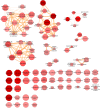
Figure 2. Semantic clustering of the GO terms enriched in the blister-causing group, compared to the annotations of all screened genes.
Enriched GO terms are shown as nodes (“biological processes” as octagons and “cellular components” as circles), and the top 3% of the strongest GO term pairwise similarities are designated as edges in the graph. The node radius relates to the generality of the terms, where smaller nodes imply more specific terms; the supplied p-values/enrichments are shown using color shading, where more saturated color of the node implies more over-represented GO terms. The semantic similarity between two GO terms is shown by the thickness of the edges, where thicker edge implies more semantically similar GO terms connected by this edge.
Figure 3
Figure 3. The Drosophila blisterome.
Each gene is an independent node, with edges between them being interactions of the genetic (cyan) or physical (red) nature, or being inferred from the physical interactions among their human orthologues (lilac). Nodes are color-coded and grouped into functional clusters according to their annotation terms; interactions within each functional cluster are shown by bold edges. Grey nodes represent genes which failed to be clustered. Genes not interconnected into the large blisterome network are grouped below it; however, some of them are color-coded, because they still belong to functional clusters of the major network. Nodes are given as circles if coming from our RNAi screening only, rectangles or triangles if coming solely from previous loss- or gain-of-function analysis, or other symbols if coming from both our and previous analysis; complete description of the node shape coding is given in the lower left corner of the Figure.
Figure 4
Figure 4. The network composed of human orthologues of the Drosophila blisterome components.
Physical interactions are shown in grey, phenotypic – in blue. Disease-related genes are marked by red crosses. Nodes are color-coded and grouped into separate functional clusters according to their annotation terms; interactions within each functional cluster are shown by bold edges. Grey nodes represent genes which are not enriched by any term. However, some of these nodes were placed close to existing clusters if most of their connections were with its members; the remaining nodes with promiscuous interactions were grouped in the center of the network. To reduce Figure complexity isolated nodes were removed leaving only the highly interconnected ones.
Figure 5
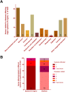
Figure 5. Human blisterome orthologues linked to diseases and disorders.
(A) Distribution of the disease-related genes among major systems of organs affected by specific maladies associated with these genes. (B) Distribution of the disease-related genes by the number of systems they affect (left bar) (see Table S9) and diseases they cause (right bar) (see Table S8). The quantity of assigned genes is indicated by numbers on the graphs on both panels (A) and (B).

References

    1. Fristrom D, Wilcox M, Fristrom J (1993) The distribution of PS integrins, laminin A and F-actin during key stages in Drosophila wing development. Development 117: 509–523. - PubMed
    1. Wilcox M, Brower DL, Smith RJ (1981) A position-specific cell surface antigen in the drosophila wing imaginal disc. Cell 25: 159–164. - PubMed
    1. Wilcox M, DiAntonio A, Leptin M (1989) The function of PS integrins in Drosophila wing morphogenesis. Development 107: 891–897. - PubMed
    1. Brower DL, Jaffe SM (1989) Requirement for integrins during Drosophila wing development. Nature 342: 285–287. - PubMed
    1. Brower DL, Piovant M, Reger LA (1985) Developmental analysis of Drosophila position-specific antigens. Developmental Biology 108: 120–130. - PubMed

Publication types

LinkOut - more resources