. 2014 Jun 26;9(6):e100748.
doi: 10.1371/journal.pone.0100748. eCollection 2014.

RNA stimulates Aurora B kinase activity during mitosis

Affiliations

RNA stimulates Aurora B kinase activity during mitosis

Ashwini Jambhekar et al. PLoS One. .

Abstract

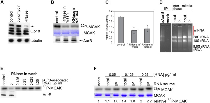
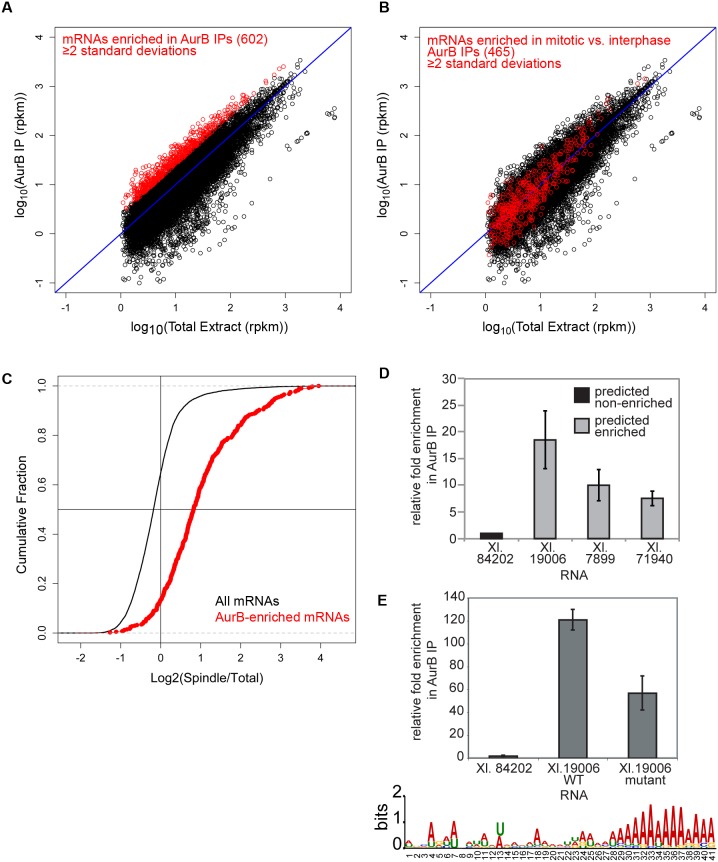
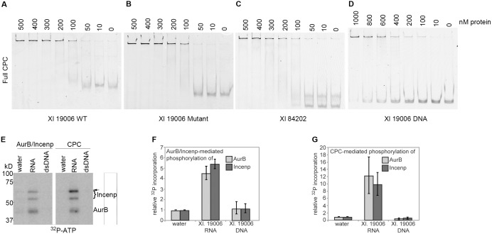
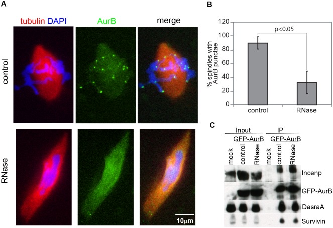
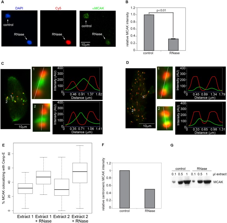
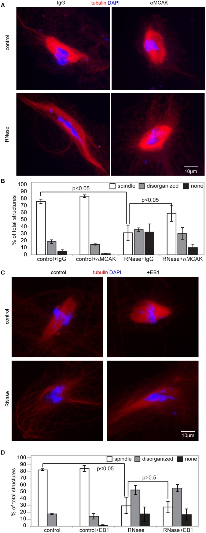
Accurate chromosome segregation is essential for cell viability. The mitotic spindle is crucial for chromosome segregation, but much remains unknown about factors that regulate spindle assembly. Recent work implicates RNA in promoting proper spindle assembly independently of mRNA translation; however, the mechanism by which RNA performs this function is currently unknown. Here, we show that RNA regulates both the localization and catalytic activity of the mitotic kinase, Aurora-B (AurB), which is present in a ribonucleoprotein (RNP) complex with many mRNAs. Interestingly, AurB kinase activity is reduced in Xenopus egg extracts treated with RNase, and its activity is stimulated in vitro by RNA binding. Spindle assembly defects following RNase-treatment are partially rescued by inhibiting MCAK, a microtubule depolymerase that is inactivated by AurB-dependent phosphorylation. These findings implicate AurB as an important RNA-dependent spindle assembly factor, and demonstrate a translation-independent role for RNA in stimulating AurB.


Conflict of interest statement

Competing Interests: The authors have declared that no competing interests exist.

Figures

Figure 1
Figure 1. AurB is an RNA-dependent kinase.
A) Hyperphosphorylation of Op18 in control extracts, or extracts treated with puromycin or RNase, containing sperm nuclei at a concentration of 5000 nuclei/µl. Arrow indicates AurB-dependent hyperphosphorylated form of Op18, as detected by western blot. Tubulin is shown as a loading control. B) Phosphorylation of MCAK N-terminus by AurB immunoprecipitated from control or RNase-treated extracts containing sperm nuclei, and washed +/− RNase following IP. MCAK substrate and AurB amounts are shown as loading controls. C) Quantitation of signals shown in (B). n = 4–5 experiments, error bars are SEM. D) RNA isolated from mock or αAurB IPs. RNA from input (total) extract is shown as a control. RNA was isolated from interphase or mitotic extracts. Red bar highlights transcripts enriched in AurB IPs. E) Rescue of RNase-washed AurB kinase activity by transcripts associated with AurB following stimulation with chromatin. Immunoprecipitations were split into aliquots containing equal amounts of beads during the final wash. One aliquot of each sample was analyzed by western blot as a loading control (lower panel), and remaining aliquots were used for kinase reactions (upper panel). F) Rescue of kinase activity as in (C) with total or AurB-binding RNAs. E-F) Gels are representative of experiments performed at least in duplicate.
Figure 2
Figure 2. Active AurB binds a distinct subset of transcripts that overlaps with RNAs found on spindles, and a degenerate primary sequence RNA motif mediates AurB recognition.
A) Scatter plot showing enrichment of a subset of transcripts (red) in mitotic AurB immunoprecipitations from biological triplicates. B) Scatter plot showing transcripts bound to AurB in interphase extracts. Transcripts specifically bound in metaphase but not in interphase are indicated with red points. C) Cumulative distribution curve showing fraction of total or AurB-bound transcripts as a function of their enrichment on spindles. D) qPCR detection of transcripts predicted by sequencing analysis to be enriched or not enriched in AurB immunoprecipitations. (n = 3 extracts). E) Bottom: Sequence logo showing a poly-A motif enriched in AurB-bound RNAs, but not in transcripts from total RNA. Top: Binding of AurB to in vitro transcribed fragments of Xl. 19006 containing the WT motif (“WT”) or harboring mutations in the 41 nt motif (“mutant”), with each purine mutated to the other purine, and each pyrimidine mutated to the other pyrimidine (see Methods). Binding to in vitro transcribed Xl. 84202 is shown as a negative control. Transcripts were added to extract, and AurB was immunoprecipitated as in (C) n = 3 extracts. (D–E) All error bars represent SEM.
Figure 3
Figure 3. The CPC binds to and is stimulated by RNA in vitro.
Binding of full, recombinant CPC to A) AurB-interacting Xl. 19006 RNA, B) Xl. 19006 RNA lacking the poly-A motif shown in Fig. 2, C) negative control Xl. 84202 RNA, D) PCR product encoding the RNA shown in (A). E) Phosphorylation of Incenp and AurB in vitro by purified AurB/Incenp or full CPC in the presence or absence of RNA or double-stranded DNA. Incenp band marked with arrow is quantified in F. Single gel image is shown, with intervening lanes omitted. F–G) Quantification of signals indicated in (E) from 3 independent experiments. Error bars represent SEM.
Figure 4
Figure 4. RNA is required for AurB localization to centromeres.
A) Localization of GFP-AurB to centromeres in control and RNase-treated spindles. Images shown are projections. B) Percentage of spindles showing chromosomal AurB punctae. Spindles were scored as positive if at least 6 punctae were visible. (n = 3 extracts, 50–100 spindles per extract, p<0.05 by paired t-test). Error bars represent SEM. C) Coprecipitation of the core CPC members–Incenp, DasraA, and Survivin–with GFP-AurB from control or RNase-treated extracts. Immunoprecipitation from extract lacking GFP-AurB (“mock”) is shown as a control. Input amounts are shown. Results are representative of experiments performed at least in triplicate.
Figure 5
Figure 5. MCAK localization to centromeres is defective in RNase-treated extracts.
A) MCAK localization was assessed on nuclei cycled through interphase and rearrested in metaphase in the presence of nocodazole. Control or RNase-treated nuclei (the latter labeled with Cy5-dUTP) were fixed, copelleted onto a single coverslip, and processed for αMCAK immunofluorescence. Pairs of control and RNase-treated nuclei in the same field of view were assayed for MCAK localization. Images shown are projections, except for Cy5, which is a single plane. B) Quantitation of centromeric MCAK levels from (A). (n = 3 extracts, 14–20 pairs of nuclei per extract, p<0.01 by single-sample t-test of average normalized values from each extract). Error bars represent SEM. C) Localization of Cenp-E (red) and MCAK (green) to sister kinetochores in control spindles. Insets show single pairs of kinetochores magnified 12x, and a line scan of Cenp-E and MCAK intensities along the indicated trajectories. D) Same as in (C), but RNase-treated. MCAK signal has been enhanced in kinetochore insets. C–D) Images shown are single planes. Kinetochore insets are magnified 12X. E) Quantitation of data shown in (C–D). Box plot showing the distribution of MCAK overlap with Cenp-E in two independent extracts. Box represents the median and upper and lower quartiles. Bars show the extent of outliers in each condition. 21–31 kinetochore pairs were measured in each extract. F) Quantitation of centromeric MCAK levels from (C–D). (n = 2 extracts, 5–6 spindles of each category per extract). G) Western blot showing MCAK amounts in control or RNase-treated extracts.
Figure 6
Figure 6. Spindle defects in the absence of RNA arise from aberrant regulation of MCAK.
A) Spindles formed in extracts (+/− RNase) in the presence of nonspecific or αMCAK antibodies. An example of a rare spindle in control RNase-treated extracts is shown. All spindle reactions contained CyclinB Δ90. Images shown are projections. B) Spindle assembly efficiency in extracts shown in (A). “None” indicates that no microtubule structures formed around nuclei. (n = 4 extracts, >100 nuclei per extract, p<0.05 by paired t-test). Error bars represent SEM. C) Examples of spindle assembly in control and RNase-treated extracts in the presence or absence of 750 nM EB1-GFP. Disorganized structures formed in RNase-treated extracts are shown. Images shown are projections. D) Spindle assembly efficiency in extracts shown in (C). (n = 3 extracts, >100 nuclei per extract p<0.05 for control vs RNase-treated p>0.5 for RNase +/− EB1 by paired t-test). Error bars represent SEM.

References

    1. Mitchison T, Kirschner M (1984) Microtubule assembly nucleated by isolated centrosomes. Nature 312: 232–237. - PubMed
    1. Heald R, Tournebize R, Blank T, Sandaltzopoulos R, Becker P, et al. (1996) Self-organization of microtubules into bipolar spindles around artificial chromosomes in Xenopus egg extracts. Nature 382: 420–425. - PubMed
    1. Karsenti E, Newport J, Kirschner M (1984) Respective roles of centrosomes and chromatin in the conversion of microtubule arrays from interphase to metaphase. J Cell Biol 99: 47s–54s. - PMC - PubMed
    1. Gadde S, Heald R (2004) Mechanisms and molecules of the mitotic spindle. Curr Biol 14: R797–805. - PubMed
    1. Nachury MV, Maresca TJ, Salmon WC, Waterman-Storer CM, Heald R, et al. (2001) Importin beta is a mitotic target of the small GTPase Ran in spindle assembly. Cell 104: 95–106. - PubMed

Publication types