. 2014 Jun 26;10(6):e1004208.
doi: 10.1371/journal.ppat.1004208. eCollection 2014 Jun.

A nucleic-acid hydrolyzing single chain antibody confers resistance to DNA virus infection in hela cells and C57BL/6 mice

Affiliations

A nucleic-acid hydrolyzing single chain antibody confers resistance to DNA virus infection in hela cells and C57BL/6 mice

Gunsup Lee et al. PLoS Pathog. .

Erratum in

  • PLoS Pathog. 2014 Jul;10(7):e1004318

Abstract

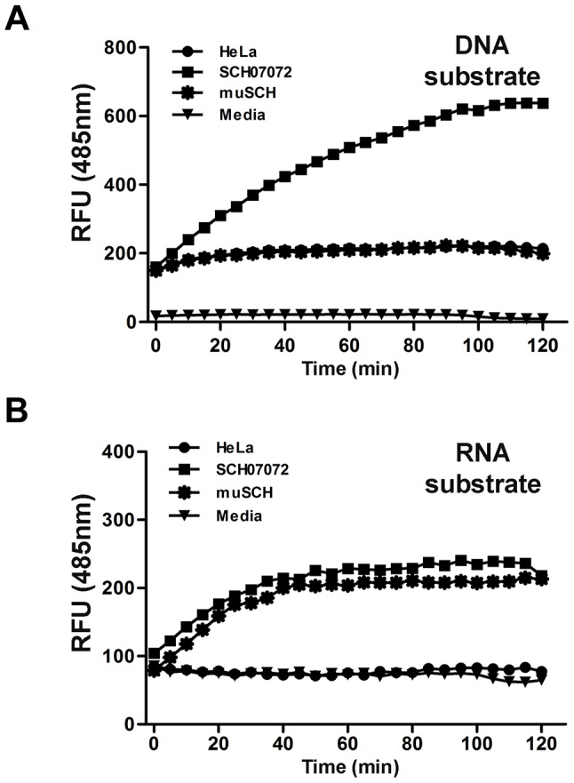
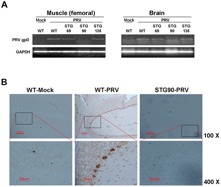
Viral protein neutralizing antibodies have been developed but they are limited only to the targeted virus and are often susceptible to antigenic drift. Here, we present an alternative strategy for creating virus-resistant cells and animals by ectopic expression of a nucleic acid hydrolyzing catalytic 3D8 single chain variable fragment (scFv), which has both DNase and RNase activities. HeLa cells (SCH07072) [corrected] expressing 3D8 scFv acquired significant resistance to DNA viruses. Virus challenging with Herpes simplex virus (HSV) in 3D8 scFv transgenic cells and fluorescence resonance energy transfer (FRET) assay based on direct DNA cleavage analysis revealed that the induced resistance in HeLa cells was acquired by the nucleic acid hydrolyzing catalytic activity of 3D8 scFv. In addition, pseudorabies virus (PRV) infection in WT C57BL/6 mice was lethal, whereas transgenic mice (STG90) that expressed high levels of 3D8 scFv mRNA in liver, muscle, and brain showed a 56% survival rate 5 days after PRV intramuscular infection. The antiviral effects against DNA viruses conferred by 3D8 scFv expression in HeLa cells as well as an in vivo mouse system can be attributed to the nuclease activity that inhibits viral genome DNA replication in the nucleus and/or viral mRNA translation in the cytoplasm. Our results demonstrate that the nucleic-acid hydrolyzing activity of 3D8 scFv confers viral resistance to DNA viruses in vitro in HeLa cells and in an in vivo mouse system.


Conflict of interest statement

The authors have declared that no competing interests exist.

Figures

Figure 1
Figure 1. Construction of 3D8 scFv-expressing HeLa cells.
A. Schematic diagrams of the plasmid constructs. Construction of the (a) pcDNA3.1/V5-HisB-3D8 scFv vector and (b) pcDNA3.1/V5-HisB-mu3D8 scFv vector. Yellow line is the mutation site (His→Ala) B. 3D8 scFv expression levels were analyzed by quantitative real-time polymerase chain reaction (PCR) in transgenic cell lines. The relative concentrations of 3D8 scFv were calculated after normalization to the GAPDH gene using the delta delta CT method. Data bars represent mean ± standard error. The expression levels of each cell line were compared to SCH07072. C. Identification of three 3D8 scFv cell lines (SCH07041, SCH07071, and SCH07072) by flow cytometry. Transgenic and wild-type HeLa cells were stained with 3D8 scFv Ab and TRITC-anti-rabbit Ig for flow cytometry. D. Localization of 3D8 scFv in transgenic and wild-type HeLa cells by immunocytochemistry. Nuclei were detected by DAPI staining (blue). 3D8 scFv expression was monitored by immunofluorescence using a polyclonal anti-3D8 scFv antibody that was visualized with TRITC (Rhodamine Red). 3D8 scFv proteins were localized in both the cytosol and nucleus of SCH07072 and muSCH cells.
Figure 2
Figure 2. 3D8 scFv expression in transgenic HeLa cells confers resistance to HSV infection.
A. Three 3D8 scFv cell lines and one mutant 3D8scFv cell line were challenged with HSV::GFP at different MOIs (0.1, 0.5, and 1). The SCH07072 line showed the lowest GFP expression compared to that of the other lines (SCH07041, SCH07071, and muSCH). B. Wild-type HeLa, SCH07072, and muSCH cells were infected with HSV::GFP at 0.1 and 0.5 MOI. Plaque assays revealed that SCH07072 cells had the lowest amount of virus progeny (0.1 MOI: HeLa = 1,343.33±139.68, SCH07041 = 266.67±31.80, SCH07071 = 483.33±43.72, SCH07072 = 163.33±29.06, muSCH = 1,356.67±131.70; 0.5 MOI: HeLa = 7,851.00±209.10, SCH07041 = 1,964.00±263.04, SCH07071 = 2,649.67±49.02, SCH07072 = 530.67±55.68, muSCH = 7,646.67±233.90). Data bars show mean ± standard error. *** indicate significant differences from HeLa cells at p<0.001 (one-way analysis of variance and Tukey's post hoc t-test). C. Western blot analysis using an anti-HSV DNA polymerase antibody demonstrated that less HSV DNA polymerase protein was present in SCH07072 cells compared to wild type HeLa cells 48 hr after virus challenge.
Figure 3
Figure 3. 3D8 scFv expression in transgenic HeLa cells confers resistance to PRV.
A. Three 3D8 scFv cell lines and one mutant 3D8 scFv cell line were challenged with PRV at two different MOIs (0.1 and 0.5). The multinuclear giant cell formation was less frequent in the SCH07072 lines than that in the other cell lines including wild type cells. Red arrow points to CPE (multinuclear giant cell formation). B. Wild-type HeLa, SCH07072, and muSCH cells were infected with PRV at 0.1 and 0.5 MOI. Plaque assay data showed the highest antiviral effects in SCH07072 cells (0.1 MOI: HeLa = 9,130.00±222.79, SCH07041 = 7,340.00±389.74, SCH07071 = 5,190.00±182.30, SCH07072 = 916.67±52.07, muSCH = 9,046.67±145.18; 0.5 MOI: HeLa = 31,833.33±3,755.14, SCH07041 = 25,866.67±4,969.35, SCH07071 = 20,700.00±2,902.30, SCH07072 = 7,466.67±762.31, muSCH = 30,400.00±2,959.73). Data bars show mean ± standard error. **, *** indicate significant differences from HeLa cells at p<0.01 and p<0.001, respectively (one-way analysis of variance and Tukey's post hoc t-test). C. Western blot analysis using an anti-PRV gpD antibody demonstrated that less PRV gpD was present in SCH07072 cells 48 hr after virus challenge than that of wild type HeLa cells.
Figure 4
Figure 4. 3D8 scFv protein have DNase and RNase activity in vitro (FRET assay).
A. DNA substrate (dsDNA) labeled with 6-carboxyfluorescein (FAM) was transferred into the different cell lines using Lipofectamine. RFUs were determined by measuring the absorbance at 485 nm. Background fluorescence was measured in wells with medium only. SCH07072 cells produced fluorescence counts up to 660 RFU 80 min after treatment. B. RNA substrate (ssRNA) labeled with 6-carboxyfluorescein (FAM) was transferred into the different cell lines using Lipofectamine. Fluorescence was determined by measuring absorbance at 485 nm. Background fluorescence was measured in wells with medium only. SCH07072 and muSCH had fluorescence levels of approximately 200 RFU in contrast to the fluorescence counts of wild-type HeLa cells and the negative control reactant, which were approximately 80 RFU.
Figure 5
Figure 5. 3D8 scFv inhibits HSV-1 encoded gene expression by DNase and RNase activity.
A. Schematic diagram for identification of viral gene expression pattern by HSV-1 challenging. B. Wild-type HeLa, SCH07072, and muSCH cells were infected with HSV::GFV at an MOI of 0.5 and then incubated for 2, 6.5, and 25 hr in the presence or absence of PAA 1 hr after virus challenge. ICP4 was used for the immediate early stage, UL9 for the early stage, and UL19 for the late stage of viral infection. Data bars show mean ± standard error. ** and *** indicate significant differences from HSV-1 viral DNA at p<0.01 and p<0.001, respectively (one-way analysis of variance and Tukey's post hoc t-test). C. Quantitative real-time reverse transcription-polymerase chain reaction (RT-PCR) was used to measure the expression of immediate early genes (ICP0 and ICP4), early genes (UL9 and UL29), and late genes (UL19 and UL38). The relative concentrations of HSV mRNAs were calculated after normalization to the GAPDH gene using the delta delta CT method. Data bars represent mean ± standard error. *, **, *** indicate significant differences from HeLa cells at p<0.05, p<0.01, and p<0.001, respectively (one-way analysis of variance and Tukey's post hoc t-test).
Figure 6
Figure 6. 3D8 scFv cannot hydrolyze methylated DNA or protein-bound DNA.
A. HeLa methylated genomic DNA and non-methylated DNA (NEB) were treated with 3D8 scFv and DNase I at four concentrations: 10×10−4 U/µl, 8.3×10⁻⁴ U/µl, 1.4×10⁻⁴ U/µl, and 0 U/µl. The DNA samples were harvested and analyzed by electrophoresis at 0, 1, 2, and 3 hr after treatment. At a concentration of 8.3×10⁻⁴ U/µl, 3D8 scFv digested more non-methylated DNA than DNase I 1 hr after treatment. B. Chromatin was prepared using the EZ-Zyme Enzymatic Chromatin Prep kit. Prepared chromatin DNA and naked DNA were treated with 3D8 scFv and DNase I (10×10−4 U, 8.3×10⁻⁴ U, and 0 U/µl). The DNA samples were harvested and analyzed by electrophoresis at 0, 1, 2, and 3 hr after treatment. Neither 3D8 scFv nor DNase I hydrolyzed histone-bound DNA at concentrations of 10×10−4 U/µl and 8.3×10⁻⁴ U/µl. When naked DNA was treated with 3D8 scFv at concentrations of 8.3×10⁻⁴ U/µl, 3D8 scFv could digest naked DNA. But, DNase I could not digest naked DNA at 8.3×10⁻⁴ U/µl. C. The relative quantitative values of each DNA of methylated, non-methylated, histone-bound, and naked HeLa genomic DNA were measured for 3 hr after each DNA was treated with 3D8 scFv (8.3×10⁻⁴ U/µl). The bar graphs indicate the average values of each sample calculated from three individual experiments.
Figure 7
Figure 7. High expression of 3D8 scFv in the muscle and brain of STG90 mice.
A. 3D8 scFv mRNA expression was verified by quantitative real-time reverse transcription polymerase chain reaction (RT-PCR) analysis in the muscle and brain of TG mice. The STG90 line expressed high levels of 3D8 scFv in both the brain and muscle, whereas the STG135 line expressed high amounts of 3D8 scFv in the brain; these strains were selected for further antiviral preventive studies. The relative expression of 3D8 scFv mRNAs was calculated after normalization to the GAPDH gene using the delta delta CT method. The primer efficiency of 3D8 scFv and GAPDH are 1.845 and 2.065, respectively. Each qRT-PCR data point is a representative example of data from three replicate experiments. Data bars represent mean ± standard error. B. Kaplan-Meier survival analysis for antiviral effects of all groups. The difference in survival between WT-PRV and STG90-PRV was statistically significant (p = 0.0042 by log-rank test). The numbers of live and dead mice were counted every 12 hr for 5 days after challenge with 10 LD50 PRV in the femoral muscle. Survival was highest in the STG90-PRV group (53%) compared to that of the other groups (WT-PRV: 11%, STG69: 40%, and STG135: 17%). C. PRV-challenged F2 mice (WT-PRV) began to exhibit symptoms of inflammation in the femoral muscle and finally died 3–5 days post-virus challenge. Red arrow points to inflammation symptoms.
Figure 8
Figure 8. STG90 exhibits antiviral effects against PRV.
A. The expression levels of PRV gpD RNA in the muscle and brain of WT-Mock, WT-PRV, STG69-PRV, STG90-PRV, and STG135-PRV mice were investigated. Only live STG90-PRV mice did not show PRV gpD expression. B. Immunohistochemistry was performed to detect PRV in PRV target organs; brain (upper panel: ×100) and femoral muscle (bottom panel: ×400). The PRV gpD protein was stained with a monoclonal anti-PRV antibody and visualized with DAB. The Purkinje layer cells in the WT-PRV group stained brown for the gpD protein, whereas no staining was observed in the other groups.
Figure 9
Figure 9. New antiviral mechanism by 3D8 scFv protein.
A. Model of the HSV-I replication cycle. Virus infection begins with binding of the virus to the cell surface. The viral envelope fuses with the cell membrane and delivers the viral capsid into the cytoplasm. Viral DNA synthesis begins shortly after the appearance of the beta proteins and the temporal program of viral gene expression ends with the appearance of the gamma or late proteins, which constitute the structural proteins of the virus. Finally, the virus undergoes a lytic cycle. B. Expression model of 3D8 scFv proteins. 3D8 scFv proteins were localized in cytosol using a vector system and targeted in the nucleus by nuclear localization signal. Therefore, 3D8 scFv proteins in SCH07072 were present in both the cytosol and nucleus. C. 3D8 scFv has a unique dual and stereoscopic protection mechanism that includes DNase activity in the nucleus and RNase activity in the cytoplasm. 3D8 scFv acts by inhibiting (1) viral DNA replication and RNA transcription in the nucleus via viral DNA degradation and (2) translation in the cytoplasm via viral RNA degradation. In other words, 3D8 scFv targets the viral DNA genome itself or its RNA transcripts spatially in two different subcellular spaces (nucleus and cytoplasm) and at two different times.

References

    1. Ding S (2008) Virology: Principles and Applications. The Yale Journal of Biology and Medicine 81: 155.
    1. Osterhaus A (2009) Timely tale of avian flu. Nature 462: 986.
    1. Yamada T, Dautry A, Walport M (2008) Ready for avian flu? Nature 454: 162. - PubMed
    1. McFarland MD, Hill HT (1987) Vaccination of mice and swine with a pseudorabies virus mutant lacking thymidine kinase activity. Can J Vet Res 51: 340–344. - PMC - PubMed
    1. Ge Q, McManus MT, Nguyen T, Shen CH, Sharp PA, et al. (2003) RNA interference of influenza virus production by directly targeting mRNA for degradation and indirectly inhibiting all viral RNA transcription. Proc Natl Acad Sci U S A 100: 2718–2723. - PMC - PubMed

Publication types

MeSH terms

LinkOut - more resources