. 2014 Jun 27:5:4244.
doi: 10.1038/ncomms5244.

Structure and mechanism of an intramembrane liponucleotide synthetase central for phospholipid biosynthesis

Affiliations
Free PMC article

Structure and mechanism of an intramembrane liponucleotide synthetase central for phospholipid biosynthesis

Xiuying Liu et al. Nat Commun. .
Free PMC article

Abstract

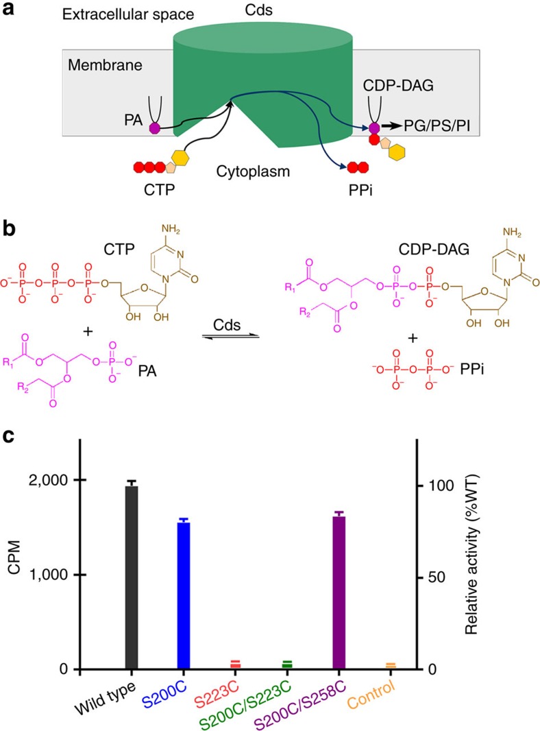
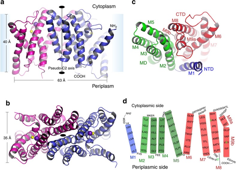
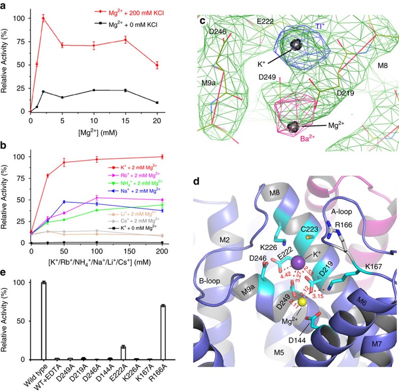
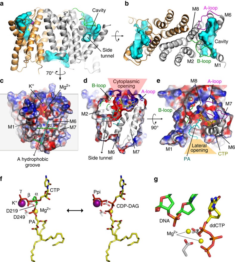
Phospholipids are elemental building-block molecules for biological membranes. Biosynthesis of phosphatidylinositol, phosphatidylglycerol and phosphatidylserine requires a central liponucleotide intermediate named cytidine-diphosphate diacylglycerol (CDP-DAG). The CDP-DAG synthetase (Cds) is an integral membrane enzyme catalysing the formation of CDP-DAG, an essential step for phosphoinositide recycling during signal transduction. Here we report the structure of the Cds from Thermotoga maritima (TmCdsA) at 3.4 Å resolution. TmCdsA forms a homodimer and each monomer contains nine transmembrane helices arranged into a novel fold with three domains. An unusual funnel-shaped cavity penetrates half way into the membrane, allowing the enzyme to simultaneously accept hydrophilic substrate (cytidine 5'-triphosphate (CTP)/deoxy-CTP) from cytoplasm and hydrophobic substrate (phosphatidic acid) from membrane. Located at the bottom of the cavity, a Mg(2+)-K(+) hetero-di-metal centre coordinated by an Asp-Asp dyad serves as the cofactor of TmCdsA. The results suggest a two-metal-ion catalytic mechanism for the Cds-mediated synthesis of CDP-DAG at the membrane-cytoplasm interface.


Figures

Figure 1
Figure 1. The fundamental role of Cds enzyme in phospholipid biosynthesis.
(a) A cartoon model illustrating the functional role of the membrane-embedded Cds enzyme in phospholipid biosynthesis. PA, phosphatidic acid; PG, phosphatidylglycerol; PS, phosphatidylserine; PI, phosphatidylinositol. (b) The chemical reaction of CDP-DAG synthesis catalysed by Cds. (c) The activities of wild-type TmCdsA and the cysteine mutants (S200C, S223C, S200C/S223C and S200C/S258C) in synthesizing 3H-labelled CDP-DAG product. CPM, count per minute of the radioactivity measured for the hydrophobic products extracted in the organic phase. The control is the parallel reaction set up without enzyme added. The error bars denote the standard error of the mean (s.e.m., n=4 except for S200C which has n=3 instead).
Figure 2
Figure 2. Electron density maps of TmCdsA.
(a) 2FoFc map (contoured at 1.2 × σ level) of the TmCdsA structure refined at 3.4 Å resolution and viewed along the membrane plane. (b) A zoom-in stereo view of the 2Fo-Fc map (contoured at 1.2 × σ level) at the transmembrane region with stick models superposed on the map.
Figure 3
Figure 3. The overall structure of TmCdsA.
(a,b) TmCdsA dimer viewed along the membrane plane (a) and along the membrane normal from cytoplasmic side (b), respectively. The protein backbone of the S200C/S223C mutant is shown as cartoon models and the Mg2+/K+ ions are shown as yellow/purple sphere models. The blue shades indicate the approximate location of the membrane. The monomer in blue (residues 4–270) is traced five residues longer at the N-terminal region than the monomer in magenta (residues 9–270). (c) Structure of a TmCdsA monomer with each individual domain coloured in blue, green or red. CTD, C-terminal domain; MD, middle domain;NTD, N-terminal domain . (d) Topological organization of the transmembrane helices within TmCdsA. The green ‘S’ characters labelled by * symbol indicate the location of S200, S223 and S258 residues, which are mutated into cysteine residues for crystallization purpose in this study.
Figure 4
Figure 4. Architecture of the active site of TmCdsA.
(a) Mg2+-dependent activity changes of TmCdsA with or without 200 mM KCl. The error bars indicate s.e.m. and n=3 for the data points with [Mg2+]=2 or 10 mM (+0 or 200 mM KCl), whereas the others are with n=4. (b) The effect of increasing monovalent cation (K+, Rb+, NH4+, Na+, Li+ or Cs+) concentration on the activity of TmCdsA. The lowest dark curve is measured with variable K+ concentration and in the absence of Mg2+ ion, whereas the other curves are measured in the presence of 2 mM Mg2+ and variable concentrations of different monovalent cations. n=3 for the following data points measured with 2 mM Mg2+: [K+]=50, 200 mM; [Na+]=200 mM; [NH4+]=0 mM; [Li+]=0, 200 mM; [Cs+]=0, 25, 50 mM; [Rb+]=0, 50 mM. n=4 for the others. (c) Isomorphous difference Fourier peaks of the FBaFMg (4.5 Å, magenta) and FTlFK (4.5 Å, blue) maps contoured at +8.0 × σ level. Green mesh is the 3.4 Å-resolution 2FoFc map (contoured at +2.0 × σ level) of the crystal containing both Mg2+ and K+ ions. The Mg2+ and K+ ions are shown as silver bullets. The peak of Ba2+ ion deviates slightly from the positions of Mg2+ site because of minor changes in the unit cell dimensions of the Ba2+-soaked crystal. (d) The key amino-acid residues surrounding the Mg2+-K+ di-metal centre in TmCdsA. (e) Mutagenesis analyses on the functional role of the charged residues around the di-metal centre. The activity of the wild-type enzyme was normalized as 100% relative activity after being subtracted with the negative control data (parallel reactions without enzyme added), whereas those of mutants and WT+EDTA are presented as relative percentage activity in comparison to the wild-type enzyme. The error bars denote s.e.m., n=3 for the wild type, D249A, D219A, D246A and K167A, or n=4 for WT+EDTA, D144A, E222A, K226A and R166A.
Figure 5
Figure 5. The substrate binding cavity of TmCdsA and its catalytic mechanism.
(a,b) The presence of two large cavities near the cytoplasmic surface of TmCdsA dimer. The cavities are shown as cyan surface models superposed on the cartoon representation of TmCdsA molecules. The front and top (from cytoplasmic side) views of TmCdsA dimer along the membrane plane and membrane normal are shown in a and b, respectively. (c) Side view of the electrostatic potential surface of TmCdsA. An elongated hydrophobic groove adjacent to the dual-opening cavity and exposed to the lipid bilayer is located on the outer surface of TmCdsA. The silver box shadow in the background indicates an estimated position of the membrane with respect to TmCdsA. The deep red and deep blue colours on the surfaces indicate electronegative and electropositive regions at −15 and 15 kT e−1, respectively. k, the Boltzmann constant; T, the temperature; e, the magnitude of the electron charge. The unit for the numbers above the dash lines is Å. (d) Sectional view of the funnel-shaped cavity within TmCdsA revealing an elongated side tunnel between the B-loop and M1 helix. The stick models of CTP (yellow) and PA (dioleoyl, cyan) are docked in the cavity through Autodock Vina programme to show their predicted binding sites in TmCdsA. (e) The cavity viewed from the cytoplasmic side. For clarity, only the region surrounding the cavity is shown. (f) A proposed model for the catalytic mechanism of CDP-DAG synthesis. The green arrows indicate the nucleophilic attack on the α-phosphate of CTP launched by the Mg2+-activated PA head group and the withdrawing of electron, which leads to the break of α-β phosphodiester bond and production of pyrophosphate in addition to CDP-DAG. (g) The dual-Mg2+ catalytic centre of human DNA polymerase in complex with both substrates, namely DNA and ddCTP (2′,3′-dideoxycytidine 5′-triphosphate). PDB code: 1BPY.

References

    1. Dowhan W., Bogdanov M. & Mileykovskaya E. inBiochemistry of Lipids, Lipoproteins and Membranes 5th Edn eds Vance D. E., Vance J. E. 1–37Elsevier (2008).
    1. Daum G. inLipid Metabolism and Membrane Biogenesis ed. Daum G. 1–3Springer (2004).
    1. Schneiter R. & Toulmay A. The role of lipids in the biogenesis of integral membrane proteins. Appl. Microbiol. Biotechnol. 73, 1224–1232 (2007). - PubMed
    1. Dowhan W. & Bogdanov M. Lipid-dependent membrane protein topogenesis. Annu. Rev. Biochem. 78, 515–540 (2009). - PMC - PubMed
    1. Bogdanov M., Xie J. & Dowhan W. Lipid-protein interactions drive membrane protein topogenesis in accordance with the positive inside rule. J. Biol. Chem. 284, 9637–9641 (2009). - PMC - PubMed

Publication types

MeSH terms