Review
. 2014:211:141-64.
doi: 10.1016/B978-0-444-63425-2.00006-4.

The multilingual nature of dopamine neurons

Affiliations
Review

The multilingual nature of dopamine neurons

Louis-Eric Trudeau et al. Prog Brain Res. 2014.

Abstract

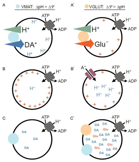
The ability of dopamine (DA) neurons to release other transmitters in addition to DA itself has been increasingly recognized, hence the concept of their multilingual nature. A subset of DA neurons, mainly found in the ventral tegmental area, express VGLUT2, allowing them to package and release glutamate onto striatal spiny projection neurons and cholinergic interneurons. Some dopaminergic axon terminals release GABA. Glutamate release by DA neurons has a developmental role, facilitating axonal growth and survival, and may determine in part the critical contribution of the ventral striatum to psychostimulant-induced behavior. Vesicular glutamate coentry may have synergistic effects on vesicular DA filling. The multilingual transmission of DA neurons across multiple striatal domains and the increasing insight into the role of glutamate cotransmission in the ventral striatum highlight the importance of analyzing DA neuron transmission at the synaptic level.

Keywords: GABA; cotransmission; dopamine; glutamate; vesicular.


Figures

FIGURE 1
FIGURE 1
Glutamate-mediated synaptic currents recorded in isolated dopamine neurons in vitro. In an isolated neuron, subsequently shown to be TH-positive, a large autaptic EPSC was recorded under voltage clamp. This was almost completely blocked by CNQX (EPSC was 4% of control; traces shown are averages of 10 stimulations; traces during drug application are shown in gray). The reversible D2 antagonist sulpiride enhanced the EPSC (117%; shown here and in subsequent traces without the initiating action current), whereas the D2 agonist quinpirole markedly attenuated the autaptic EPSC (76%). This suggests that concomitant DA release modulates the glutamate-mediated EPSC.
FIGURE 2
FIGURE 2
Summary diagram of the cellular heterogeneity within the A10 region. Differential distribution of VGLUT2-only neurons, VGLUT2–TH neurons, and TH-only neurons within each subdivision of the A10 region. (A–D) Distribution of VGLUT2-only neurons (neurons expressing VGLUT2 mRNA but lacking TH immunoreactivity). These VGLUT2-only neurons are present throughout the rostrocaudal levels of each subdivision of the A10 region with a lateromedial increasing gradient of concentration. VGLUT2-only neurons are infrequent in the most lateral region of the PBP, a region with a high concentration of TH-only neurons (A″, B″, and C″). (A′–D′) Distribution of VGLUT2–TH neurons (neurons coexpressing VGLUT2 mRNA and TH immunoreactivity). VGLUT2–TH neurons are restricted to the most rostromedial aspects of the PBP and PN; however, they are present at all rostrocaudal and mediolateral levels of the RLi, CLi, and IF. (A″–D″) Distribution of TH-only neurons (neurons lacking expression of VGLUT2 mRNA but containing TH immunoreactivity). The TH-only neurons are present throughout the rostrocaudal levels of each subdivision of the A10 region, including the lateral aspects of the PBP. Each panel represents the average number of labeled neurons found in three sections, each section from a different rat. PIF, parainterfascicular nucleus; mtg, mammillotegmental tract; fr, fasciculus retroflexus; mp, mammillary peduncle; RRF, retrorubral field. Taken from Yamaguchi et al. (2007).
FIGURE 3
FIGURE 3
Schematic representation of the synaptic and nonsynaptic axon terminals established by dopamine neurons. DA neurons are known to establish two morphologically distinguishable axon terminals: some that are nonsynaptic and others that are synaptic. The nonsynaptic terminals (varicose-like structures) display no obvious pre- and postsynaptic specializations (see lower illustration showing a magnified view of a single nonsynaptic terminal), contain tyrosine hydroxylase (TH), and could be specialized for the release of DA. The synaptic terminals display a more classical active zone, postsynaptic density, and synaptic cleft and could be the site of VGLUT2 expression and of glutamate (Glu) release (see upper illustration showing a magnified view of a synaptic axon terminal). Although not shown, some of the terminals can also contain both TH and VGLUT2. Taken from Trudeau and Gutierrez (2007).
FIGURE 4
FIGURE 4
Functional connectome analysis of DA neuron connections in striatum. Mice are generated with ChR2 expressed selectively in DA neurons. DA neuron cell bodies are shown in the ventral midbrain (left) with their axons projecting into the median forebrain bundle (mfb) toward the striatum. In the striatum (right), spiny projection neurons (SPNs) and cholinergic interneurons (ChIs) are recorded and DA neuron inputs activated by wide-field photostimulation (larger circle around cells and terminals). With repeated measurements, the incidence and strength of DA neuron inputs to SPNs and ChIs are quantitated to determine functional connectivity.
FIGURE 5
FIGURE 5
Vesicular coloading of glutamate with dopamine leads to synergistic effects on vesicle filling. Vesicular neurotransmitter loading is dependent on both the ΔpH and ΔΦ established by the vATPase. However, due to both the stoichiometry of the transporters and the opposite charges of the substrates, (A) dopamine loading through VMAT is dependent mostly on ΔpH, while (A′) glutamate uptake through VGLUT depends principally on ΔΦ. Although the efficient filling of vesicles with dopamine relies on ΔpH—in the absence of a counterion (B)—the transport of H+ by the vATPase sets up a large ΔΦ that impedes vesicle acidification. The generation of a large ΔpH therefore requires dissipation of ΔΦ, for example, by the (B′) entry of a permeant anion. Chloride anions are abundant at the nerve terminal and serve this function. However, glutamate anions are also present, and their vesicular entry through VGLUT can facilitate acidification (i.e., increase ΔpH) of dopamine-containing synaptic vesicles. (C) This model predicts increased dopamine quantal content for a subset of (C′) VGLUT2- and VMAT2-cocontaining vesicles. Abbreviations: ΔpH, vesicular proton gradient; ΔΦ, vesicular membrane potential; H+, protons; A, anion; DA, dopamine; Glu, glutamate.

References

    1. Alsiö J, Nordenankar K, Arvidsson E, Birgner C, Mahmoudi S, Halbout B, Smith C, Fortin GM, Olson L, Descarries L, Trudeau LE, Kullander K, Levesque D, Wallén-Mackenzie Å. Enhanced sucrose and cocaine self-administration and cue-induced drug seeking after loss of VGLUT2 in midbrain dopamine neurons in mice. J Neurosci. 2011;31:12593–12603. - PMC - PubMed
    1. Amilhon B, Lepicard E, Renoir T, Mongeau R, Popa D, Poirel O, Miot S, Gras C, Gardier AM, Gallego J, Hamon M, Lanfumey L, Gasnier B, Giros B, El Mestikawy S. VGLUT3 (vesicular glutamate transporter type 3) contribution to the regulation of serotonergic transmission and anxiety. J Neurosci. 2010;30:2198–2210. - PMC - PubMed
    1. Antonopoulos J, Dori I, Dinopoulos A, Chiotelli M, Parnavelas JG. Postnatal development of the dopaminergic system of the striatum in the rat. Neuroscience. 2002;110:245–256. - PubMed
    1. Arbuthnott GW, Wickens J. Space, time and dopamine. Trends Neurosci. 2007;30:62–69. - PubMed
    1. Beaudet A, Descarries L. The monoamine innervation of rat cerebral cortex: synaptic and nonsynaptic axon terminals. Neuroscience. 1978;3:851–860. - PubMed

Publication types

MeSH terms