. 2014 Nov 15;23(22):5976-88.
doi: 10.1093/hmg/ddu321. Epub 2014 Jun 26.

An integrated computational approach can classify VHL missense mutations according to risk of clear cell renal carcinoma

Affiliations

An integrated computational approach can classify VHL missense mutations according to risk of clear cell renal carcinoma

Lucy Gossage et al. Hum Mol Genet. .

Abstract

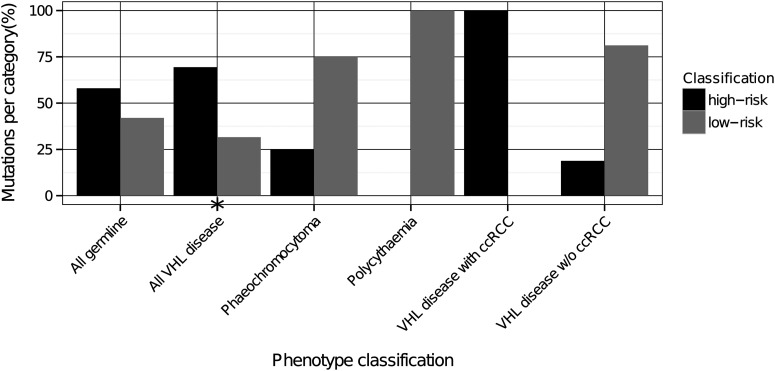
Mutations in the von Hippel-Lindau (VHL) gene are pathogenic in VHL disease, congenital polycythaemia and clear cell renal carcinoma (ccRCC). pVHL forms a ternary complex with elongin C and elongin B, critical for pVHL stability and function, which interacts with Cullin-2 and RING-box protein 1 to target hypoxia-inducible factor for polyubiquitination and proteasomal degradation. We describe a comprehensive database of missense VHL mutations linked to experimental and clinical data. We use predictions from in silico tools to link the functional effects of missense VHL mutations to phenotype. The risk of ccRCC in VHL disease is linked to the degree of destabilization resulting from missense mutations. An optimized binary classification system (symphony), which integrates predictions from five in silico methods, can predict the risk of ccRCC associated with VHL missense mutations with high sensitivity and specificity. We use symphony to generate predictions for risk of ccRCC for all possible VHL missense mutations and present these predictions, in association with clinical and experimental data, in a publically available, searchable web server.


Figures

Figure 1.
Figure 1.
Workflow for predicting ccRCC risk for missense mutations in VHL. For a given mutation, computational methods from different paradigms are used to quantitatively assess its effects on protein stability and protein interactions with other proteins or ligands, all of which could affect function. These results are combined in optimized predictors via a regression model tree (using the M5 algorithm) (48) as a way to leverage the best of each method as well as to generate a consensus prediction. Experimental data were used to label each mutation in a training set according to ccRCC risk. The stability and affinity predictions are then used as evidence to train and test a binary classifier, using the ensemble learning method Random Forest (49), that outputs the predicted risk of ccRCC in a binary classification scheme (high or low risk). We named this integrated computational approach symphony.
Figure 2.
Figure 2.
Exposure classification of mutations in different categories of disease. Type 2 mutations are more likely to involve surface residues than Type 1 mutations. ccRCC-associated VHL disease mutations are less likely to involve surface residues than non-ccRCC-associated VHL disease mutations. A high proportion of polycythaemia-associated mutations involves surface residues. Statistical significance (P < 0.05) indicated by asterisk.Inter., interface; Surf., surface; W/O, without.
Figure 3.
Figure 3.
Binary classification of ccRCC risk for germline VHL mutations associated with different phenotypes. High-risk mutations were more likely to be associated with VHL disease than low-risk mutations. All ccRCC-associated VHL disease mutations were high risk. All polycythaemia-associated mutations were low risk. Statistical significance (P < 0.05) indicated by asterix. w/o, without.
Figure 4.
Figure 4.
Predictions for risk of ccRCC in sporadic tumours. The proportion of high-risk mutations is significantly higher for mutations which have been described several times in sporadic disease compared with those that have been described only once and is significantly higher in sporadic ccRCC compared with other somatic tumour types.
Figure 5.
Figure 5.
Wall-eye stereograms representing examples of the diverse mechanisms of the effects of VHL mutations at the molecular level. (A) Mutations that disrupt the HIFα-hydroxyproline-binding site. Mutations of H115 (A) cause loss of the tetracoordination of a buried water molecule, as well as the loss of an acceptor group for the hydroxyl donor group in the HYP residue in hydroxylated HIF. They also disrupt up to four H-bonds within the buried hydrogen bond network that recognizes the HYP. Mutations at S111 can cause the loss of the hydrogen donor for the HYP hydroxyl, with similar effects as above. Mutations at other depicted residues that participate in the HYP hydrogen-bonding recognition network can have similar outcomes. Mutations at secondary HIFα-binding sites, such as the loop G104-R108 (B) could also impair HIFα binding. Side chains of L562 and Y565 in HIF have not been represented for clarity in A. (B) Mutations that disrupt the hydrophobic core. Mutation in the two VHL hydrophobic cores can disrupt VHL subunit conformation, such as mutations at F76 (beta domain, B) and V170 (alpha domain, C). (C) Mutations that disrupt H-bond networks. Mutation of N78 (B) disrupts a significant buried H-bond network that stabilizes a region of VHL connecting two loops that are key for interaction with HIF and elongin C. Mutation of residues that tightly interact in this network, such as S80 and T105 have the same effect. (D) Mutations that disrupt the conformation of pVHL. Mutations of conserved glycines (e.g. G93, A) and prolines can directly disrupt and destabilize pVHL. (E) Mutations of residues within the elongin C- and elongin B-binding sites. These mutations disrupt interaction with VHL-binding partners through disruption of H-bonding networks, such as mutations at R82 and its neighbouring residues (B) and R161, or through disruption of hydrophobic cores formed by the interacting subunits in the VCB complex, such as mutations at V155, L158, K159, C162, V165, V166, V170, L178 and L184 (C). (F) Mutations that disrupt long-range electrostatic interactions. These mutations can alter the charge complementarity of the subunits in the VCB complex and destabilize protein–protein long- and short-range interaction, such as mutations at R79, R82, R107, D121, D126 (B), K159 and D187 (C). In this figure, VHL is coloured in green, elongin C in cyan, elongin B in yellow, HIFα in magenta, Cullin 2 in pink-orange and water oxygen atoms are represented as red balls.
Figure 6.
Figure 6.
A model to explain diverse phenotypes associated with VHL missense mutations. Frameshift/nonsense VHL mutations are likely to prevent formation of a functional VCB complex and result in severe disruption of HIFα regulation, thereby explaining the high risk of ccRCC in Type 1 VHL disease. Missense VHL mutations may destabilize the VCB complex through a variety of mechanisms. Mutations which are severely destabilizing are likely to severely disrupt HIFα regulation and are associated with a high risk of ccRCC. Less severely destabilizing mutations have a milder effect on HIFα regulation resulting in a lower risk of ccRCC. A few mutations do not destabilize the VCB complex as a whole but instead directly disrupt the HIFα-hydroxyproline-binding site, thereby affecting HIFα regulation. These mutations may be associated with a low risk of PCC. Some missense VHL mutations are not destabilizing and would not be predicted to affect the HIFα-hydroxyproline-binding site and may represent passenger mutations.

Similar articles

Cited by

References

    1. Maher E.R., Neumann H.P., Richard S. von Hippel-Lindau disease: a clinical and scientific review. Eur. J. Hum. Genet. 2011;19:617–623. - PMC - PubMed
    1. Beroud C., Joly D., Gallou C., Staroz F., Orfanelli M.T., Junien C. Software and database for the analysis of mutations in the VHL gene. Nucleic Acids Res. 1998;26:256–258. - PMC - PubMed
    1. Nordstrom-O'Brien M., van der Luijt R.B., van Rooijen E., van den Ouweland A.M., Majoor-Krakauer D.F., Lolkema M.P., van Brussel A., Voest E.E., Giles R.H. Genetic analysis of von Hippel-Lindau disease. Hum. Mutat. 2010;31:521–537. - PubMed
    1. Ang S.O., Chen H., Hirota K., Gordeuk V.R., Jelinek J., Guan Y., Liu E., Sergueeva A.I., Miasnikova G.Y., Mole D., et al. Disruption of oxygen homeostasis underlies congenital Chuvash polycythemia. Nat. Genet. 2002;32:614–621. - PubMed
    1. Pastore Y.D., Jelinek J., Ang S., Guan Y., Liu E., Jedlickova K., Krishnamurti L., Prchal J.T. Mutations in the VHL gene in sporadic apparently congenital polycythemia. Blood. 2003;101:1591–1595. - PubMed

Publication types

MeSH terms

Substances