. 2015 Oct;25(10):3369-80.
doi: 10.1093/cercor/bhu140. Epub 2014 Jun 26.

Supraspinal Control of Urine Storage and Micturition in Men--An fMRI Study

Affiliations

Supraspinal Control of Urine Storage and Micturition in Men--An fMRI Study

Lars Michels et al. Cereb Cortex. 2015 Oct.

Abstract

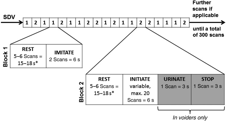
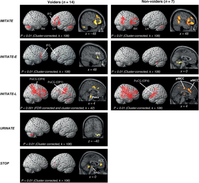
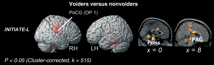
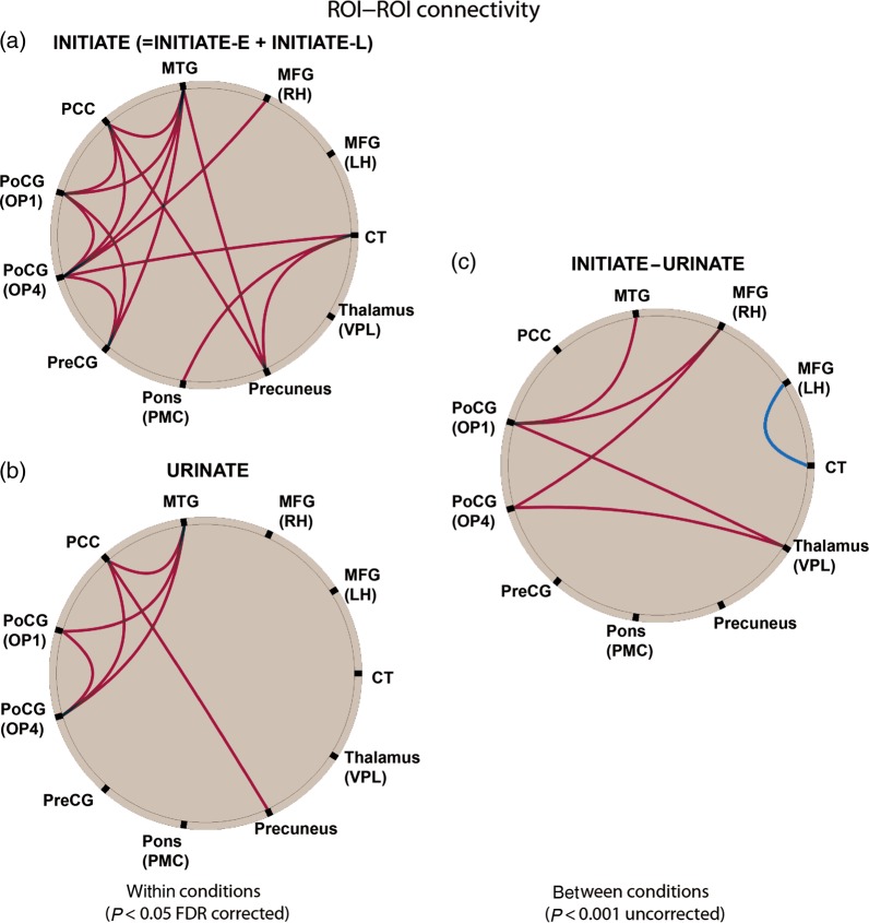
Despite the crucial role of the brain in the control of the human lower urinary tract, little is known about the supraspinal mechanisms regulating micturition. To investigate the central regulatory mechanisms activated during micturition initiation and actual micturition, we used an alternating sequence of micturition imitation/imagination, micturition initiation, and actual micturition in 22 healthy males undergoing functional magnetic resonance imaging. Subjects able to micturate (voiders) showed the most prominent supraspinal activity during the final phase of micturition initiation whereas actual micturition was associated with significantly less such activity. Initiation of micturition in voiders induced significant activity in the brainstem (periaqueductal gray, pons), insula, thalamus, prefrontal cortex, parietal operculum and cingulate cortex with significant functional connectivity between the forebrain and parietal operculum. Subjects unable to micturate (nonvoiders) showed less robust activation during initiation of micturition, with activity in the forebrain and brainstem particularly lacking. Our findings suggest that micturition is controlled by a specific supraspinal network which is essential for the voluntary initiation of micturition. Once this network triggers the bulbospinal micturition reflex via brainstem centers, micturition continues automatically without further supraspinal input. Unsuccessful micturition is characterized by a failure to activate the periaqueductal gray and pons during initiation.

Keywords: functional magnetic resonance imaging; lower urinary tract; micturition; pontine micturition center; supraspinal control.


Figures

Figure 1.
Figure 1.
The scan paradigm consisted of 2 randomly alternating blocks. Block (1) comprised 2 conditions: REST (visual fixation) and IMITATE (subjects visually imagine starting micturition). Block (2) comprised 4 conditions: REST (visual fixation), INITIATE (subjects should start micturition), URINATE (actual micturition, urine is flowing), and STOP (=interruption of micturition). The experimental onsets for REST, IMITATE, INITIATE, and STOP were defined by the visual cues presented for each condition, while the onset for URINATE was determined by the actual onset of micturition, when urine flow was first detected by the flow detector. If micturition could not be started during the INITIATE condition, which was limited to a maximum duration of 60 s, URINATE and STOP were skipped and the next block followed immediately, starting with REST. Each block was randomly repeated at least 8 times and as often as possible depending on the duration of the INITIATE condition. The conditions highlighted in gray, that is, URINATE and STOP, were only applicable when actual micturition could be initiated. In nonvoiders, the INITIATE condition was always followed by the REST condition of the subsequent block. SDV, strong desire to void. *REST conditions following INITIATE (in the case of inability to initiate actual micturition) or IMITATE had a duration of 18 s. REST conditions following STOP had a duration of 15 s.
Figure 2.
Figure 2.
Rendered brain displaying BOLD-signal peaks during different conditions (IMITATE, INITIATE-E, INITIATE-L, URINATE, and STOP) compared with REST in subjects who were able (voiders) and those unable (nonvoiders) to void during scanning. ACC, anterior cingulate cortex; LH, left hemisphere; RH, right hemisphere; IFG, inferior frontal gyrus; PAG, periaqueductal gray; PoCG, postcentral gyrus; OP, parietal operculum; aMCC, anterior midcingulate cortex; pMCC, posterior midcingulate cortex.
Figure 3.
Figure 3.
Rendered brain displaying BOLD-signal peaks of voiders versus nonvoiders. LH, left hemisphere; RH, right hemisphere; PoCG, postcentral gyrus; OP, parietal operculum; PAG, periaqueductal gray.
Figure 4.
Figure 4.
BOLD-signal changes in the pons (6, −30, −24) of voiders during different micturition-related conditions, that is, IMITATE (gray line), INITIATE-E (dashed line), INITIATE-L (black line), URINATE (dotted line) (a). Fitted BOLD responses in the pons (6, −30, −24) during INITIATE-L of a single subject who was able to micturate 15 times over the course of the experiment (b). Fitted BOLD responses in the pons (6, −30, −24) during INITIATE-L of a single subject who was able to micturate 6 times in the course of the experiment (c).
Figure 5.
Figure 5.
Summary of the functional connectivity (FC) analysis for voiders. FC during INITIATE (=INITIATE-E + INITIATE-L, P < 0.05, FDR corrected) (a). FC during URINATE (P < 0.05, FDR corrected) (b). FC for the contrast “INITIATE–URINATE” (P < 0.001, uncorrected) (c). The red lines indicate that coupling between connected areas was significantly stronger for the contrasts INITIATE versus REST, URINATE versus REST, and INITIATE versus URINATE. The blue line indicates a significantly stronger coupling between left MFG and CT for the contrast URINATE versus INITIATE. CT, cerebellar tonsil; LH, left hemisphere; MFG, middle frontal gyrus; MTG, middle temporal gyrus; OP, parietal operculum; PCC, posterior cingulate cortex; PMC, pontine micturition center; PoCG, postcentral gyrus; PreCG, precentral gyrus; RH, right hemisphere; VPL, ventral posterolateral nucleus.

References

    1. Andrew J, Nathan PW. 1964. Lesions on the anterior frontal lobes and disturbances of micturition and defaecation. Brain. 87:233–262. - PubMed
    1. Barrington F. 1925. The effect of lesions of the hind- and midbrain on micturition in the cat. Q J Exp Physiol Cogn Med. 15:81–102.
    1. Behzadi Y, Restom K, Liau J, Liu TT. 2007. A component based noise correction method (CompCor) for BOLD and perfusion based fMRI. Neuroimage. 37:90–101. - PMC - PubMed
    1. Blok BF. 2002. Central pathways controlling micturition and urinary continence. Urology. 59:13–17. - PubMed
    1. Blok BF, Holstege G. 1999. The central control of micturition and continence: implications for urology. BJU Int. 83(Suppl 2):1–6. - PubMed

Publication types