. 2014;106(2):149-55.
doi: 10.1159/000362433. Epub 2014 Jun 26.

GABA, resting-state connectivity and the developing brain

Affiliations

GABA, resting-state connectivity and the developing brain

Soo Hyun Kwon et al. Neonatology. 2014.

Abstract

Background: Preclinical data demonstrate that gamma-aminobutyric acid (GABA) interneurons initiate connectivity in the developing brain.

Objectives: The goal of this study was to compare GABA concentration and its relationship to functional connectivity in the brains of term and preterm infants at term-equivalent age.

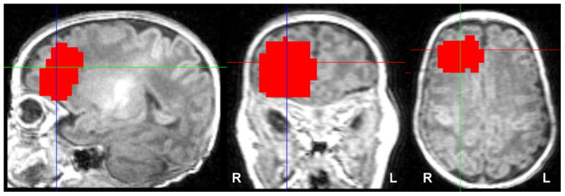
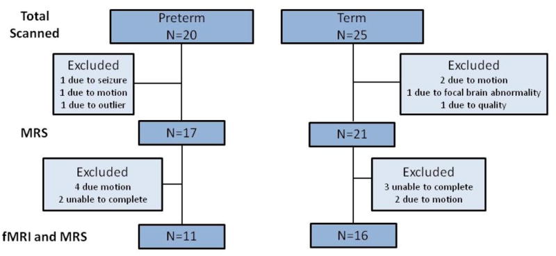
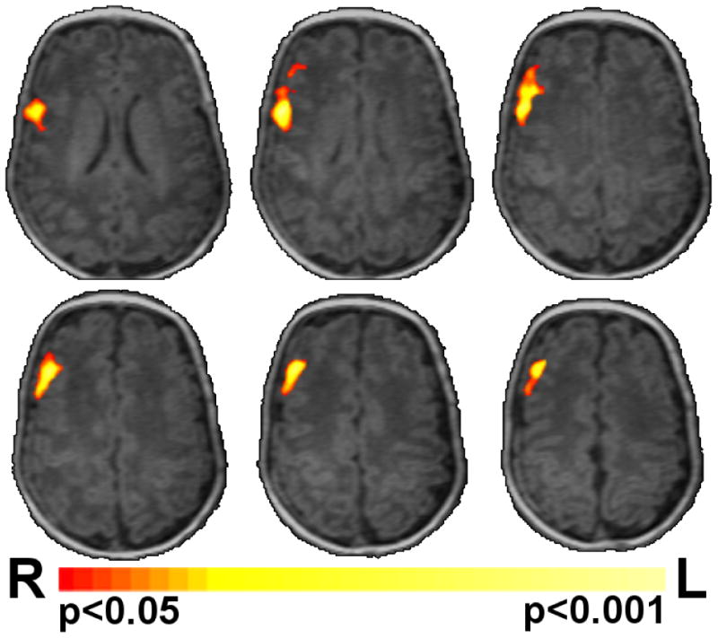
Methods: Infants received both magnetic resonance spectroscopy (MRS) and functional magnetic resonance imaging (fMRI) scans at term-equivalent age. Whole brain functional connectivity MRI data using intrinsic connectivity distribution maps were compared to identify areas with differences in resting-state functional connectivity between the preterm and term control groups. MRS measured concentrations of GABA, glutamate, N-acetyl-aspartate (NAA) and choline; NAA/choline was then calculated for comparison between the 2 groups.

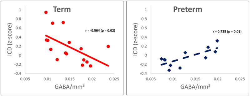
Results: Preterm infants had lower right frontal GABA and glutamate concentrations than term controls and showed a significantly different relationship between connectivity and GABA concentration in the right frontal lobe. Preterm infants had a positive correlation between GABA concentration and connectivity, while term controls demonstrated a negative correlation between these two developmentally regulated parameters.

Conclusion: These results suggest that regional GABA concentrations are associated with normal and altered neonatal resting-state connectivity.


Figures

Figure 1
Figure 1
Location of GABA voxel. GABA spectra were collected from a single 2×3×3 cm3 region of interest localized over the R frontal region. The red area shows a composite of all individuals' regions overlaid on common space template. Images are displayed in radiological convention.
Figure 2
Figure 2
Chart of usable MRS and fMRI data. This flow chart shows total subjects scanned and number of subjects excluded.
Figure 3
Figure 3
Correlation of connectivity and GABA. A) Preterm infants showed a significantly (p<0.05 corrected) different relationship between connectivity and GABA in the right frontal lobe when compared to term controls (Figure 3A). This region of significantly different correlations between the experimental groups is shown in red/yellow colors on a template brain. B) Scatter plots of the data from this region are shown in Panels B and C. Term controls showed a negative correlation between connectivity and GABA (Figure 3B), whereas preterm infants showed a positive correlation between connectivity and GABA (Figure 3C). Images are displayed in radiological convention.
Figure 3
Figure 3
Correlation of connectivity and GABA. A) Preterm infants showed a significantly (p<0.05 corrected) different relationship between connectivity and GABA in the right frontal lobe when compared to term controls (Figure 3A). This region of significantly different correlations between the experimental groups is shown in red/yellow colors on a template brain. B) Scatter plots of the data from this region are shown in Panels B and C. Term controls showed a negative correlation between connectivity and GABA (Figure 3B), whereas preterm infants showed a positive correlation between connectivity and GABA (Figure 3C). Images are displayed in radiological convention.

References

    1. Volpe JJ. Brain injury in premature infants: A complex amalgam of destructive and developmental disturbances. Lancet Neurol. 2009;8:110–124. - PMC - PubMed
    1. Ment LR, Constable RT. Injury and recovery in the developing brain: Evidence from functional MRI studies of prematurely born children. Nat Clin Pract Neurol. 2007;3:558–571. - PMC - PubMed
    1. Petanjek Z, Kostovic I, Esclapez M. Primate-specific origins and migration of cortical GABAergic neurons. Front Neuroanat. 2009;3:26. - PMC - PubMed
    1. Xu G, Broadbelt KG, Haynes RL, Folkerth RD, Borenstein NS, Belliveau RA, Trachtenberg FL, Volpe JJ, Kinney HC. Late development of the GABAergic system in the human cerebral cortex and white matter. J Neuropathol Exp Neurol. 2011;70:841–858. - PMC - PubMed
    1. Stevens HE, Su T, Yanagawa Y, Vaccarino FM. Prenatal stress delays inhibitory neuron progenitor migration in the developing neocortex. Psychoneuroendocrinology. 2013;38:509–521. - PMC - PubMed

Publication types

MeSH terms