. 2014 Jun 27;344(6191):1506-10.
doi: 10.1126/science.1252480.

Virus entry. Lassa virus entry requires a trigger-induced receptor switch

Affiliations

Virus entry. Lassa virus entry requires a trigger-induced receptor switch

Lucas T Jae et al. Science. .

Abstract

Lassa virus spreads from a rodent to humans and can lead to lethal hemorrhagic fever. Despite its broad tropism, chicken cells were reported 30 years ago to resist infection. We found that Lassa virus readily engaged its cell-surface receptor α-dystroglycan in avian cells, but virus entry in susceptible species involved a pH-dependent switch to an intracellular receptor, the lysosome-resident protein LAMP1. Iterative haploid screens revealed that the sialyltransferase ST3GAL4 was required for the interaction of the virus glycoprotein with LAMP1. A single glycosylated residue in LAMP1, present in susceptible species but absent in birds, was essential for interaction with the Lassa virus envelope protein and subsequent infection. The resistance of Lamp1-deficient mice to Lassa virus highlights the relevance of this receptor switch in vivo.


Figures

Figure 1
Figure 1. Human LAMP1 is an α-DG-independent host factor for Lassa virus and bypasses an infection barrier in avian cells
(A) Haploid genetic screen for host factors required for infection with rVSV-GP-LASV in cells lacking α-DG. The y axis indicates the significance of enrichment of gene-trap insertions in particular genes as compared to a non-selected control cell population. Filled circles represent genes and their size corresponds to the number of insertion sites identified in the virus-selected cell population. Hits were colored if they passed the statistical criteria described in the supplementary experimental procedures. Significant hits were grouped by function horizontally and data is displayed until –log(P value) = 0.01. (B) HAP1 cell lines with nuclease-generated mutations in the corresponding genes were exposed to wild-type Lassa virus and stained with antibodies specific for viral antigens to measure infected cells. LAMP1-deficient cells were complemented with human LAMP1 (L1) cDNA. (C) Chicken fibroblasts were transduced with retroviruses expressing chicken (c) or human (h) LAMP1 and challenged with rVSV-GP-LASV. Average number (±SD) of infected cells per field (eGFP-positive) is indicated. (D) Wild-type or LAMP1-deficient HAP1 cells transduced with retroviruses expressing cLAMP1 or hLAMP1 were exposed to rVSV-G or rVSV-GP-LASV. Percentage (±SD) of infected cells (eGFP-positive) is indicated. Scale bars: 50μm.
Figure 2
Figure 2. Lassa-GP undergoes a pH-induced switch to engage LAMP1
(A) Flag-tagged Lassa-GP was immobilized on beads and incubated with cell lysates from HEK-293T cells at the indicated pH. Bound proteins were subjected to immunoblot analysis and uncoated beads served as a control. IP = immunoprecipitation. (B) Flag-tagged Lassa-GP was immobilized on beads and incubated with lysates from human, mouse and chicken cells at the indicated pH. Bound proteins were subjected to immunoblot analysis. (C) Electron micrographs of wild-type and LAMP1-deficient HEK-293T cells that were infected with rVSV-GP-LASV. LAMP1-deficient cells show an accumulation of the bullet-shaped viral particles (arrows) in intracellular vesicles. Scale bars: 100nm. (D) Flag-tagged Lassa-GP was immobilized on beads and incubated with purified LAMP1-Fc at the indicated pH. Complexes (IP) were precipitated and subjected to immunoblot analysis. The supernatant (Sup) was analyzed for the release of Lassa-GP1. (E) LAMP1-deficient (top) or LAMP1d384 expressing HEK-293T cells (bottom) were transfected with expression vectors for Lassa-GP and GFP and exposed to pH 5.5. Cell boundaries were visualized with fluorescent wheat germ agglutinin (red). Enlarged homogenous green fluorescent areas result from Lassa-GP-induced syncytia formation (yellow outline). Scale bar: 50μm.
Figure 3
Figure 3. Binding of Lassa-GP to LAMP1 depends on ST3GAL4 and LAMP1-Asn76 is critical for host factor function
(A) Haploid genetic screen pointing out genetic interactions between ST3GAL4 and other Lassa entry factors. ST3GAL4-deficient cells were mutagenized and exposed to rVSV-GP-LASV. Gene-trap insertion sites were mapped in the resistant cell population and data was analyzed as in Fig. 1A. (B) Flag-tagged Lassa-GP was immobilized on beads and incubated with cell lysates from wild-type and ST3GAL4-deficient HAP1 cells at pH 5.5. Bound proteins were subjected to immunoblot analysis. (C) Wild-type and LAMP1-deficient (ΔL1) HAP1 cells complemented with cDNAs expressing the distal domain of LAMP1 containing mutations at the indicated glycosylation sites were exposed to rVSV-GP-LASV. Percentage (±SD) of infected cells (eGFP-positive) is indicated. (D) Comparison of LAMP1 polypeptides from indicated species highlights Asn76 as a marker of susceptibility to Lassa virus infection. (E) Flag-tagged Lassa-GP was immobilized on beads and incubated with lysates from LAMP1-deficient HEK-293T cells expressing human LAMP1 or ‘chickenized’ LAMP1 carrying the N76S substitution.
Figure 4
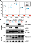
Figure 4. Lamp1 knockout mice are resistant to wild-type Lassa virus and both host factors require distinct glycosyltransferases
(A) Lassa virus propagation in Lamp1+/+, Lamp1+/− and Lamp−/− mice. Mice were injected intraperitoneally with wild-type Lassa virus and viral titers were determined after 6 days in the indicated tissues. The horizontal line marks the detection limit. (B) Flag-tagged Lassa-GP was immobilized on beads and incubated with cell lysates from wild-type, TMEM5- or ST3GAL4-deficient cells at the indicated pH. The glycosyltransferase TMEM5 is needed to generate an epitope on α-DG that is recognized by Lassa-GP (4). Bound proteins were subjected to immunoblot analysis. * = non-specific background band.

References

    1. Kunz S, et al. J Virol. 2005 Nov;79:14282. - PMC - PubMed
    1. Cao W, et al. Science. 1998 Dec 11;282:2079. - PubMed
    1. Lukashevich IS, Maryankova RF, Fidarov FM. Acta Virol. 1983 May;27:282. - PubMed
    1. Jae LT, et al. Science. 2013 Apr 26;340:479. - PubMed
    1. Saito F, et al. FEBS Lett. 2005 Apr 25;579:2359. - PubMed

Publication types

MeSH terms

Substances

Associated data