

Development and Application of Multidimensional HPLC Mapping Method for O-linked Oligosaccharides
- PMID: 24970123
- PMCID: PMC4030830
- DOI: 10.3390/biom1010048
Development and Application of Multidimensional HPLC Mapping Method for O-linked Oligosaccharides
Abstract
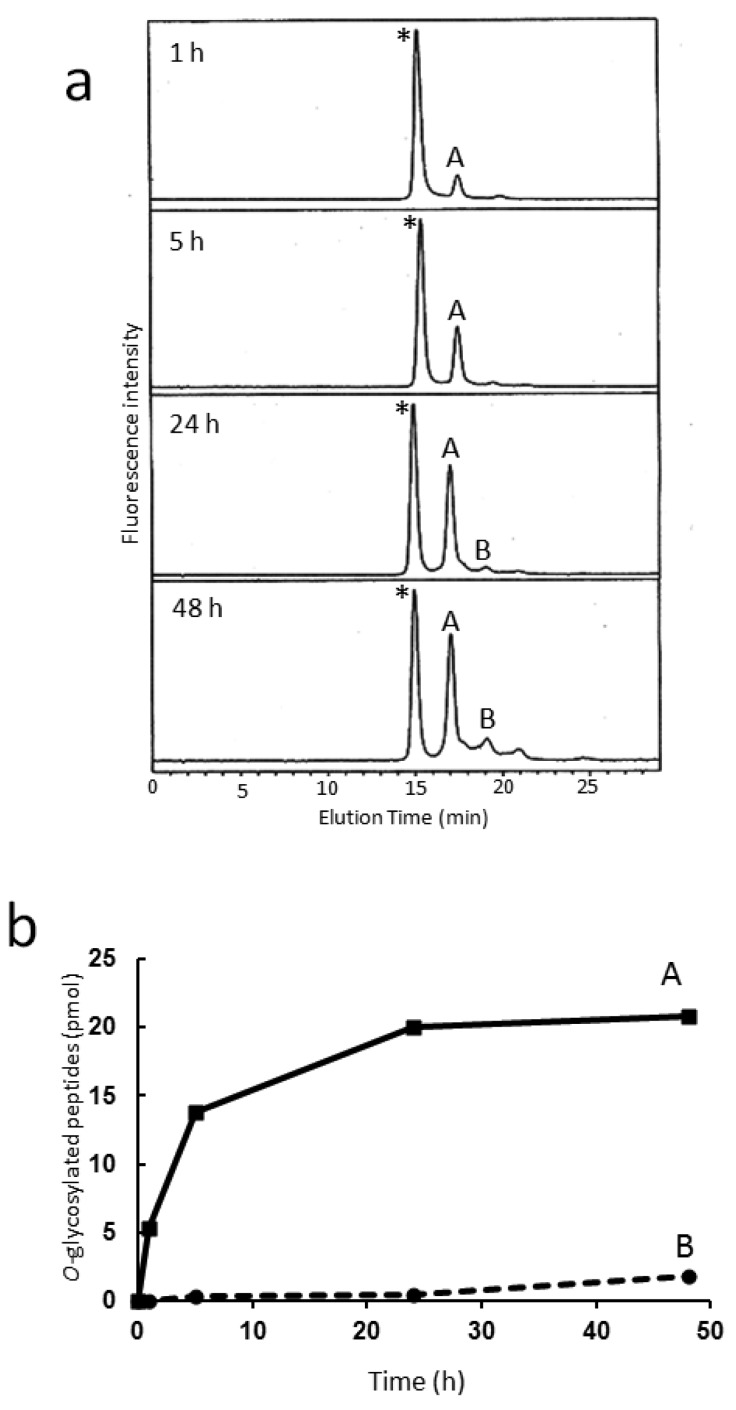
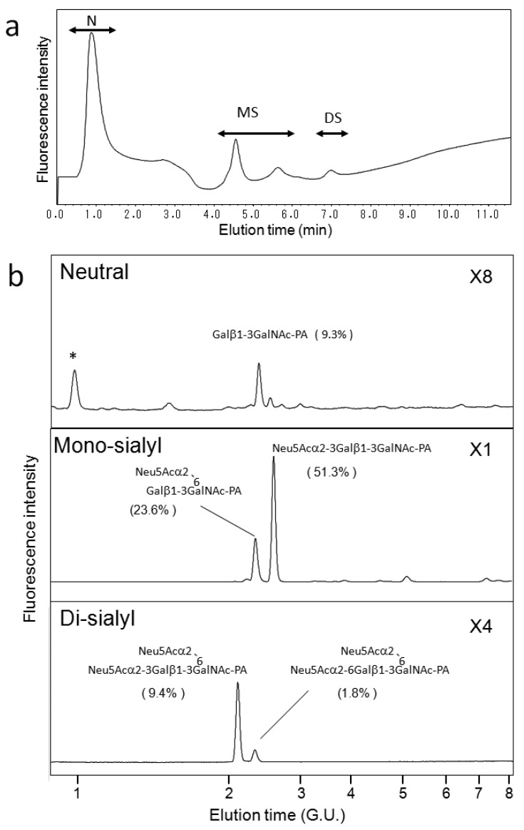
Glycosylation improves the solubility and stability of proteins, contributes to the structural integrity of protein functional sites, and mediates biomolecular recognition events involved in cell-cell communications and viral infections. The first step toward understanding the molecular mechanisms underlying these carbohydrate functionalities is a detailed characterization of glycan structures. Recently developed glycomic approaches have enabled comprehensive analyses of N-glycosylation profiles in a quantitative manner. However, there are only a few reports describing detailed O-glycosylation profiles primarily because of the lack of a widespread standard method to identify O-glycan structures. Here, we developed an HPLC mapping method for detailed identification of O-glycans including neutral, sialylated, and sulfated oligosaccharides. Furthermore, using this method, we were able to quantitatively identify isomeric products from an in vitro reaction catalyzed by N-acetylglucosamine-6O-sulfotransferases and obtain O-glycosylation profiles of serum IgA as a model glycoprotein.
Figures
Similar articles
-
Development of structural analysis of sulfated N-glycans by multidimensional high performance liquid chromatography mapping methods.Glycobiology. 2005 Oct;15(10):1051-60. doi: 10.1093/glycob/cwi092. Epub 2005 Jun 15. Glycobiology. 2005. PMID: 15958418
-
Precise structural analysis of O-linked oligosaccharides in human serum.Glycobiology. 2014 Jun;24(6):542-53. doi: 10.1093/glycob/cwu022. Epub 2014 Mar 24. Glycobiology. 2014. PMID: 24663386
-
Glycosylation of a CNS-specific extracellular matrix glycoprotein, tenascin-R, is dominated by O-linked sialylated glycans and "brain-type" neutral N-glycans.Glycobiology. 1999 Aug;9(8):823-31. doi: 10.1093/glycob/9.8.823. Glycobiology. 1999. PMID: 10406848
-
Current Methods for the Characterization of O-Glycans.J Proteome Res. 2020 Oct 2;19(10):3890-3905. doi: 10.1021/acs.jproteome.0c00435. Epub 2020 Sep 19. J Proteome Res. 2020. PMID: 32893643 Review.
-
Advances toward mapping the full extent of protein site-specific O-GalNAc glycosylation that better reflects underlying glycomic complexity.Curr Opin Struct Biol. 2019 Jun;56:146-154. doi: 10.1016/j.sbi.2019.02.007. Epub 2019 Mar 16. Curr Opin Struct Biol. 2019. PMID: 30884379 Review.
Cited by
-
Comparison of analytical methods for profiling N- and O-linked glycans from cultured cell lines : HUPO Human Disease Glycomics/Proteome Initiative multi-institutional study.Glycoconj J. 2016 Jun;33(3):405-415. doi: 10.1007/s10719-015-9625-3. Epub 2015 Oct 28. Glycoconj J. 2016. PMID: 26511985 Free PMC article.
References
-
- Sharon N. Lectins: Carbohydrate-specific reagents and biological recognition molecules. J. Biol. Chem. 2007;282:2753–2764. - PubMed
-
- Kawashima H., Petryniak B., Hiraoka N., Mitoma J., Huckaby V., Nakayama J., Uchimura K., Kadomatsu K., Muramatsu T., Lowe J.B., Fukuda M. N-acetylglucosamine-6-O-sulfotransferases 1 and 2 cooperatively control lymphocyte homing through L-selectin ligand biosynthesis in high endothelial venules. Nat. Immunol. 2005;6:1096–1104. - PubMed
LinkOut - more resources
Full Text Sources
Miscellaneous
