. 2012 Dec 21;3(1):1-17.
doi: 10.3390/biom3010001.

Molecular Insights into Poly(ADP-ribose) Recognition and Processing

Affiliations

Molecular Insights into Poly(ADP-ribose) Recognition and Processing

Roko Zaja et al. Biomolecules. .

Abstract

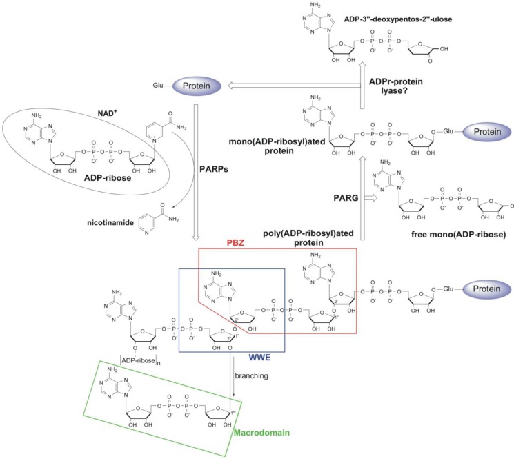
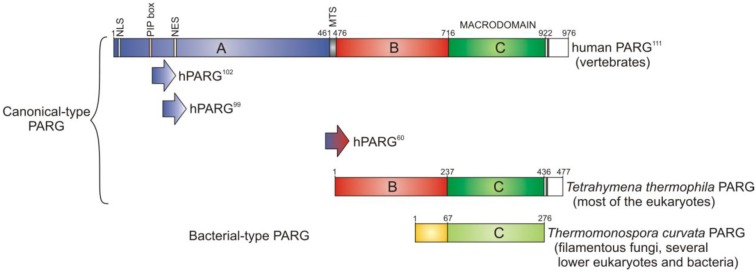
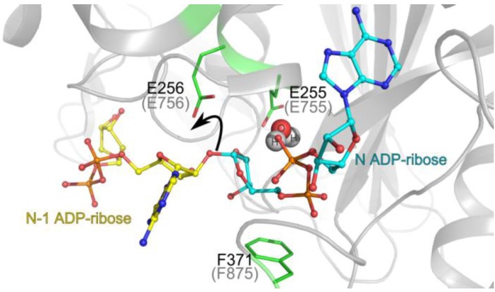
Poly(ADP-ribosyl)ation is a post-translational protein modification involved in the regulation of important cellular functions including DNA repair, transcription, mitosis and apoptosis. The amount of poly(ADP-ribosyl)ation (PAR) in cells reflects the balance of synthesis, mediated by the PARP protein family, and degradation, which is catalyzed by a glycohydrolase, PARG. Many of the proteins mediating PAR metabolism possess specialised high affinity PAR-binding modules that allow the efficient sensing or processing of the PAR signal. The identification of four such PAR-binding modules and the characterization of a number of proteins utilising these elements during the last decade has provided important insights into how PAR regulates different cellular activities. The macrodomain represents a unique PAR-binding module which is, in some instances, known to possess enzymatic activity on ADP-ribose derivatives (in addition to PAR-binding). The most recently discovered example for this is the PARG protein, and several available PARG structures have provided an understanding into how the PARG macrodomain evolved into a major enzyme that maintains PAR homeostasis in living cells.


Figures

Figure 1
Figure 1
Schematic representation of poly(ADP-ribosyl)ation (PAR) metabolism. PARPs catalyse the transfer of an ADP-ribose moiety from NAD+ to a target protein with the concomitant release of nicotinamide. The first ADP-ribose is attached via an ester linkage to the glutamate on the target protein. The subsequent transfer of additional ADP-ribose molecules through the 2',1''- and 2'',1'''-O-glycosidic bond leads to the synthesis of linear and branched PAR polymers, respectively. PARG cleaves PAR chains and releases mono(ADP-ribose), whilst the proximal ADP-ribose is removed by ADP-ribosyl protein lyase. Regions of PAR recognised by Poly(ADP-Ribose)-Binding Zinc Finger (PBZ), WWE and macrodomain PAR-binding modules are boxed.
Figure 2
Figure 2
Domain structure of different PARG proteins. The full-length human PARG111 contains an N-terminal regulatory region (A-domain; blue) which includes a nuclear localisation signal (NLS), PCNA binding motif (PIP box) and a nuclear export signal (NES). Full length human PARG also contains a mitochondrial targeting sequence (MTS), a highly structured and conserved B-domain (red), and the essential catalytic macrodomain (C-domain; green). Two shorter isoforms of human PARG, PARG102 and PARG99, lack the NLS and PIP box, while PARG60 lacks the entire regulatory region. Human and Tetrahymena thermophila PARGs represent canonical-type PARGs and share high conservation in their B-domain and macrodomain. Highly divergent bacterial-type PARG represented by Thermomonospora curvata PARG contains the catalytic macrodomain and an N-terminal accessory element (yellow).
Figure 3
Figure 3
PARG catalytic mechanism. The positions of the reaction product N ADP-ribose (teal backbone; observed in the crystal structure) and a model of N-1 ADP-ribose (yellow) are represented in the Tetrahymena thermophila PARG active site. The key O-glycosydic ribose-ribose bond is positioned in direct hydrogen bonding contact with the catalytic glutamate 256 (Glu756 in human PARG; grey brackets). The 2’-OH leaving group of n-1 ADP-ribose’ is protonated (black arrow) by the catalytic glutamate. A subsequently formed oxocarbenium intermediate is stabilised by the close proximity of the terminal diphosphate group, which is restrained by the conserved phenylalanine 371 (Phe875 in human PARG). A water molecule (shown in spheres) is ideally positioned to attack this oxocarbenium intermediate. This leads to release of ADP-ribose.

References

    1. Hassa P.O., Haenni S.S., Elser M., Hottiger M.O. Nuclear ADP-ribosylation reactions in mammalian cells: where are we today and where are we going? Microbiol. Mol. Bio. Rev. 2006;7:789–829. - PMC - PubMed
    1. Yates S.P., Jørgensen R., Andersen G.R., Merrill R. Stealth and mimicry by deadly bacterial toxins. Trends Biochem. Sci. 2006;31:123–133. doi: 10.1016/j.tibs.2005.12.007. - DOI - PubMed
    1. Holbourn K.P., Shone C.C., Acharya K.R. A family of killer toxins. Exploring the mechanism of ADP-ribosylating toxins. FEBS J. 2006;273:4579–4593. doi: 10.1111/j.1742-4658.2006.05442.x. - DOI - PubMed
    1. Di Girolamo M., Dani N., Stilla A., Corda D. Physiological relevance of the endogenous mono(ADP-ribosyl)ation of cellular proteins. FEBS J. 2005;272:4565–4575. doi: 10.1111/j.1742-4658.2005.04876.x. - DOI - PubMed
    1. Laing S., Unger M., Koch-Nolte F., Haag F. ADP-ribosylation of arginine. Amino acids. 2011;41:257–269. doi: 10.1007/s00726-010-0676-2. - DOI - PMC - PubMed

LinkOut - more resources