. 2013 Jul 25;3(3):435-48.
doi: 10.3390/biom3030435.

Altered sphingolipid metabolism in patients with metastatic pancreatic cancer

Affiliations

Altered sphingolipid metabolism in patients with metastatic pancreatic cancer

Yixing Jiang et al. Biomolecules. .

Abstract

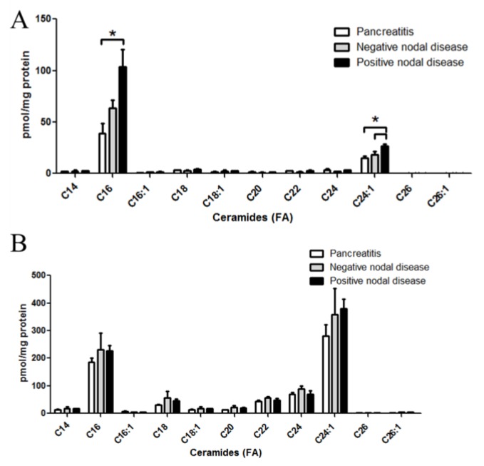
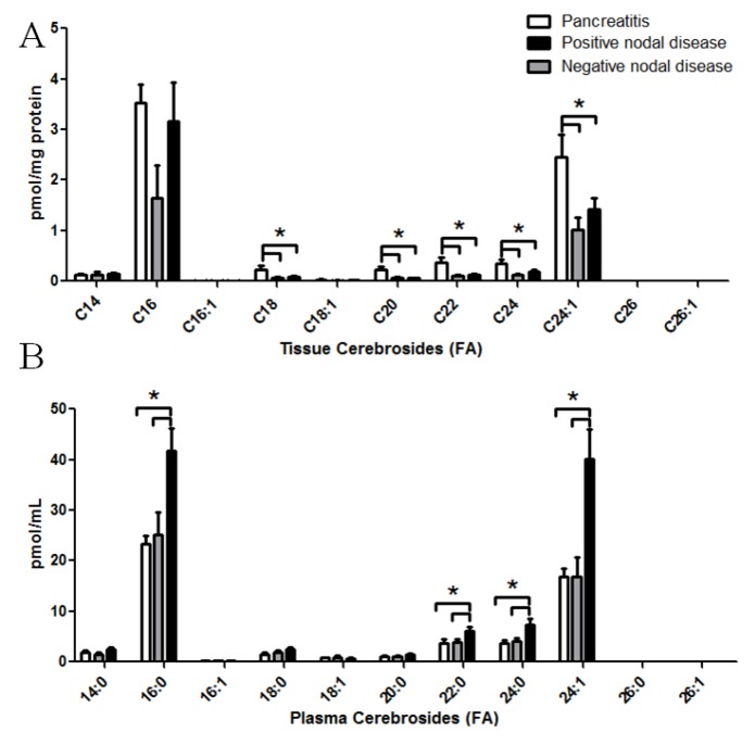
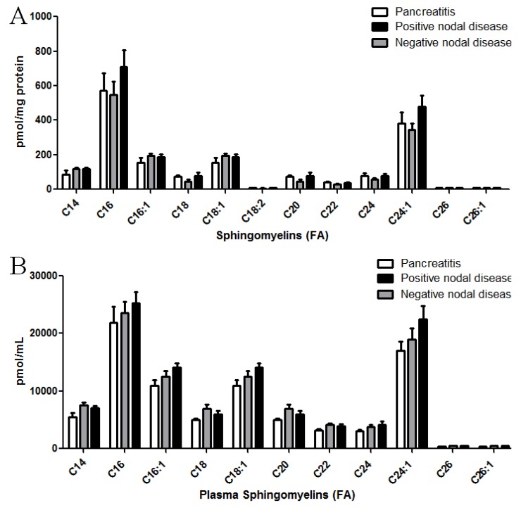
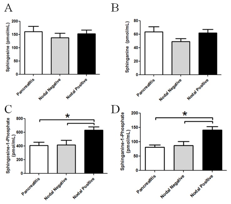
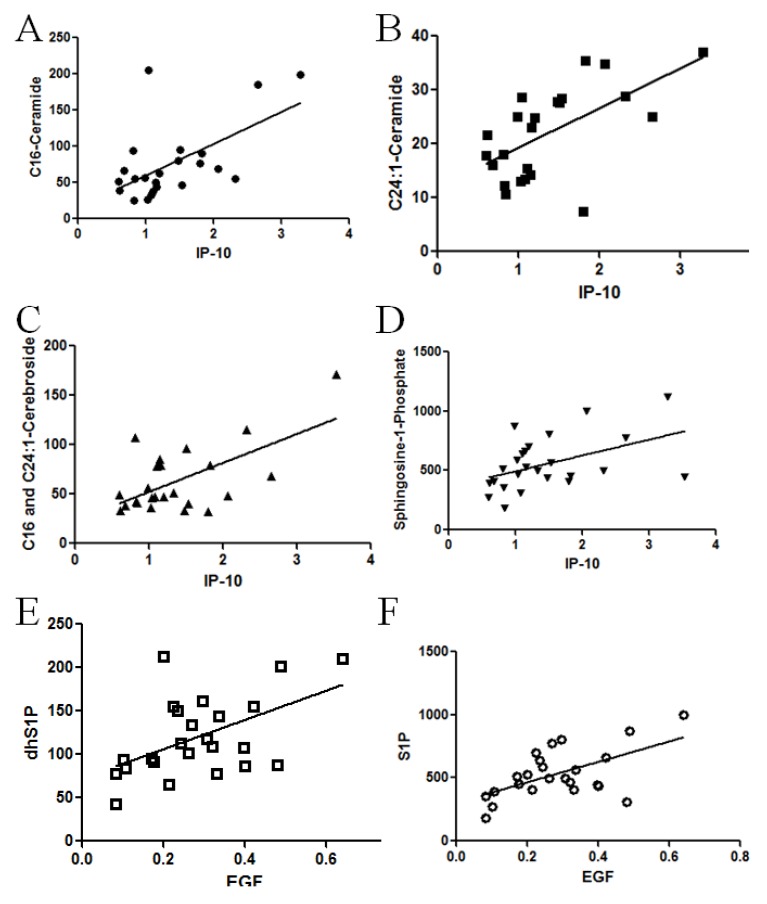
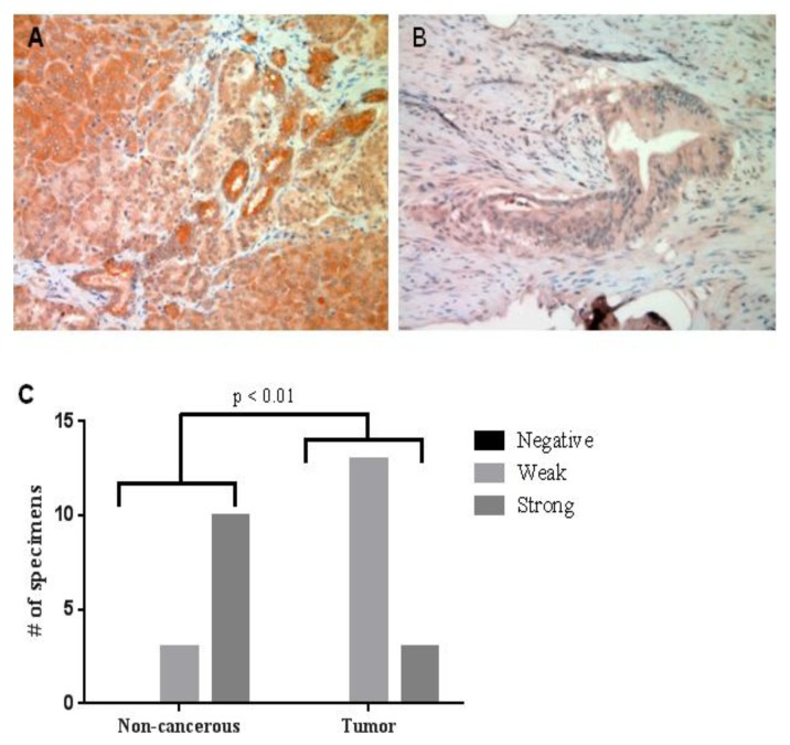
Although numerous genetic mutations and amplifications have been identified in pancreatic cancer, much of the molecular pathogenesis of the disease remains undefined. While proteomic and transcriptomic analyses have been utilized to probe and characterize pancreatic tumors, lipidomic analyses have not been applied to identify perturbations in pancreatic cancer patient samples. Thus, we utilized a mass spectrometry-based lipidomic approach, focused towards the sphingolipid class of lipids, to quantify changes in human pancreatic cancer tumor and plasma specimens. Subgroup analysis revealed that patients with positive lymph node metastasis have a markedly higher level of ceramide species (C16:0 and C24:1) in their tumor specimens compared to pancreatic cancer patients without nodal disease or to patients with pancreatitis. Also of interest, ceramide metabolites, including phosphorylated (sphingosine- and sphinganine-1-phosphate) and glycosylated (cerebroside) species were elevated in the plasma, but not the pancreas, of pancreatic cancer patients with nodal disease. Analysis of plasma level of cytokine and growth factors revealed that IL-6, IL-8, CCL11 (eotaxin), EGF and IP10 (interferon inducible protein 10, CXCL10) were elevated in patients with positive lymph nodes metastasis, but that only IP10 and EGF directly correlated with several sphingolipid changes. Taken together, these data indicate that sphingolipid metabolism is altered in human pancreatic cancer and associated with advanced disease. Assessing plasma and/or tissue sphingolipids could potentially risk stratify patients in the clinical setting.


Figures

Figure 1
Figure 1
Altered ceramide levels in patient specimens with pancreatic cancer. LC-MS/MS was utilized to quantify ceramides from patient specimens. Ceramides with a d18:1 backbone with fatty acids from 14 to 26 carbons were assessed. Molecular species of ceramides were quantified from pancreas tissues (A); and corresponding plasma samples (B) from patients with pancreatitis, non-metastatic (nodal negative) pancreatic cancer, or metastatic (nodal positive) pancreatic cancer. * = p < 0.05 assessed by t-test analysis.
Figure 2
Figure 2
Altered cerebroside levels in patient specimens with pancreatic cancer. LC-MS/MS was utilized to quantify cerebrosides from patient specimens. Cerobrosides with a d18:1 backbone with fatty acids from 14 to 26 carbons were assessed. Molecular species of cerebrosides were quantified from pancreas tissues (A); and corresponding plasma samples (B) from patients with pancreatitis, non-metastatic (nodal negative) pancreatic cancer, or metastatic (nodal positive) pancreatic cancer. * p < 0.05 assessed by t-test analysis.
Figure 3
Figure 3
Sphingomyelin levels in patient specimens with pancreatic cancer. LC-MS/MS was utilized to quantify sphingomyelin levels from patient specimens. Sphingomyelins with a d18:1 backbone with fatty acids from 14 to 26 carbons were assessed. Molecular species of sphingomyelins were quantified from pancreas tissues (A) and corresponding plasma samples (B) from patients with pancreatitis, non-metastatic (nodal negative) pancreatic cancer, or metastatic (nodal positive) pancreatic cancer. * p < 0.05 assessed by t-test analysis.
Figure 4
Figure 4
Altered plasma phosphorylated long chain sphingoid bases in patient samples with pancreatic cancer. LC-MS/MS was utilized to quantify perturbation in the long chain bases from plasma. Graphical representations of the data for (A) sphingosine; (B) sphinganine; (C) sphingosine-1-phosphate; and (D) sphinganine-1-phosphate are shown from the plasma from patients with pancreatitis, nodal negative pancreatic cancer or nodal positive pancreatic cancer. * p < 0.05 assessed by t-test analysis.
Figure 5
Figure 5
Correlation between cytokines and sphingolipid alterations in patient specimens with pancreatic cancer. To determine if a statistical relationship exists between cytokines and sphingolipids in patient specimens, Pearson correlation analysis was performed. (A) Tissue C16-ceramide and plasma IP-10 levels (r = 0.583, p = 0.0035); and (B) tissue C24:1-ceramide and plasma IP-10 levels from tissue demonstrate a positive correlation (r = 0.6012, p = 0.0024); (C) as did plasma C16:0 and C24:1 cerebroside mass (r = 0.6174, p = 0.0013), with plasma IP10; and (D) plasma S1P with IP10 (r = 0.4655, p = 0.019). Plasma EGF levels demonstrated positive correlated with both plasma; (E) S1P (r = 0.6928, p = 0.0001) and (F) dhS1P (r = 0.6787, p = 0.0002) levels.
Figure 6
Figure 6
IP10 expression in pancreatic tumor specimens. IP 10 immunohistochemistry staining shows IP10 expression was down-regulated in tumor tissue (B) compared with non-cancerous, tissue (A). (C) Fisher’s exact test shows the quantitative staining intensity difference between tumor and normal tissue is significant (p < 0.01).

References

    1. Tholey R., Sawicki J.A., Brody J.R. Molecular-based and alternative therapies for pancreatic cancer: Looking “out of the box”. Cancer J. 2012;18:665–673. doi: 10.1097/PPO.0b013e3182793ff6. - DOI - PubMed
    1. Pan S., Brentnall T.A., Kelly K., Chen R. Tissue proteomics in pancreatic cancer study: Discovery, emerging technologies and challenges. Proteomics. 2013;13:710–721. doi: 10.1002/pmic.201200319. - DOI - PMC - PubMed
    1. Lopez-Casas P.P., Lopez-Fernandez L.A. Gene-expression profiling in pancreatic cancer. Expert Rev. Mol. Diagn. 2010;10:591–601. doi: 10.1586/erm.10.43. - DOI - PubMed
    1. Ogretmen B., Hannun Y.A. Biologically active sphingolipids in cancer pathogenesis and treatment. Nat. Rev. Cancer. 2004;4:604–616. doi: 10.1038/nrc1411. - DOI - PubMed
    1. Hannun Y.A., Obeid L.M. Principles of bioactive lipid signalling: Lessons from sphingolipids. Nat. Rev. Mol. Cell. Biol. 2008;9:139–150. doi: 10.1038/nrm2329. - DOI - PubMed

LinkOut - more resources