. 2013 Nov 11;3(4):943-66.
doi: 10.3390/biom3040943.

Control of cell differentiation by mitochondria, typically evidenced in dictyostelium development

Affiliations

Control of cell differentiation by mitochondria, typically evidenced in dictyostelium development

Yasuo Maeda et al. Biomolecules. .

Abstract

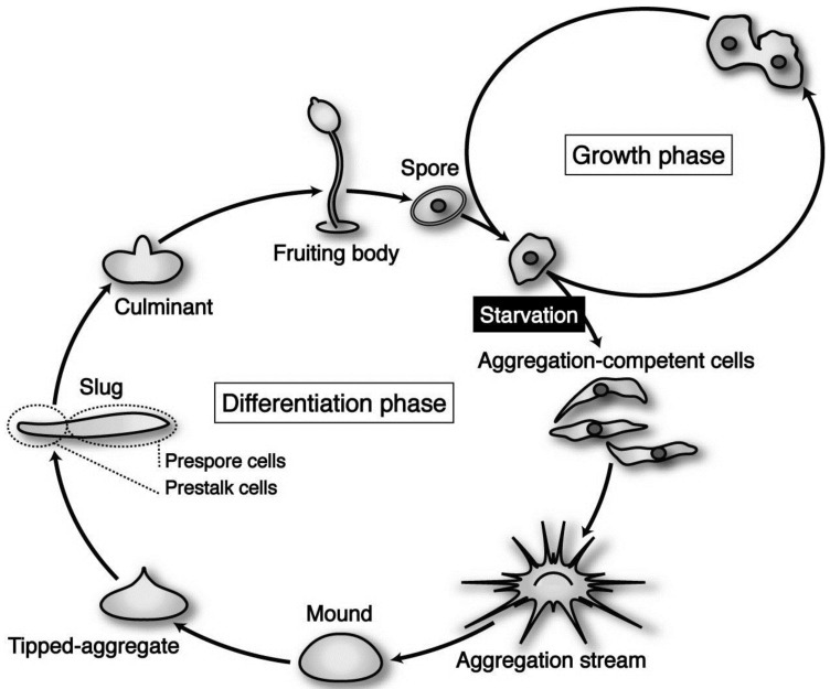
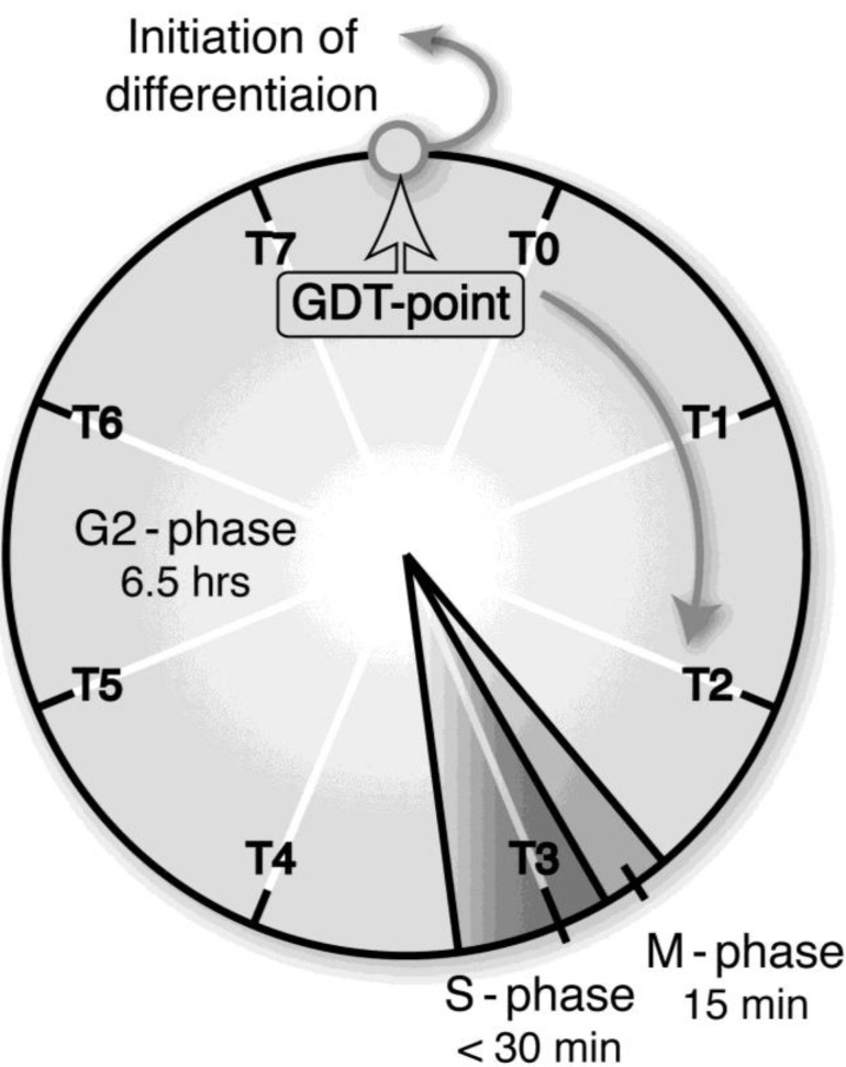
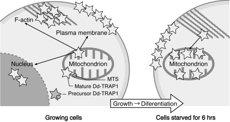
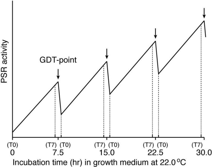
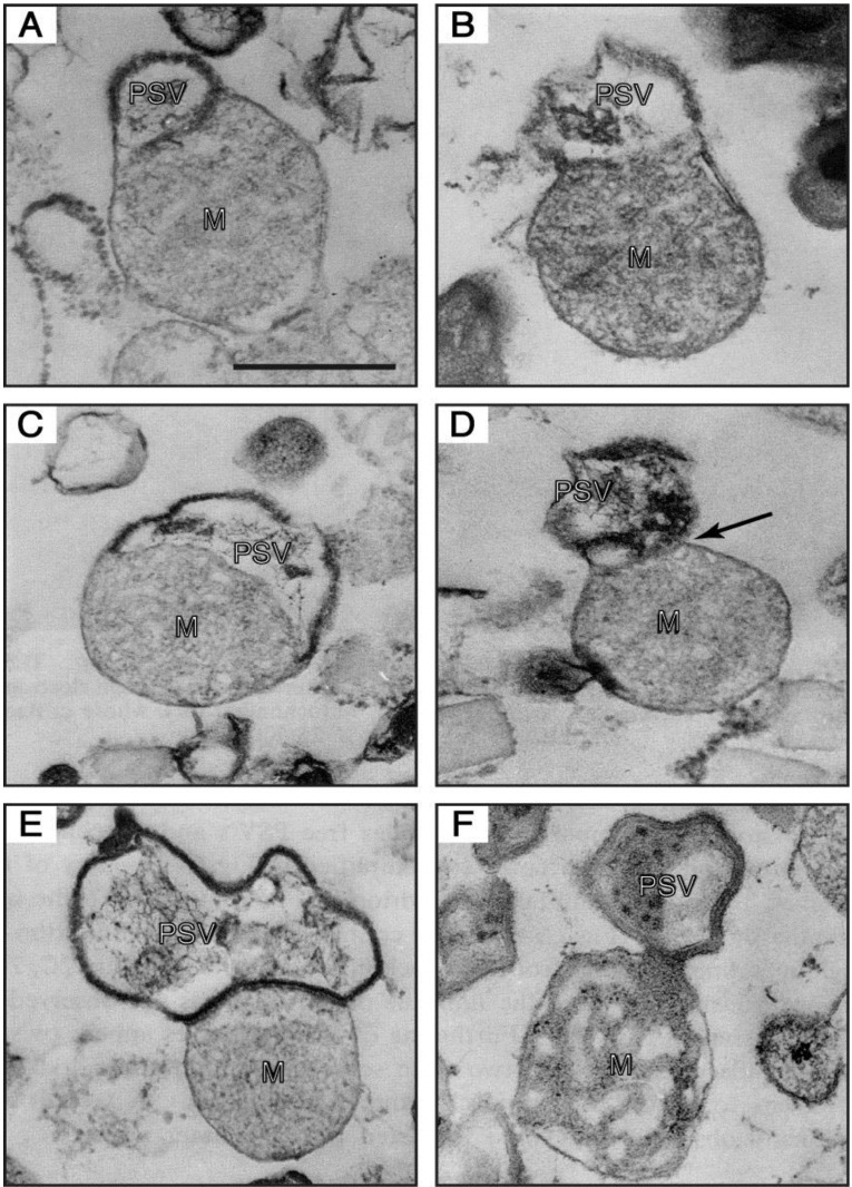
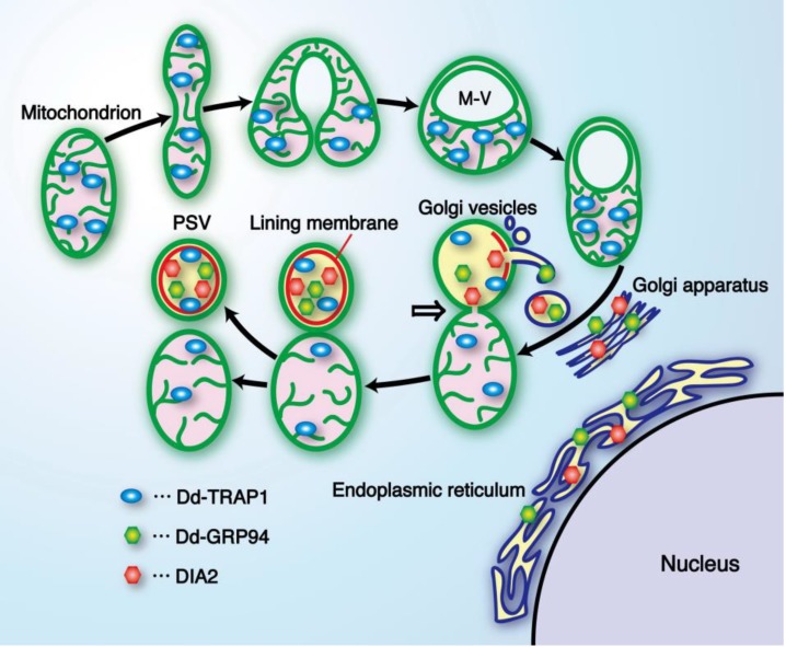
In eukaryotic cells, mitochondria are self-reproducing organelles with their own DNA and they play a central role in adenosine triphosphate (ATP) synthesis by respiration. Increasing evidence indicates that mitochondria also have critical and multiple functions in the initiation of cell differentiation, cell-type determination, cell movement, and pattern formation. This has been most strikingly realized in development of the cellular slime mold Dictyostelium. For example, the expression of the mitochondrial ribosomal protein S4 (mt-rps4) gene is required for the initial differentiation. The Dictyostelium homologue (Dd-TRAP1) of TRAP-1 (tumor necrosis receptor-associated protein 1), a mitochondrial molecular chaperone belonging to the Hsp90 family, allows the prompt transition of cells from growth to differentiation through a novel prestarvation factor (PSF-3) in growth medium. Moreover, a cell-type-specific organelle named a prespore-specific vacuole (PSV) is constructed by mitochondrial transformation with the help of the Golgi complex. Mitochondria are also closely involved in a variety of cellular activities including CN-resistant respiration and apoptosis. These mitochondrial functions are reviewed in this article, with special emphasis on the regulation of Dictyostelium development.


Figures

Figure 1
Figure 1
The life cycle of Dictyostelium discoideum axenic strain Ax-2. The vegetative cells are usually grown in liquid medium, by means of pinocytotic incorporation of external nutrients. Under natural conditions, its parental strain D. discoideum NC-4 grows and multiplies by mitosis at the vegetative phase, phagocytosing nearby bacteria such as Escherichia coli and Klebsiella aerogenes. Upon exhaustion of nutrients, however, starving cells initiate differentiation, form multicellular structures (aggregates; mounds), and undergo a series of well-organized morphogenesis to construct fruiting bodies, each of which is consisting of a mass of spores (sorus) and a supporting cellular stalk. (see the text for details).
Figure 2
Figure 2
A growth/differentiation checkpoint (GDT point) in the cell cycle of a Dictyostelium discoideum Ax-2 cell. The doubling time of axenically growing Ax-2 cells is about 7.2 h and most of their cell cycle is composed of G2-phase with little or no G1-phase and a short period of M- and S-phases. A specific checkpoint (referred to as the GDT point) of GDT is located at the mid–late G2-phase (just after T7 and just before T0). Ax-2 cells progress through their cell cycle to the GDT point, irrespective of the presence or absence of nutrients, and enter the differentiation phase from this point under starvation conditions [2]. T0, T1, and T7 indicates 0, 1, and 7 h, respectively, after a temperature shift from 11.5 °C to 22.0 °C for cell synchrony. The absence of G1 phase in the Dictyostelium cell cycle is not so strange, because there is little or no G1 phase in rapidly dividing cells such as animal cells at the cleavage stage, and also in the true slime mold Physalum and Hydra. (Basically from Maeda [3]).
Figure 3
Figure 3
Strategy for creating rps4-null cells and their phenotypes. (a) As a starting material, LpCSfo cells in which pCoxIV (MTS)-SfoI is expressed under the tetracycline-minus (−Tet) condition, were prepared. Subsequently, the mutant rps4 gene (Mut-mtDNA for homologous recombination), in which the upstream SfoI-site and a 5'-half of rps4 coding region were deleted, was introduced into LpCSfo cells to obtain heteroplasmic transformants (LpCSfoHR cells) with mitochondria consisting of the Mut-mtDNA and wild-type mtDNA (Wt-mtDNA). Coupled with removal of Tet from growth medium, the fusion protein MTS-SfoI synthesized in the cytoplasm is exclusively transferred into mitochondria of LpCSfoHR cells and selectively digests Wt-mtDNA but not Mut-mtDNA. Since the digested Wt-mtDNA is not duplicated, the Mut-mtDNA becomes dominant during the course of growth under the −Tet condition, thus eventually giving rps4-null cells. (b) These cells were grown in growth medium with (+Tet) or without (−Tet) teteracyclin. Membrane potential of mitochondria was visualized by staining of cells with MitoTracker Orange. As was expected, the staining of mitochondria was almost completely vanished in LpCSfo cells grown without Tet, because their mtDNA with an intact SfoI site would be cleaved by SfoI eventually to become a ρ0 state devoid of mitochondrial DNA. Bars, 200 nm. (c) Development of starved MB35 cells and LpCSfoHR cells on agar. MB35 cells and LpCSfoHR cells grown with (+Tet) or without (−Tet) tetracycline were washed twice in BSS and plated on 1.5% non-nutrient agar at a density of 5 × 10⁶ cells/cm2. This was followed by incubation for the indicated time at 22 °C. Bars, 0.5 mm. (Basically from Chida et al. [22]).
Figure 4
Figure 4
Schematic drawing showing the behavior of Dd-TRAP1 during the prestarvation response (PSR) and the initiation of differentiation in Dictyostelium cells (See the text for details).
Figure 5
Figure 5
A working hypothesis for explaining the existence of the GDT-point in the Dictyostelium cell-cycle, by assuming a temporally oscillating PSR activity during synchronized cell growth. This model is based on the fact that there is a good correlation between the expression patterns of the prestarvation (PS) genes and the GDT point-specific genes, and is also constructed from the presumption that the amount of PSFs (i.e., PSF activity) in an ideally synchronized cell population may oscillate in a cell-cycle dependent manner during growth, as shown here. The cell-cycle positions of T0 and T7 offer themselves repeatedly during the course of a completely synchronized growth (see Figure 2). The PSR activity reaches the maximum value at the GDT-point (arrow between T7 and T0) in each cell cycle. Although it is presently unknown whether the expressions of Dd-TRAP1 and/or the amount of PSF-3 actually oscillate in a cell-cycle dependent manner, increasing their basal levels during the progression of synchronous cell growth, this issue remains to be examined in future. The horizontal axis of this figure represents incubation time (h) of an ideally synchronized D. discoideum (Ax-2) cell population in growth medium at 22.0 °C. T0 and T7 indicates 0 and 7 h, respectively, after a temperature shift from 11.5 °C to 22.0 °C for cell synchrony, and these are repeatedly represented during a prolonged time of synchronized culture.
Figure 6
Figure 6
Electron-micrographs of PSV-mitochondrion complexes present in a 47.5%–50% fraction (the intermediate fraction between a lighter pure mitochondria and a heavier pure PSV fractions) of a multilayered sucrose gradient for cell fractionation. (A,B) the outer membrane of a mitochondrion (M) is continuous with the unit membrane of a PSV. (C) a PSV and a mitochondrion are partitioned by a single membrane derived from either the PSV or the mitochondrion, and the electron-dense lining membrane of the PSV is not observed at the contact region. (D) a PSV appears twisted at the contact region (arrow). (E) a completely formed PSV is in close contact with a mitochondrion. (F) a mitochondrial part itself loses its inner structural integrity and seems to be transformed into the PSV. Bar, 500 nm. (From Maeda [49]).
Figure 7
Figure 7
A diagrammatic representation showing formation of the prespore-specific vacuole (PSV) from a mitochondrion and Golgi vesicles. Prior to PSV formation, mitochondria in differentiating prespore cells undergo drastic transformation to form a sort of vacuole (M vacuole), and some Dd-TRAP1 molecules translocate into the M vacuole. Subsequently, Golgi vesicles containing DIA2, Dd-GRP94 and other materials required for PSV formation fuse with the M vacuole, thus resulting in formation of the lining membrane (red) and the internal fibrous structure in the M vacuole. The mitochondrion-PSV complex is eventually twisted at the junction (arrow) and detached to form the respective organelles. Interestingly, almost all of the DIA2 molecules are selectively translocated to PSVs (possibly M vacuoles) in differentiating prespore cells, and seem to be required for exocytotic secretion of PSVs to form the outer-most membrane of spore cell wall [15]. M-V, M vacuole; GRP94, glucose-regulated protein 94 (endoplasmic reticulum Hsp90). (Slightly modified from Maeda [12]).

Similar articles

Cited by

References

    1. Kuspa A., Loomis W. Tagging developmental genes in Dictyostelium by restriction enzyme-mediated integration of plasmid DNA. Proc. Natl. Acad. Sci. USA. 1992;89:8803–8807. doi: 10.1073/pnas.89.18.8803. - DOI - PMC - PubMed
    1. Maeda Y., Ohmori T., Abe T., Abe F., Amagai A. Transition of starving Dictyostelium cells to differentiation phase at a particular position of the cell cycle. Cell Differ. 1989;41:169–175. doi: 10.1111/j.1432-0436.1989.tb00744.x. - DOI - PubMed
    1. Maeda Y. Cell-cycle checkpoint for transition from cell division to differentiation. Dev. Growth Differ. 2011;53:463–481. doi: 10.1111/j.1440-169X.2011.01264.x. - DOI - PubMed
    1. Hartwell L.H., Weinert T.A. Checkpoints: Controls that ensure the order of cell cycle events. Science. 1989;246:629–634. - PubMed
    1. Chen R.H., Shevchenko A., Mann M., Murray A.W. Spindle checkpoint protein Xmad1 recruits Xmad2 to unattached kinetochores. J. Cell Biol. 1998;143:283–295. doi: 10.1083/jcb.143.2.283. - DOI - PMC - PubMed

LinkOut - more resources