. 2014 Jul 15;5(13):5029-39.
doi: 10.18632/oncotarget.2078.

Sulfatase 1 (hSulf-1) reverses basic fibroblast growth factor-stimulated signaling and inhibits growth of hepatocellular carcinoma in animal model

Affiliations

Sulfatase 1 (hSulf-1) reverses basic fibroblast growth factor-stimulated signaling and inhibits growth of hepatocellular carcinoma in animal model

Gaoya Xu et al. Oncotarget. .

Abstract

The human sulfatase 1 (hSulf-1) gene encodes an endosulfatase that functions to inhibit the heparin-binding growth factor signaling, including the basic fibroblast growth factor (bFGF)-mediated pathway, by desulfating the cell surface heparan sulfate proteoglycans (HSPGs). bFGF could stimulate cell cycle progression and inhibit cell apoptosis, this biological effect can be reversed by hSulf-1. However, molecular mechanisms have not been fully reported. In the current study, by reactivation of hSulf-1 expression and function in the hSulf-1-negative hepatocellular carcinoma (HCC) cell lines and HCC xenograft tumors, we found that hSulf-1 blocked the bFGF effect on the promotion of cell cycle and inhibition of apoptosis. The bFGF-stimulated activation of protein kinase B (AKT) and extracellular signal-regulated kinase (ERK) pathways was suppressed by hSulf-1, which led to a decreased expression of the target genes Cyclin D1 and Survivin, then finally induced cell cycle arrest and apoptosis in HCC cells. Our data suggested that hSulf-1 may be a suitable target for cancer therapy.


Figures

Figure 1
Figure 1. hSulf-1 reactivation reversed the bFGF-stimulated cell cycle progression and apoptosis inhibition in HCC cells
(A) SMMC-7721 cells were cultured in 6-well plates at 1×10⁶ cells/well with or without bFGF, and infected with adenovirus Ad5-hSulf1 at a multiplicity of infection (MOI) of 100 pfu/cell. Forty-eight h later, the parental cells, cells cultured in media containing bFGF and cells infected with adenovirus were harvested and the cell lysates were prepared. The expression of hSulf-1 was examined by Western blot. Glyceraldehydes 3-phosphate dehydrogenase (GAPDH) was used as the loading control. (B, C) The harvested cells were stained by propidium iodide (PI) and Annexin V/PI, cell cycle (B) and apoptosis (C) were examined by flow cytometry. The data of all cell lines were showed and analyzed in Table 1.
Figure 2
Figure 2. Suppression of bFGF-induced AKT and ERK signaling by hSulf-1 in HCC cells
MHCC97H and SMMC-7721 cells were cultured in 6-well plates at 1×10⁶ cells/well with bFGF, and infected with adenovirus Ad5-hSulf1 at an MOI of 100 pfu/cell. Forty-eight h later, the expression of the indicated factors were examined by Western blot. GAPDH was used as the loading control. The densitometry analysis of p-AKT and p-ERK levels was performed, normalized with their corresponding total proteins (t-AKT and t-ERK, respectively). Columns, mean of three separate analyses; bars, standard deviation (SD); **P<0.01 and ***P<0.001.
Figure 3
Figure 3. Downegulation of Cyclin D1 and Survivin by hSulf-1 in HCC cells
MHCC97H and SMMC-7721 cells were cultured in 6-well plates at 1×10⁶ cells/well with or without bFGF, and infected with adenovirus Ad5-hSulf1 at an MOI of 100 pfu/cell. Forty-eight h later, the expression of the indicated factors were examined by Western blot. GAPDH was used as the loading control. The densitometry analysis of Cyclin D1 and Survivin levels was performed, normalized with GAPDH content. Columns, mean of three separate analyses; bars, standard deviation (SD); *P<0.05, **P<0.01 and ***P<0.001.
Figure 4
Figure 4. Cyclin D1 and Survivin was downregulated after silencing AKT and ERK expression in HCC cells
MHCC97H and SMMC-7721 cells were cultured in 6-well plates at 1×10⁶ cells/well with bFGF, and transfected with AKT-siRNA or ERK-siRNA at a concentration of 10 μg/well. Cells were harvested at 48 h later and examined the expression of the indicated factors by Western blot. GAPDH was used as the loading control. The densitometry analysis of every factor was performed, normalized with GAPDH content. Columns, mean of three separate analyses; bars, standard deviation (SD); *P<0.05, **P<0.01 and ***P<0.001.
Figure 5
Figure 5. hSulf-1 produces a synergic inhibitory effect on HCC cell proliferation and signaling combined with rapamycin
(A) SMMC-7721 cells were cultured in media containing 0 to 1,000 nmol/L rapamycin together with or without infection of adenovirus Ad5-hSulf1 at MOI of 100 pfu/cell. Cell viability was measured by Cell Proliferation Kit I after cultured for 48 h. (B) SMMC-7721 cells treated with 1,000 nmol/L rapamycin or/and 100 pfu/cell Ad5-hSulf1 were harvested and detected for the expression of AKT, ERK, Cyclin D1 and Survivin by Western blot. GAPDH was used as the loading control.
Figure 6
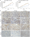
Figure 6. hSulf-1-mediated antitumor efficacy in HCC xenografts in nude mice
(A) The xenograft tumors in nude mice were established by subcutaneously injection of the parental SMMC-7721 cells and the bFGF-cultured SMMC-7721 cells, respectively, 106 cells per mouse. Every model was separated 3 groups, 5 mice per group. After treated with adenoviruses Ad5-hSulf1 and Ad5-EGFP, tumor volume was measured according to the previously reported method and formula [13,39]. Lines, mean of tumor volume (n=5); bars, standard deviation (SD); ***P<0.001. (B) Immunohistochemical examination of the indicated factor expression in the xenograft tumors, original magnification ×200. The data of all the quantitative analyses in every group were showed in Table 2.

References

    1. Wang TR, Yan LY, Yan J, Lu CL, Xia X, Yin TL, Zhu XH, Gao JM, Ding T, Hu WH, Guo HY, Li R, Qiao J. Basic fibroblast growth factor promotes the development of human ovarian early follicles during growth in vitro. >Hum Reprod. 2014. doi: 10.1093/humrep/det465. - PubMed
    1. Lau MT, So WK, Leung PC. Correction: Fibroblast Growth Factor 2 Induces E-Cadherin Down-Regulation via PI3K/Akt/mTOR and MAPK/ERK Signaling in Ovarian Cancer Cells. PLoS One. 2013;8(3):e59083. - PMC - PubMed
    1. Park DJ, Yoon C, Thomas N, Ku GY, Janjigian YY, Kelsen DP, Ilson DH, Goodman KA, Tang LH, Strong VE, Coit DG, Yoon SS. Prognostic Significance of Targetable Angiogenic and Growth Factors in Patients Undergoing Resection for Gastric and Gastroesophageal Junction Cancers. Ann Surg Oncol. 2013. doi: 10.1245/s10434-013-3429-0. - PubMed
    1. Eroglu Z, Tagawa T, Somlo G. Human Epidermal Growth Factor Receptor (HER) Family-Targeted Therapies in the Treatment of HER2-Overexpressing Breast Cancer. Oncologist. 2014. doi: 10.1634/theoncologist.2013-0283. - PMC - PubMed
    1. Chiorean EG, Sweeney C, Youssoufian H, Qin A, Dontabhaktuni A, Loizos N, Nippgen J, Amato R. A phase I study of olaratumab, an anti-platelet-derived growth factor receptor alpha (PDGFRα) monoclonal antibody, in patients with advanced solid tumors. Cancer Chemother Pharmacol. 2014. doi: 10.1007/s00280-014-2389-9. - PubMed

Publication types

MeSH terms