. 2014 Jul 15;5(13):5113-24.
doi: 10.18632/oncotarget.2089.

FoxD3-regulated microRNA-137 suppresses tumour growth and metastasis in human hepatocellular carcinoma by targeting AKT2

Affiliations

FoxD3-regulated microRNA-137 suppresses tumour growth and metastasis in human hepatocellular carcinoma by targeting AKT2

Li-Li Liu et al. Oncotarget. .

Abstract

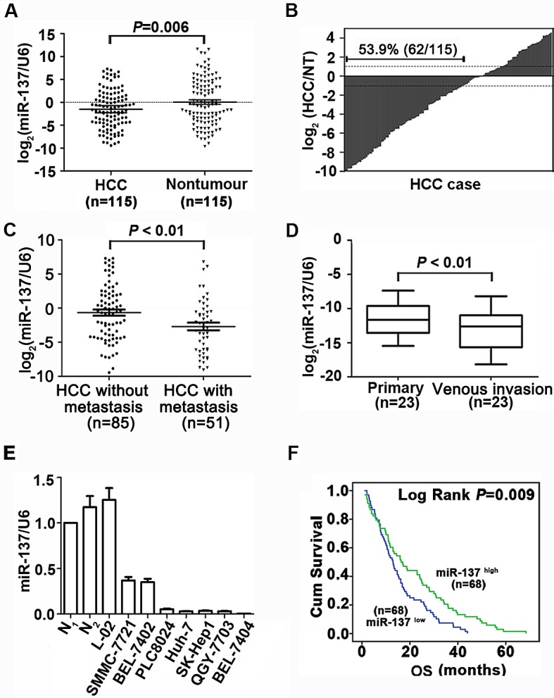
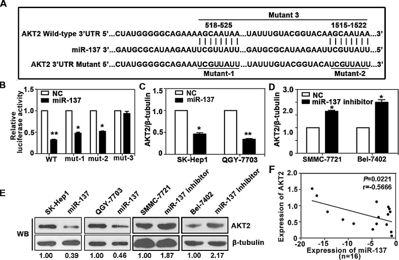
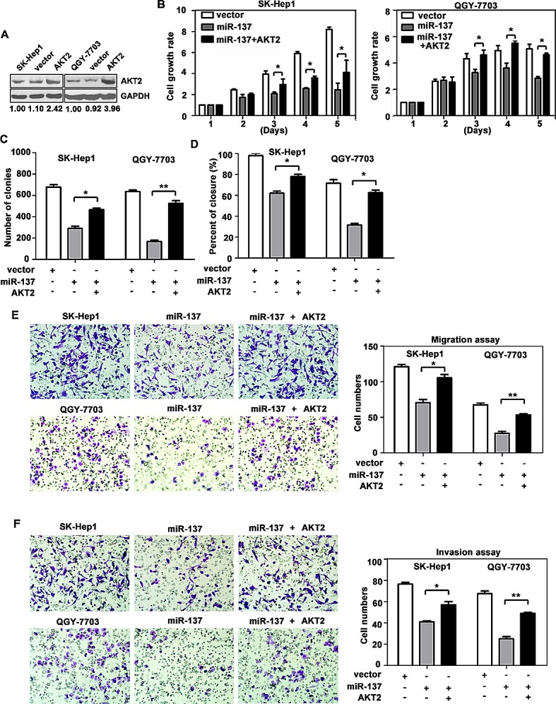
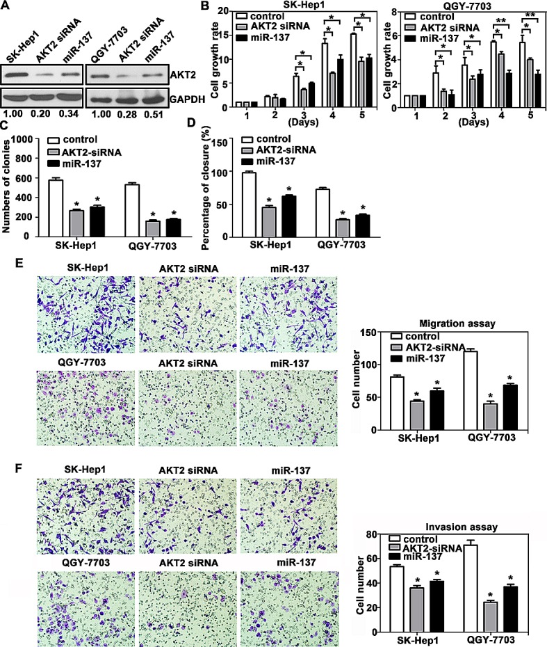
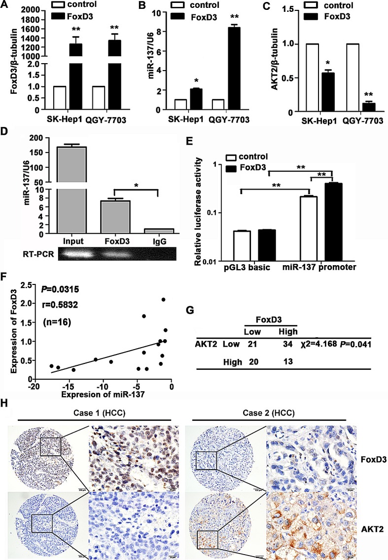
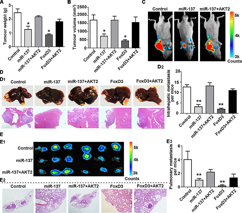
microRNAs, frequently deregulated in human cancer, have been implicated in the progression of hepatocarcinogenesis. Here, we show that microRNA (miR)-137 is significantly down-regulated in hepatocellular carcinoma (HCC). Its decreased expression is associated with vein invasion, incomplete Involucrum, and distant metastasis. Multivariate analysis suggests that miR-137 is an independent indicator for poor survival. We next show that over-expression of miR-137 suppresses cell proliferation, migration and invasion in vitro. Conversely, miR-137 inhibition promotes HCC cell growth. We also identify AKT2 as a key target of miR-137 in this context. Statistical data reveal a reverse correlation of AKT2 and miR-137 expression in HCC patients. Silencing of AKT2 phenotypically copied miR-137-induced phenotypes, whereas re-expression of AKT2 reversed the suppressive effects of miR-137. Further investigations showed that miR-137 exerted its anti-tumour activity via inhibiting the AKT2/mTOR pathway. Moreover, we demonstrate that FoxD3 directly binds to the promoter of miR-137 and activates its transcription. In vivo studies confirm that FoxD3-regulated miR-137 inhibited HCC growth and metastasis via targeting AKT2. Together, our findings indicate that miR-137 is a valuable biomarker for HCC prognosis and the FoxD3/miR-137/AKT2 regulatory network plays an important role in HCC progression.


Conflict of interest statement

All authors declare no conflict of interest.

Figures

Figure 1
Figure 1. miR-137 is frequently down-regulated in HCC and associates with poor outcome
(A, B) miR-137 expression was significantly decreased in HCC compared to the corresponding non-tumourous (NT) livers using qRT-PCR analyses. Expression was shown as a log2-fold change. (C) Scatter plots of the relative expression of miR-137 in patients without metastasis or with metastasis. 136 cases (venous invasion was observed in 29 patients and distant metastasis was observed in 22 patients) were subjected to qRT-PCR. Data were the mean ± SEM. (D) miR-137 expression was down-regulated in metastatic tumours (venous metastases) compared to primary HCC. Twenty-three cases were subjected to qRT-PCR. (E) miR-137 expression in 2 normal liver tissues, the immortalized liver cell line, L-02, and HCC cell lines. miR-137 expression was lower in HCC cell lines compared to normal liver (N1). Data were the mean ± SEM. (F) Decrease in miR-137 was significantly associated with the overall survival of 136 HCC patients. The median was used as the cut-off value to divide patients into low and high expression groups. All *P<0.05, **P<0.01.
Figure 2
Figure 2. miR-137 inhibits HCC cell proliferation, migration and invasion in vitro
(A) miR-137 re-expression was confirmed in SK-Hep1 and QGY-7703 cells using qRT-PCR. (B) miR-137 reduced cell viabilities in HCC. Cells were seeded onto 96-well plates and transfected with miR-137 mimics for 5 days. Cell viabilities were determined using MTT assays. (C) miR-137 suppressed colony formation in HCC. Cells stably expressing miR-137 were cultured with 400 μg/ml G418 for two weeks. The number of colonies was calculated and depicted by the histogramme. Data were represented as the mean + SEM of three independent experiments. (D, E) miR-137 inhibited cell migration and invasion in HCC. Transwell and invasion assays were performed to determine the effect of miR-137 on HCC cell migration and invasion. Representative images (left panel) and the quantification of three randomly selected fields (right panel) are shown. (F) The protein expression levels of E-cadherin, N-cadherin, vimentin, and MMP2 in SK-Hep1 and QGY-7703 cells transfected with miR-137 mimics for 24 h were examined. All *P < 0.05; ** P < 0.01.
Figure 3
Figure 3. AKT2 is a direct target of miR-137 in HCC
(A) Schematic of the construction of wild-type or mutant pGL3-AKT2 3'UTR vectors is indicated. (B) Relative luciferase activities were analysed in QGY-7703 cells. Renilla luciferase vector was used as an internal control. (C) Decrease in AKT2 mRNA expression by miR-137 was determined using qRT-PCR. (D) Increase in AKT2 mRNA expression by miR-137 inhibitor was determined using qRT-PCR. (E) Related expression of AKT2 protein in cells treated with miR-137 or its inhibitor was determined by western blot. (F) AKT2 protein expression was inversely correlated with miR-137 level in 16 pairs of HCC samples using linear regression models. The values of miR-137 were presented by the log2-fold change (HCC/NT). All *P<0.05, **P<0.01.
Figure 4
Figure 4. Reintroduction of AKT2 abrogates miR-137-induced suppression of HCC growth and metastasis
(A) Re-expression AKT2 in HCC cells was examined using western blotting analyses. (B) AKT2 significantly repressed the miR-137-induced reduction of cell viability. Cells were transfected with miR-137 and AKT2 for specific time periods. The cell growth rates were determined using MTT assays. (C) A colony formation assay was performed to confirm the AKT2-mediated abrogation of miR-137-induced growth arrest in HCC. Cells expressing miR-137 and/or AKT2 were cultured for 14 days. Colonies were stained with 0.5% crystal violet and quantified. (D) Wound healing assays were performed on cells transfected with miR-137 and AKT2. The related percentages of wound closure are shown. (E, F) AKT2 reversed the inhibitory effects of miR-137 on cell migration and invasion. Cells transfected with miR-137 and/or AKT2 were transferred to transwell chambers in the absence (E) or presence (F) of Matrigel coating and incubated for 24 h. All *P < 0.05; ** P < 0.01.
Figure 5
Figure 5. AKT2 silencing recapitulates the effects of miR-137 on HCC cells
(A) AKT2 expression was silenced using AKT2 siRNA and miR-137 mimics. (B-F) The experiments described in Figure 4 were re-performed using AKT2 siRNA.
Figure 6
Figure 6. miR-137 is directly regulated by the transcription factor FoxD3
(A-C) Levels of FoxD3 mRNA, miR-137 and AKT2 mRNA were determined. HCC cells were transfected with FoxD3 and an empty vector for 24 h. The related expression of FoxD3 (A), miR-137 (B) and AKT2 (C) were examined using qRT-PCR. (D) FoxD3 directly bound to the promoter of miR-137. ChIP assay was performed in QGY-7703 cells transfected with a vector expressing FoxD3. qRT-PCR was performed using primers specific for −1485 to −1496 of the miR-137 promoter. (E) Luciferase activity of miR-137-luc construct after transfection of FoxD3 plasmid in QGY-7703. (F) FoxD3 protein expression was positively correlated with miR-137 in 16 pairs of HCC patients using linear regression models. The values of miR-137 were presented by the log2-fold change (HCC/NT). (G) FoxD3 expression was inversely correlated with AKT2 in HCC patients. The expression levels of FoxD3 and AKT2 in 88 HCC samples were determined using IHC. The median of IHC score was used as the cut-off value to divide patients into low and high expression groups. (H) The representative imagines were shown that IHC staining of FoxD3 and AKT2 in HCC. *P<0.05, **P<0.01.
Figure 7
Figure 7. miR-137 inhibits HCC growth and metastasis via targeting AKT2 in vivo
(A-C) miR-137 inhibits tumour growth via targeting AKT2 in HCC.1 X 107 QGY-7703 cells of each group were injected into the right flank of nude mice. After 30 days, the tumours were weighed (A) and measured (B). The bioluminescence images of the liver tumours were obtained in the three indicated groups (C). (D, E) miR-137 inhibits tumour metastasis via targeting AKT2 in HCC. Representative images of the liver with metastatic nodules and H&E staining are shown for each group (D1). The average of the nodules in every liver was quantified (D2). Lung metastasis of HCC was identified using bioluminescence and H&E staining (E1-2). The mean of the metastatic nodules in the lung were measured (E3). All *P<0.05, **P<0.01.

References

    1. Jemal A, Bray F, Center MM, Ferlay J, Ward E, Forman D. Global cancer statistics. CA: a cancer journal for clinicians. 2011;61(2):69–90. - PubMed
    1. Forner A, Hessheimer AJ, Isabel Real M, Bruix J. Treatment of hepatocellular carcinoma. Crit Rev Oncol Hematol. 2006;60(2):89–98. - PubMed
    1. El-Serag HB, Rudolph KL. Hepatocellular carcinoma: epidemiology and molecular carcinogenesis. Gastroenterology. 2007;132(7):2557–2576. - PubMed
    1. Bartel DP. MicroRNAs: genomics, biogenesis, mechanism, and function. Cell. 2004;116(2):281–297. - PubMed
    1. Chen CZ. MicroRNAs as oncogenes and tumor suppressors. The New England journal of medicine. 2005;353(17):1768–1771. - PubMed

Publication types

MeSH terms