. 2014 Jul 15;5(13):5153-64.
doi: 10.18632/oncotarget.2095.

The subcellular distribution and function of MTA1 in cancer differentiation

Affiliations

The subcellular distribution and function of MTA1 in cancer differentiation

Jian Liu et al. Oncotarget. .

Abstract

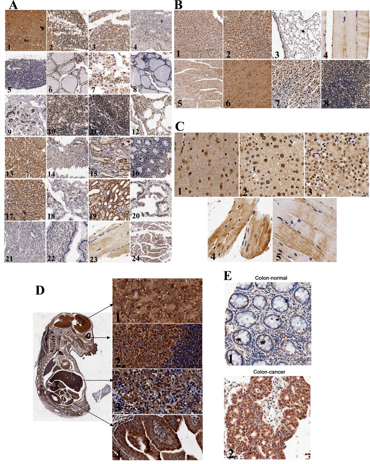
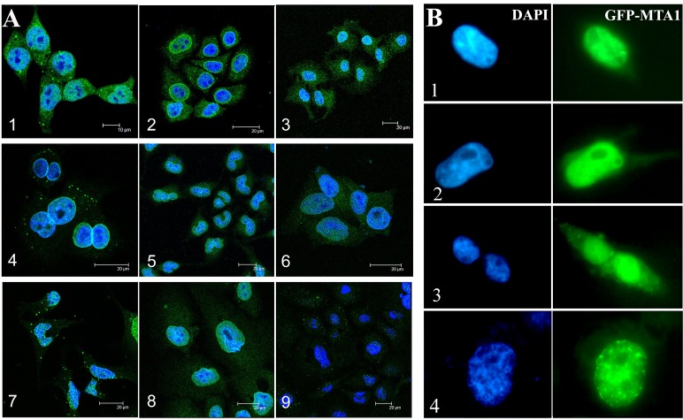
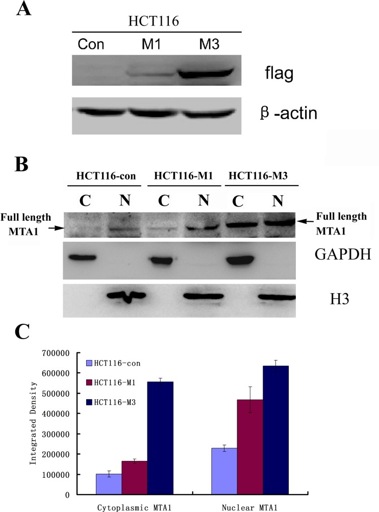
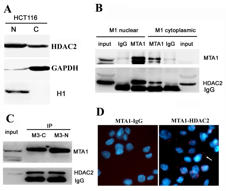
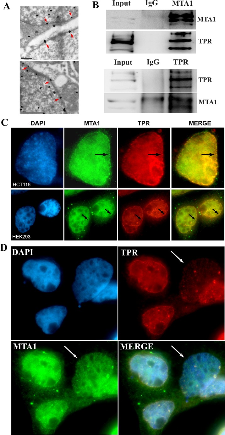
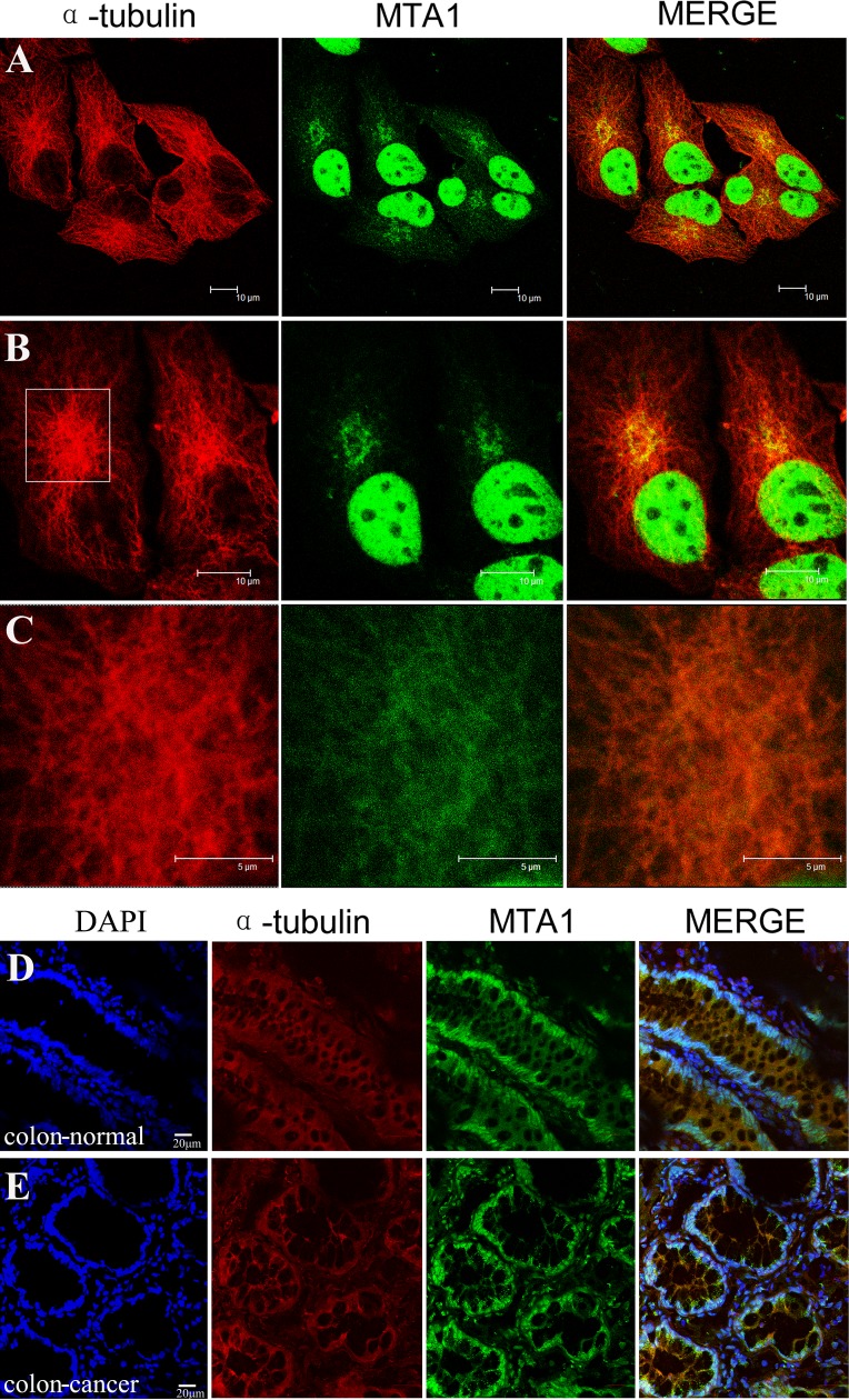
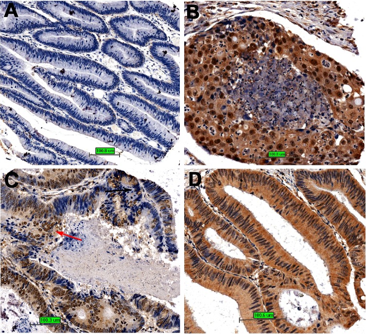
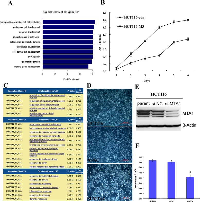
The functions and mechanisms of metastasis-associated protein 1 (MTA1) in cancer progression are still unclear due to a lagged recognition of the subcellular localization. In the present study, using multiple molecular technologies we confirmed for the first time that MTA1 localizes to the nucleus, cytoplasm and nuclear envelope. MTA1 is primarily localized in the nucleus of normal adult tissues but in the cytoplasm of embryonic tissues. While in colon cancer, both distributions have been described. Further investigation revealed that MTA1 localizes on the nuclear envelope in a translocated promoter region (TPR)-dependent manner, while in the cytoplasm, MTA1 shows an obvious localization on microtubules. Both nuclear and cytoplasmic MTA1 are associated with cancer progression. However, these functions may be associated with different mechanisms because only nuclear MTA1 has been associated with cancer differentiation. Overexpression of MTA1 in HCT116 cells inhibited differentiation and promoted proliferation, whereas MTA1 knockdown resulted in cell differentiation and death. Theses results not only suggest that nuclear MTA1 is a good marker for cancer differentiation diagnosis and a potential target for the treatment of cancers but also reveal the necessity to differentially examine the functions of nuclear and cytoplasmic MTA1.


Figures

Figure 1
Figure 1. Expression and localization of endogenous MTA1 in tissues detected using immunohistochemistry
A, Expression of MTA1 in normal adult human tissues: 1. brain, 2. cerebellum, 3. adrenal gland, 4. ovary, 5. pancreas, 6. parathyroid gland, 7. pituitary gland, 8. thyroid, 9. mammary gland, 10.spleen, 11. tonsil, 12. lung, 13. cardiac muscle, 14. esophagus, 15. stomach, 16. colon, 17. liver, 18. salivary gland, 19. kidney, 20. prostate, 21. Endometrium, 22. cervix, 23. skeletal muscle, and 24. throat; B, Expression of MTA1 in normal adult mouse tissues: 1. liver, 2. kidney, 3. lung, 4. skeletal muscle, 5. cardiac muscle, 6. brain, 7. stomach, and 8. spleen; C, Subcellular distribution of MTA1: 1. brain, 2. liver, 3. kidney, 4. cardiac muscle, and 5. skeletal muscle; D, Left, expression of MTA1 in the mouse embryo (day 14); Right, subcellular localization of MTA1 in original tissues: 1. brain, 2. eyes, 3. liver, and 4. intestines; E, Expression and subcellular localization of MTA1 in normal (1) and cancerous (2) colon tissues.
Figure 2
Figure 2. Subcellular localization of MTA1 in cell lines detected through IF
(A) and GFP-tag tracing (B). A, 1. HCT116, 2. NCI-H446, 3. Ishikawa, 4. SF-767, 5. Hep3B, 6. HCT8, 7. HEK293, 8. HaCaT, and 9. Caski; B, Distributions of GFP-MTA1 expression in HEK293 cells. 1 and 2 show cells with low GFP-MTA1 expression in the cytoplasm, 3. cells with high cytoplasmic GFP-MTA1 expression, 4. a cell with apparent GFP-MTA1 fluorescence on the nuclear envelope.
Figure 3
Figure 3. Both nuclear and cytoplasmic MTA1 are enhanced with the increasing expression of exogenous MTA1 in HCT116 cells
A, Western blot analysis of the different levels of flag-tagged MTA1 expression in stably transfected overexpressing HCT116 cell clones, HCT116-M1 and HCT116-M3, using an anti-Flag antibody. B, The expression of full-length MTA1 to the cytoplasm (C) and nucleus (N) were detected through Western blot analysis using an anti-MTA1 antibody. C, Quantitive analysis of the nuclear and cytoplasmic MTA1 in B using Image J software. Results represent the mean±S.D. of triplicate experiments.
Figure 4
Figure 4. MTA1 interacts with HDAC2 at both the nucleus and cytoplasm
A, Western blot analysis showing the localization of HDA2 to both the nucleus (N) and cytoplasm (C) of HCT116 cells; B, In vitro immunoprecipitation analysis of the MTA1-HDAC2 interaction in the nuclear and cytoplasmic fractions of HCT116-M1 cells; C, In vitro immunoprecipitation analysis of the MTA1-HDAC2 interaction in the nuclear and cytoplasmic fractions of HCT116-M3 cells; D, In situ PLA analysis. left, the control interaction detected using mouse antibody against MTA1 and rabbit IgG; right, in situ PLA visualization of the MTA1-HDAC2 interaction in both the nucleus and cytoplasm of HCT116 cells. The white arrow shows the positive signal in the cytoplasm.
Figure 5
Figure 5. MTA1 interacts and colocalizes with TPR at the nuclear envelope
A. The localization of MTA1 on the nuclear envelope (indicated with red arrows) using immuno-electron microscopy; B. Co-IP analysis of the interaction between MTA1 and TPR; C. Immuno-colocalization of MTA1 and TPR at the nuclear envelope of HCT116 and HEK293 cells is indicated with black arrows; D. The knock down of TPR in HEK293 cells impairs the nuclear envelope localization of MTA1. The white arrow indicates a typical TPR knocked down cell.
Figure 6
Figure 6. Localization of MTA1 on microtubules in the cytoplasm
NCI-H446 cells and colon tissues were immuno-stained with specific antibodies against MTA1 and α-tubulin, followed by detection using a laser scanning confocal microscope. The localization of MTA1 on microtubules in NCI-H446 cells is shown in A, B, and C. An enlargement of the rectangular region in B is shown in C. D and E show the localization of MTA1 on microtubules in the cytoplasm of normal and cancerous colon tissues respectively.
Figure 7
Figure 7. The relationship between MTA1 and cancer differentiation
A. Cancer tissue is well differentiated with little MTA1 expression; B, The cancer tissue is poorly differentiated with high nuclear MTA1 expression; C, Different regions with different nuclear MTA1 expression in colon cancer tissue show different differentiation states; D, The cancer tissue is well differentiated with high cytoplasmic but low nuclear MTA1 expression.
Figure 8
Figure 8. Role of MTA1 in cancer differentiation
A. The genes down-regulated in response to MTA1 overexpression are highly enriched on gut development and differentiation; B. MTA1 promotes the proliferation of cancer cells; C. The genes up-regulated in response to MTA1 knockdown show the highest enrichment score on the functional annotation cluster for development and differentiation; D. Transient knock down of MTA1 using siRNA causes the floating and death of HCT116 colon cancer cells; E. Western blot analysis showing the effect of MTA1 siRNA (si-MTA1) on HCT116 cells; F. At 72 h after transfection, the number of survival si-MTA1 transfected cells was 2/3 that of control cells (P<0.05).

Similar articles

Cited by

References

    1. Toh Y, Pencil SD, Nicolson GL. A novel candidate metastasis-associated gene, mta1, differentially expressed in highly metastatic mammary adenocarcinoma cell lines. cDNA cloning, expression, and protein analyses. J Biol Chem. 1994;269:22958–22963. - PubMed
    1. Toh Y, Pencil SD, Nicolson GL. Analysis of the complete sequence of the novel metastasis-associated candidate gene, mta1, differentially expressed in mammary adenocarcinoma and breast cancer cell lines. Gene. 1995;159:97–104. - PubMed
    1. Pencil SD, Toh Y, Nicolson GL. Candidate metastasis-associated genes of the rat 13762NF mammary adenocarcinoma. Breast Cancer Res Treat. 1993;25:165–174. - PubMed
    1. Toh Y, Nicolson GL. The role of the MTA family and their encoded proteins in human cancers: molecular functions and clinical implications. Clin Exp Metastasis. 2009;26:215–227. - PubMed
    1. Xue Y, Wong J, Moreno GT, Young MK, Cote J, Wang W. NURD, a novel complex with both ATP-dependent chromatin-remodeling and histone deacetylase activities. Mol Cell. 1998;2:851–861. - PubMed

Publication types

MeSH terms