. 2014 Sep 1;117(5):440-51.
doi: 10.1152/japplphysiol.00084.2014. Epub 2014 Jun 26.

Surface tension in situ in flooded alveolus unaltered by albumin

Affiliations

Surface tension in situ in flooded alveolus unaltered by albumin

Angana Banerjee Kharge et al. J Appl Physiol (1985). .

Abstract

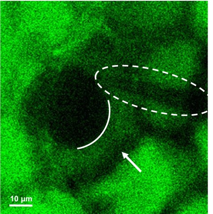
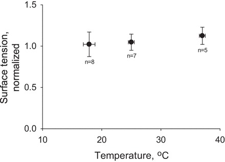
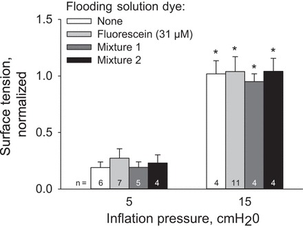
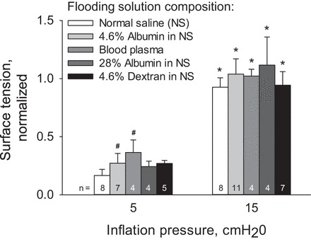
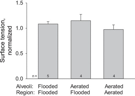
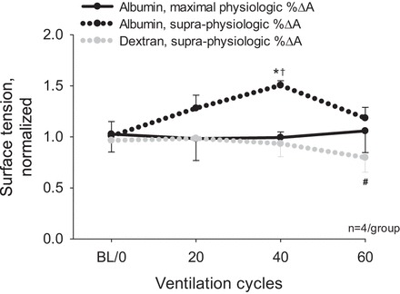
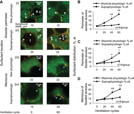
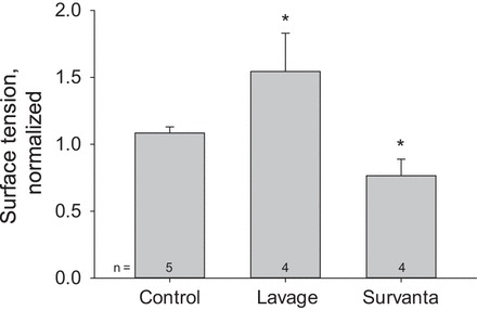
In the acute respiratory distress syndrome, plasma proteins in alveolar edema liquid are thought to inactivate lung surfactant and raise surface tension, T. However, plasma protein-surfactant interaction has been assessed only in vitro, during unphysiologically large surface area compression (%ΔA). Here, we investigate whether plasma proteins raise T in situ in the isolated rat lung under physiologic conditions. We flood alveoli with liquid that omits/includes plasma proteins. We ventilate the lung between transpulmonary pressures of 5 and 15 cmH2O to apply a near-maximal physiologic %ΔA, comparable to that of severe mechanical ventilation, or between 1 and 30 cmH2O, to apply a supraphysiologic %ΔA. We pause ventilation for 20 min and determine T at the meniscus that is present at the flooded alveolar mouth. We determine alveolar air pressure at the trachea, alveolar liquid phase pressure by servo-nulling pressure measurement, and meniscus radius by confocal microscopy, and we calculate T according to the Laplace relation. Over 60 ventilation cycles, application of maximal physiologic %ΔA to alveoli flooded with 4.6% albumin solution does not alter T; supraphysiologic %ΔA raise T, transiently, by 51 ± 4%. In separate experiments, we find that addition of exogenous surfactant to the alveolar liquid can, with two cycles of maximal physiologic %ΔA, reduce T by 29 ± 11% despite the presence of albumin. We interpret that supraphysiologic %ΔA likely collapses the interfacial surfactant monolayer, allowing albumin to raise T. With maximal physiologic %ΔA, the monolayer likely remains intact such that albumin, blocked from the interface, cannot interfere with native or exogenous surfactant activity.

Keywords: albumin; alveolar edema; plasma proteins; surface tension; surfactant.


Figures

Fig. 1.
Fig. 1.
Surface tension determination method. Image (24-μm subpleural depth) of flooded alveolus at transpulmonary pressure, PALV, of 15 cmH2O, obtained with a ×10 air objective. Meniscus (solid arc) separates alveolar liquid phase [native liquid phase plus injected 4.6% albumin solution with 2′,7′-bis-(2-carboxyethyl)-5-(and-6)-carboxyfluorescein (BCECF); green] and air (black). Alveolar walls are also black (arrow). Tip of micropipette (dashed oval) inserted in alveolar liquid phase for pressure measurement by servo-nulling system. Following pipette removal, Z-stack of confocal images of meniscus is obtained (×20 water immersion objective) for determination of meniscus radius.
Fig. 2.
Fig. 2.
Temperature control chamber. Plexiglass steam chamber (10-cm diameter × 3.5-cm height, 5.5-cm diameter hole in top surface) designed to maintain lung at 37°C. Steam is imported from an enclosed water bath through insulated silicone tubing. The silicone tubing leads to one inlet of a Y-connector. Steam escapes through the free port of the Y-connector and an additional port on the far side of the chamber. Pivoting the Y-connector regulates chamber temperature. A plexiglass guard between the steam entry point and the lung protects the lung from direct steam exposure. A thermometer inserted through a port in the chamber wall monitors temperature. Plastic wrap layers cover chamber's top hole and lung surface. Central holes in each plastic wrap layer allow access to lung surface for micropuncture and imaging. During micropuncture, plastic wrap layers are in contact with one another. At all other times, including during imaging with the ×20 objective, the top plastic layer seals the top the of chamber.
Fig. 3.
Fig. 3.
Interfacial curvature in flooded and aerated alveoli at PALV of 15 cmH2O. Solid arcs mark interface in representative X–Y and Y–Z sections. Dashed lines indicate X-locations of Y–Z sections. A: meniscus of a flooded alveolus. Liquid is labeled with green fluorescence (fluorescein); air is nonfluorescent. Meniscus radius, determined from a three-dimensional set of points marked along the interface in sections between 39 and 51 μm in subpleural depth, is 15.6 μm. B: air-liquid interface in corner of an alveolus in the aerated lung. Perfusate is labeled with fluorescein such that vasculature and liquid lining layer exhibit high (circle) and low (triangle) degrees, respectively, of green fluorescence intensity. Air is nonfluorescent. Interface radius, determined from sections between 6 and 12 μm in subpleural depth, is 17.7 μm.
Fig. 4.
Fig. 4.
Temperature effect on surface tension in flooded alveoli at PALV of 15 cmH2O. Flooding is with 4.6% albumin solution containing fluorescein. Ventilation is according to protocol IA. Surface tension is normalized by average value of 12.0 mN/m at PALV of 15 cmH2O.
Fig. 5.
Fig. 5.
Inflation pressure and dye effects on surface tension in alveoli flooded with 4.6% albumin solution. Mixture 1 contains 31 μM BCECF, 19 μM calcein red-orange AM, 95 nM lysotracker red, and 476 nM FM 4–64. Mixture 2 contains 31 μM fluorescein, 40 μM calcein AM, 263 nM lysotracker red, and 439 nM FM 1–43. Ventilation according to protocol I. Surface tension at both PALV normalized by average value of 12.0 mN/m at PALV of 15 cmH2O. Inflation increases surface tension (*P < 0.01 vs. PALV of 5 cmH2O data point in same group). Dye inclusion does not alter surface tension.
Fig. 6.
Fig. 6.
Alveolar liquid composition effect on surface tension in flooded alveoli at two inflation pressures. Flooding solution, of indicated composition, is labeled with fluorescein. Ventilation according to protocol I. Inflation increases surface tension: *P < 0.01 vs. PALV of 5 cmH2O data point in same group. At PALV of 5 cmH2O: #P < 0.05 vs. normal saline group. At PALV of 15 cmH2O, liquid composition does not alter surface tension.
Fig. 7.
Fig. 7.
Surface tension, T, in aerated and flooded alveoli at PALV of 15 cmH2O. In a lung region flooded with 4.6% albumin solution containing fluorescein, T is determined in both flooded and aerated alveoli. In the aerated lung, the liquid lining layer is labeled with fluorescein via the perfusate; perfusion is paused for T determination. Ventilation according to protocol IA.
Fig. 8.
Fig. 8.
Ventilation pattern and alveolar liquid protein content effects on surface tension in flooded alveoli. Flooding solutions contain fluorescein. Ventilation according to protocol II between PALV of 5 and 15 or 1 and 30 cmH2O applying maximal physiologic or supraphysiologic surface area compression, %ΔA, respectively. Surface tension is determined at 15 cmH2O (on deflation with supraphysiologic %ΔA ventilation). Statistical differences: *P < 0.01 vs. baseline (BL/0) data point in same experimental group; †P < 0.01 vs. other two data groups at same time point; #P < 0.05 vs. albumin/supraphysiologic %ΔA ventilation at same time point.
Fig. 9.
Fig. 9.
Ventilation protocol effect on surfactant distribution in aerated and flooded alveoli of a flooded lung region. Region flooded with 4.9% albumin solution containing BCECF and FM 4–64. Ventilation according to protocol II. A: confocal microscopy images show alveolar liquid (green) and alveolar surfactant (yellow/orange) before and after maximal physiologic or supraphysiologic %ΔA ventilation. Arrowheads mark example surfactant locations at the perimeters (white) of aerated (A) and liquid-flooded (L) alveoli or at the menisci (blue) of flooded alveoli. Supraphysiologic %ΔA ventilation sometimes causes fluid movement between alveoli. In Aii, over 60 ventilation cycles, an originally flooded alveolus (#2) clears and an originally aerated alveolus (#1) becomes flooded. Group data demonstrate that supraphysiologic %ΔA ventilation causes greater surfactant spreading than maximal physiologic %ΔA ventilation at the aerated alveolar perimeter (B), the flooded alveolar perimeter (C), and the flooded alveolar meniscus (D). Statistical differences: *P < 0.01 vs. baseline; #P < 0.01 vs. maximal physiologic %ΔA ventilation at same time point.
Fig. 10.
Fig. 10.
Detection of increased and decreased surface tension at PALV of 15 cmH2O. Control: alveoli flooded with 4.6% albumin solution containing fluorescein. Lavage: lung lavaged with normal saline solution (see text for details). Survanta: alveoli flooded with 4.6% albumin solution containing 0.9% Survanta and fluorescein. Ventilation according to protocol IA. Statistical differences: *P < 0.01 vs. control.

Comment in

Similar articles

Cited by

References

    1. Ashino Y, Ying X, Dobbs LG, Bhattacharya J. [Ca2+]i oscillations regulate type II cell exocytosis in the pulmonary alveolus. Am J Physiol Lung Cell Mol Physiol 279: L5–L13, 2000. - PubMed
    1. Bachofen H, Schürch S, Michel RP, Weibel ER. Experimental hydrostatic pulmonary edema in rabbit lungs. Morphology. Am Rev Respir Dis 147: 989–996, 1993. - PubMed
    1. Bachofen H, Schürch S, Urbinelli M, Weibel ER. Relations among alveolar surface tension, surface area, volume, and recoil pressure. J Appl Physiol 62: 1878–1887, 1987. - PubMed
    1. Bhattacharya J, Gropper MA, Staub NC. Interstitial fluid pressure gradient measured by micropuncture in excised dog lung. J Appl Physiol 56: 271–277, 1984. - PubMed
    1. Brower RG, Fessler HE. Another “negative” trial of surfactant. Time to bury this idea? Am J Respir Crit Care Med 183: 966–968, 2011. - PubMed

Publication types