. 2014 Aug 15;289(33):23233-23245.
doi: 10.1074/jbc.M114.550046. Epub 2014 Jun 26.

Dissection of the adenoviral VA RNAI central domain structure reveals minimum requirements for RNA-mediated inhibition of PKR

Affiliations

Dissection of the adenoviral VA RNAI central domain structure reveals minimum requirements for RNA-mediated inhibition of PKR

Jo L Wilson et al. J Biol Chem. .

Abstract

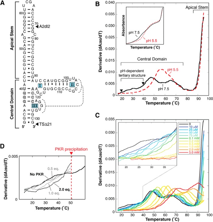
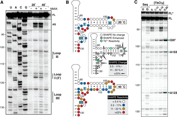
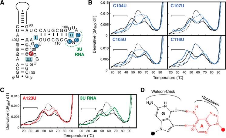
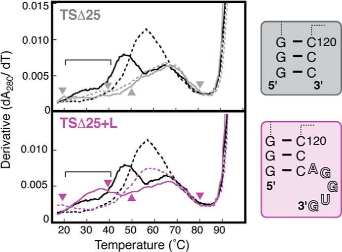
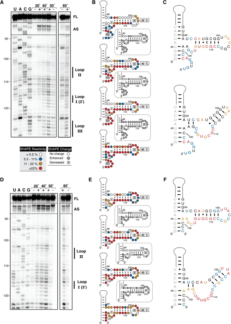
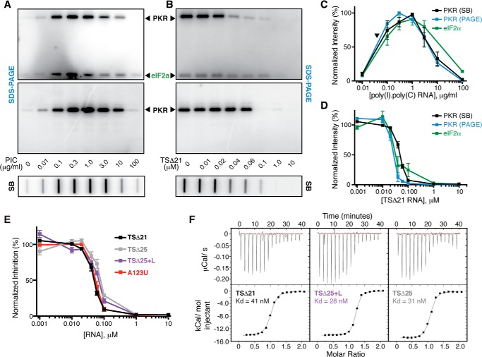
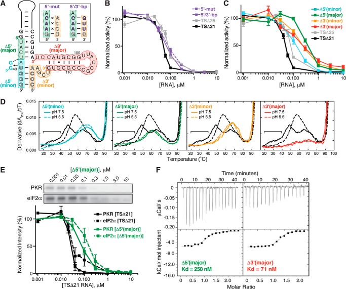
Virus-associated RNA I (VA RNAI) is a short (∼160-nucleotide) non-coding RNA transcript employed by adenoviruses to subvert the innate immune system protein double-stranded RNA-activated protein kinase (PKR). The central domain of VA RNAI is proposed to contain a complex tertiary structure that contributes to its optimal inhibitory activity against PKR. Here we use a combination of VA RNAI mutagenesis, structural analyses, as well as PKR activity and binding assays to dissect this tertiary structure and assess its functional role. Our results support the existence of a pH- and Mg(2+)-dependent tertiary structure involving pseudoknot formation within the central domain. Unexpectedly, this structure appears to play no direct role in PKR inhibition. Deletion of central domain sequences within a minimal but fully active construct lacking the tertiary structure reveals a crucial role in PKR binding and inhibition for nucleotides in the 5' half of the central domain. Deletion of the central domain 3' half also significantly impacts activity but appears to arise indirectly by reducing its capacity to assist in optimally presenting the 5' half sequence. Collectively, our results identify regions of VA RNAI critical for PKR inhibition and reveal that the requirements for an effective RNA inhibitor of PKR are simpler than appreciated previously.

Keywords: Adenovirus; Isothermal Titration Calorimetry (ITC); Mutagenesis; Protein Kinase RNA-activated (PKR); RNA Structure; RNA-Protein Interaction; SHAPE Structure Probing; Translation Control; UV Melting.


Figures

FIGURE 1.
FIGURE 1.
The VA RNAI central domain contains putative tertiary interactions that are stabilized by low pH, Mg2+, and PKR. A, TSΔ21, the truncated form of adenovirus type 2 VA RNAI, used as the starting point in this study. This RNA construct lacks all terminal stem nucleotides (TSΔ21) and has a six-nucleotide deletion in the apical stem (A2dl2). The proposed pseudoknot interaction between loop II and loop III (dashed lines) and conserved complementary tetranucleotide sequences (outline font) are highlighted. B, UV melting analysis of VA RNAI with combined TSΔ21 and A2dl2 deletions. Melting profiles depict the first derivative of the UV melting curve (inset) at pH 5.5 (red) and pH 7.5 (black). Apparent transitions (“peaks”) in the melting profile correspond to unfolding of structures within the VA RNAI apical stem (partly visible at >90 °C) and central domain (three at pH 7.5 and two at pH 5.5 over the range of 20–80 °C) are indicated. Of the central domain transitions, the first is assigned to the putative RNA tertiary structure and is stabilized dramatically at low pH (rightward shift of profile in red dashed line). Arrowheads denote temperatures used for SHAPE probing of this RNA. The first apparent transition is also stabilized by both Mg2+ ion (C) and PKR (D). The concentration of divalent ion or molar equivalents (eq.) of PKR included in the experiment is indicated.
FIGURE 2.
FIGURE 2.
Identification of tertiary interactions and Mg2+ binding sites within the VA RNAI central domain. A, representative gel showing SHAPE probing of TSΔ21 RNA below (20 °C) and above (40 °C) the apparent transition corresponding to the central domain tertiary structure unfolding (marked with arrowheads in Fig. 1B). Lanes with (+) and without (−) SHAPE reagent (NMIA) and RT reactions with dideoxynucleotides (U, A, C, G) for RNA sequencing are indicated. VA RNAI nucleotide numbering is shown on the left, and bands corresponding to central domain loops I-III, the apical stem (AS) loop, and full-length product (FL) are indicated on the right. B, quantification of SHAPE reactivity at 20 (top panel) and 40 °C (bottom panel) and changes between these temperatures (center panel). Normalized band intensities were averaged from three sets of reactions and categorized according to the reactivity keys shown. C, Tb3+ probing of the VA RNAI central domain with sites of RNA cleavage enhanced by increasing concentrations of Tb3+ (0–0.25 mm) indicated (right), and shown on the secondary structure map in B (center panel). Sequencing lanes (Seq) contain the complementary dideoxynucleotide. FL*, full-length product with contrast reduced 5×.
FIGURE 3.
FIGURE 3.
A123 is the protonated nucleotide within the VA RNAI central domain. A, central domain secondary structure highlighting the four individual single-stranded C-to-U mutants (blue circles), mutation of all three loop II C nucleotides to U (3U RNA, green), and the A123-to-U point mutant (red circle). B and C, UV melting profiles of C104U, C105U, C107U, C116U (blue), A123U (red), and 3U RNA (green). The melting profile for each RNA is shown at pH 7.5 (solid line) and pH 5.5 (dashed line) and compared with those of TSΔ21 RNA (black). The region corresponding to unfolding of the TSΔ21 RNA pH-dependent tertiary structure at pH 7.5 is marked with a horizontal bar. D, the cis Watson-Crick/Hoogsteen A+·G base pair proposed for A123+·G97, highlighting the available base edges for potential additional interactions as discussed in the text.
FIGURE 4.
FIGURE 4.
Loop III nucleotides 123–127 are sufficient to form the central domain pseudoknot structure. Melting profiles of TSΔ25+L (purple) and TSΔ25 (gray) RNAs shown at pH 7.5 (solid line) and pH 5.5 (dashed line) and compared with those of TSΔ21 RNA (black). The region corresponding to unfolding of the pH-dependent tertiary structure present in TSΔ25+L RNA, but absent in TSΔ25 RNA, is marked with a horizontal bar. Arrowheads on each profile denote temperatures used for SHAPE probing of the corresponding RNA. Loop III nucleotides are shown in outline font in the structure of TSΔ25+L RNA (right panel).
FIGURE 5.
FIGURE 5.
SHAPE analysis of TSΔ25+L and TSΔ25 RNAs. A, representative gel showing SHAPE probing of TSΔ25+L RNA at temperatures below (20 °C), between (40 and 50 °C), and above (85 °C) the apparent transitions corresponding to the unfolding of the central domain structure. Lanes with (+) and without (−) SHAPE reagent (NMIA) and RT reactions with dideoxynucleotides (U, A, C, and G) for RNA sequencing are indicated. VA RNAI nucleotide numbering is shown on the left, and bands corresponding to central domain loops I-III, the apical stem (AS) loop, and full-length product (FL) are indicated on the right. B, quantification of SHAPE reactivity from low to high temperature (top to bottom) with relative changes shown between each pair of temperatures (dashed boxes). Normalized band intensities were averaged from three sets of reactions and categorized according to the reactivity key shown. C, nucleotide flexibility at 20 °C mapped on the nucleotide sequence of TSΔ25+L RNA in the currently established central domain secondary structure (top panel) and redrawn on the basis of observed SHAPE reactivities. D, E, and F, as A–C for TSΔ25 RNA.
FIGURE 6.
FIGURE 6.
The intact VA RNAI central domain tertiary structure is not required for PKR inhibition or binding. A, comparison of representative assays of phosphorylation activation by poly(I)·poly(C) dsRNA in the presence of [γ32P]ATP with PKR/eIF2α (top panel) and PKR alone (center panel) using SDS-PAGE and with PKR alone (bottom panel) using the newly established slot blot method (SB). B, as A but for inhibition of phosphorylation by TSΔ21 RNA. C and D, quantification of replicate experiments shown in A and B, respectively, demonstrating the reproducibility of the approach and correlation between PKR autophosphorylation (blue, SDS-PAGE; black, slot blot) and phosphorylation of the substrate eIF2α (green). The arrowhead in C indicates the concentration of PKR used in all subsequent kinase inhibition assays. E, quantification of [γ32P]ATP slot blot PKR autophosphorylation inhibition assays using the VA RNAI variants: TSΔ21, TSΔ25, TSΔ25+L, and A123U RNAs at a fixed concentration of poly(I)·poly(C) dsRNA activator. F, representative ITC experiments for titration of PKR into (left to right): TSΔ21, TSΔ25+L, and TSΔ25 RNAs.
FIGURE 7.
FIGURE 7.
Role of the central domain 5′-strand nucleotides and three-helix junction in optimal PKR inhibition. A, VA RNAI central domain mutants made in the TSΔ25 context: two terminus sequence variants, 5′-mut and 5′/3′-bp RNAs, and four deletion mutants, Δ5′(minor), Δ5′(major), Δ3′(minor), and Δ3′(major) RNAs. B, quantification of slot blot PKR autophosphorylation inhibition assays for the 5′-mut (dashed purple line) and 5′/3′-bp (solid purple line) sequence variants in the presence of [γ32P]ATP and poly(I)·poly(C) dsRNA activator. TSΔ21 (black) and TSΔ25 (gray) are shown for comparison. C, as B but for the deletion mutants Δ5′(minor) RNA (cyan), Δ5′(major) RNA (green), Δ3′(minor) RNA (orange), and Δ3′(major) RNA (red). D, UV melting profiles of each deletion mutant (colored as in A and C). The melting profile for each RNA is shown at pH 7.5 (solid line) and pH 5.5 (dashed line) and compared with those of TSΔ21 RNA (black). The region corresponding to unfolding of the TSΔ21 RNA pH-dependent tertiary structure at pH 7.5 is marked with a horizontal bar. E, representative SDS-PAGE analysis (top panel) and quantification of replicate (bottom panel) PKR (solid line) and eIF2α (dashed line) phosphorylation inhibition experiments with Δ5′(major) RNA (green) in the presence of poly(I)·poly(C) dsRNA activator and [γ32P]ATP. Equivalent TSΔ21 RNA data shown for comparison are the same as in Fig. 6D. F, representative ITC experiments for titration of PKR into Δ5′(major) (left) and Δ3′(major) (right) RNAs.

References

    1. Mathews M. B., Shenk T. (1991) Adenovirus virus-associated RNA and translation control. J. Virol. 65, 5657–5662 - PMC - PubMed
    1. García M. A., Gil J., Ventoso I., Guerra S., Domingo E., Rivas C., Esteban M. (2006) Impact of protein kinase PKR in cell biology: from antiviral to antiproliferative action. Microbiol. Mol. Biol. Rev. 70, 1032–1060 - PMC - PubMed
    1. Andersson M. G., Haasnoot P. C., Xu N., Berenjian S., Berkhout B., Akusjärvi G. (2005) Suppression of RNA interference by adenovirus virus-associated RNA. J. Virol. 79, 9556–9565 - PMC - PubMed
    1. Lu S., Cullen B. R. (2004) Adenovirus VA1 noncoding RNA can inhibit small interfering RNA and microRNA biogenesis. J. Virol. 78, 12868–12876 - PMC - PubMed
    1. Aparicio O., Carnero E., Abad X., Razquin N., Guruceaga E., Segura V., Fortes P. (2010) Adenovirus VA RNA-derived miRNAs target cellular genes involved in cell growth, gene expression and DNA repair. Nucleic Acids Res. 38, 750–763 - PMC - PubMed

Publication types

LinkOut - more resources