. 2014 Aug 22;289(34):23596-608.
doi: 10.1074/jbc.M114.569061. Epub 2014 Jun 26.

Inhibition of plasma kallikrein by a highly specific active site blocking antibody

Affiliations

Inhibition of plasma kallikrein by a highly specific active site blocking antibody

Jon A Kenniston et al. J Biol Chem. .

Abstract

Plasma kallikrein (pKal) proteolytically cleaves high molecular weight kininogen to generate the potent vasodilator and the pro-inflammatory peptide, bradykinin. pKal activity is tightly regulated in healthy individuals by the serpin C1-inhibitor, but individuals with hereditary angioedema (HAE) are deficient in C1-inhibitor and consequently exhibit excessive bradykinin generation that in turn causes debilitating and potentially fatal swelling attacks. To develop a potential therapeutic agent for HAE and other pKal-mediated disorders, we used phage display to discover a fully human IgG1 monoclonal antibody (DX-2930) against pKal. In vitro experiments demonstrated that DX-2930 potently inhibits active pKal (Ki = 0.120 ± 0.005 nM) but does not target either the zymogen (prekallikrein) or any other serine protease tested. These findings are supported by a 2.1-Å resolution crystal structure of pKal complexed to a DX-2930 Fab construct, which establishes that the pKal active site is fully occluded by the antibody. DX-2930 injected subcutaneously into cynomolgus monkeys exhibited a long half-life (t½ ∼ 12.5 days) and blocked high molecular weight kininogen proteolysis in activated plasma in a dose- and time-dependent manner. Furthermore, subcutaneous DX-2930 reduced carrageenan-induced paw edema in rats. A potent and long acting inhibitor of pKal activity could be an effective treatment option for pKal-mediated diseases, such as HAE.

Keywords: Antibody Engineering; Enzyme Inhibitor; Inflammation; Kallikrein; Protease.


Figures

FIGURE 1.
FIGURE 1.
Activation cycle of the kallikrein-kinin enzymatic pathway. Autoactivation of the proenzyme form of factor XII to XIIa (step 1) initiates a proteolytic cascade that activates pKal (step 2), which in turn cleaves HWMK to release bradykinin (step 3) (in addition to the positive feedback activation of more FXII). Bradykinin subsequently binds and activates the bradykinin B2 receptor (step 4), thereby initiating an intracellular signaling cascade that ultimately results in swelling and pain in the affected tissues. Exoprotease activity on bradykinin generates des-Arg9bradykinin, which signals through the bradykinin B1 receptor (step 5). Individuals with HAE are deficient in C1-INH (step 6), the major inhibitor of active pKal and FXIIa, and subsequently exhibit unpredictable attacks of bradykinin-mediated pain and edema.
FIGURE 2.
FIGURE 2.
DX-2930 potently inhibits pKal proteolytic activity. A, increasing concentrations of DX-2930 were incubated with full-length pKal until equilibrium was achieved, and the rate of pKal-mediated proteolysis of the peptide PFR-AMC was measured by fluorescence changes over time (F/s). Inhibition data were fit to Equation 1. The inset demonstrates the linear increase in Kiapp for DX-2930 interaction with pKal alone as a function of increasing substrate concentration. This behavior is consistent with a competitive inhibition mechanism (41), yielding an average Ki = 0.120 ± 0.005 nm from Equation 2 (see under “Experimental Procedures”). A similar Ki value was generated for pKal that was preincubated with 600 nm HMWK (Ki = 0.115 ± 0.003 nm), an excess concentration of HMWK that promotes near-complete complex formation with pKal as shown in B, a plot of equilibrium biosensor response units (RU) with increasing concentrations of pKal flowed over a biosensor surface bearing immobilized HMWK. C, ELISA detection of DX-2930 binding to pKal complexed with HWMK that is HUVEC cell membrane-bound. Unlike C1-INH, DX-2930 can effectively target membrane-anchored pKal. D, DX-2930 also potently inhibits cleavage of the native pKal substrate HMWK and consequently prevents the release of detectable bradykinin, as assessed by a semi-quantitative ELISA that detects the bradykinin nonapeptide in solution. Error bars shown above are the standard deviation between replicates. E, characterization of DX-2930 binding specificity by surface plasmon resonance. The binding of 500 nm pKal, prekallikrein, and pKal-AEBSF to a DX-2930-coated biosensor surface was monitored over time. The loss of binding to both the prekallikrein and pKal-AEBSF samples suggests DX-2930 interacts with the pKal active site. F, Coomassie-stained SDS-PAGE analysis of DX-2930 IgG or Fab construct (both at 2 μm) incubated with 2 μm pKal for 30 min at 30 °C. No proteolysis of DX-2930 IgG or Fab is observed in either reducing (5 mm DTT) or nonreducing PAGE (no new fragments generated), confirming that the intimate binding of the DX-2930 HV-CDR3 loop with the pKal active site does not result in cleavage of the antibody. Lane 1, pKal only; lane 2, pKal and DX-2930; lane 3, pKal and DX-2930 Fab; lane 4, DX-2930 only; lane 5, DX-2930 Fab only.
FIGURE 3.
FIGURE 3.
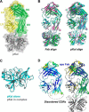
X-ray crystallographic analysis of the pKal catalytic domain complexed with the DX-2930 Fab. A, ribbon structure of the pKal·DX-2930 Fab complex (PDB 4OGX) with transparent surface rendering. pKal ribbon is colored in gray with cyan marking the catalytic triad residues, and the DX-2930 Fab is in green (heavy chain) and yellow (light chain). B, all three resolved DX-2930·pKal complex structures from this study overlaid in ribbon (orientation as in A) with alignment on the Fab domain or pKal catalytic domain. Complex from PDB 4OGX is colored in magenta, and the two distinct complexes from PDB 4OGY are in cyan and green. C, overlay of pKal catalytic domain structure alone (cyan, PDB 1ANY) with the pKal catalytic domain crystallized in complex with DX-2930 (gray, from PDB 4OGX) reveals minimal changes in the bound/unbound forms (r.m.s.d. 0.36 Å). D, left, crystal structure of the unbound DX-2930 Fab (PDB 4PUB) in ribbon with color reflecting B-factor values reveals disordered heavy chain CDR loops but an intact light chain CDR2. Right, overlay of the apo-DX-2930 Fab (now all in blue) overlaid on the pKal·DX-2930 Fab complex structure (colored as in A).
FIGURE 4.
FIGURE 4.
Structural analysis of DX-2930 CDR interactions with the pKal catalytic domain. A, surface rendering of the pKal catalytic domain highlighting complex interface residues, with heavy chain-interacting residues in green and light chain-interacting ones in yellow. Detailed views of critical side-chain interactions between pKal and the DX-2930 Fab residues are shown as follows: B, heavy chain FR1 and CDR1 (HV FR1/CDR1); C, light chain CDR2 (LV CDR2); and D, heavy chain CDR3 (HV CDR3). E, overlay of the benzamidine molecule from PDB 2ANY reveals a bidentate interaction with pKal residue Asp-572 (chymotrypsin Asp-195) in a manner analogous to Arg-106 of the DX-2930 Fab.
FIGURE 5.
FIGURE 5.
Pharmacokinetic and pharmacodynamic properties of DX-2930 in cynomolgus monkey. A, 20 mg/kg DX-2930 was dosed by either intraperitoneal (IP) or subcutaneous (SC) injection. Citrated plasma was collected at indicated times (hours) post-dose, and the concentration of DX-2930 was measured by a quantitative ELISA utilizing an antibody specific for DX-2930 (see “Experimental Procedures”). The s.c. dosing results in a maximal DX-2930 concentration reached by 96 h and a prolonged serum half-life of ∼12.5 days. Noncompartmental pharmacokinetic analyses yield the parameters shown in Table 3. Error bars shown are the standard deviation between replicates (n = 3). B, inhibition of pKal-mediated HMWK cleavage by DX-2930 in activated plasma. The KKS in plasma collected from cynomolgus monkeys subcutaneously treated with 25 mg/kg or 50 mg/kg DX-2930 was activated by kaolin addition and samples analyzed by Western blots detecting intact 1-chain or proteolyzed 2-chain HMWK. A light image of included molecular mass standards (in kDa) is shown adjacent to the Western blot image. Purified human 1- and 2-chain HMWK at 5 nm were included as controls. Addition of DX-2930 reduces HMWK proteolysis relative to nondosed samples (day −12) even when sampled 28 days post-injection.
FIGURE 6.
FIGURE 6.
DX-2930 inhibits carrageenan-induced paw edema in the rat. DX-2930 was administered via a s.c. injection 24 h prior to carrageenan injection into the rat hind paw, although the vehicle (Formulation Buffer) and indomethacin (5 mg/kg) controls were administered by i.p. injection 30 min prior to carrageenan treatment. Paw volume was measured at the indicated time points following carrageenan injection and averaged for each treatment group (n = 10). Error calculated using the standard deviation.
FIGURE 7.
FIGURE 7.
Comparison of inhibitor interactions with serine protease active site pockets. Surface rendering of serine proteases are shown docked with stick models of their respective ligands just above the schematic models of these interactions with canonical protease docking sites highlighted. A, small molecule benzamidine docks into the deep S1 binding pocket of KLK4 (PDB 2BDG). B, peptide sequence from the small protein BPTI binds matriptase in a “substrate-like” mode (PDB 1EAW). C, part of the HV-CDR3 of DX-2930 binds the S1-S3 sites of pKal and then abruptly turns away from the catalytic serine. N and C refer to N- and C-terminal ends of docked schematic peptides. Inhibitor stick color scheme by atom: blue, nitrogen; red, oxygen; green, carbon. Red in surface representation and red asterisk in schematic representation denote the catalytic serine residue.

Similar articles

Cited by

References

    1. Colman R. W., Schmaier A. H. (1997) Contact system: a vascular biology modulator with anticoagulant, profibrinolytic, antiadhesive, and proinflammatory attributes. Blood 90, 3819–3843 - PubMed
    1. Leeb-Lundberg L. M., Marceau F., Müller-Esterl W., Pettibone D. J., Zuraw B. L. (2005) International union of pharmacology. XLV. Classification of the kinin receptor family: from molecular mechanisms to pathophysiological consequences. Pharmacol. Rev. 57, 27–77 - PubMed
    1. Oschatz C., Maas C., Lecher B., Jansen T., Björkqvist J., Tradler T., Sedlmeier R., Burfeind P., Cichon S., Hammerschmidt S., Müller-Esterl W., Wuillemin W. A., Nilsson G., Renné T. (2011) Mast cells increase vascular permeability by heparin-initiated bradykinin formation in vivo. Immunity 34, 258–268 - PubMed
    1. Maas C., Govers-Riemslag J. W., Bouma B., Schiks B., Hazenberg B. P., Lokhorst H. M., Hammarström P., ten Cate H., de Groot P. G., Bouma B. N., Gebbink M. F. (2008) Misfolded proteins activate factor XII in humans, leading to kallikrein formation without initiating coagulation. J. Clin. Invest. 118, 3208–3218 - PMC - PubMed
    1. van der Meijden P. E., Munnix I. C., Auger J. M., Govers-Riemslag J. W., Cosemans J. M., Kuijpers M. J., Spronk H. M., Watson S. P., Renné T., Heemskerk J. W. (2009) Dual role of collagen in factor XII-dependent thrombus formation. Blood 114, 881–890 - PubMed