. 2014 Jun 12:5:253.
doi: 10.3389/fpls.2014.00253. eCollection 2014.

Can vessel dimension explain tolerance toward fungal vascular wilt diseases in woody plants? Lessons from Dutch elm disease and esca disease in grapevine

Affiliations

Can vessel dimension explain tolerance toward fungal vascular wilt diseases in woody plants? Lessons from Dutch elm disease and esca disease in grapevine

Jérôme Pouzoulet et al. Front Plant Sci. .

Abstract

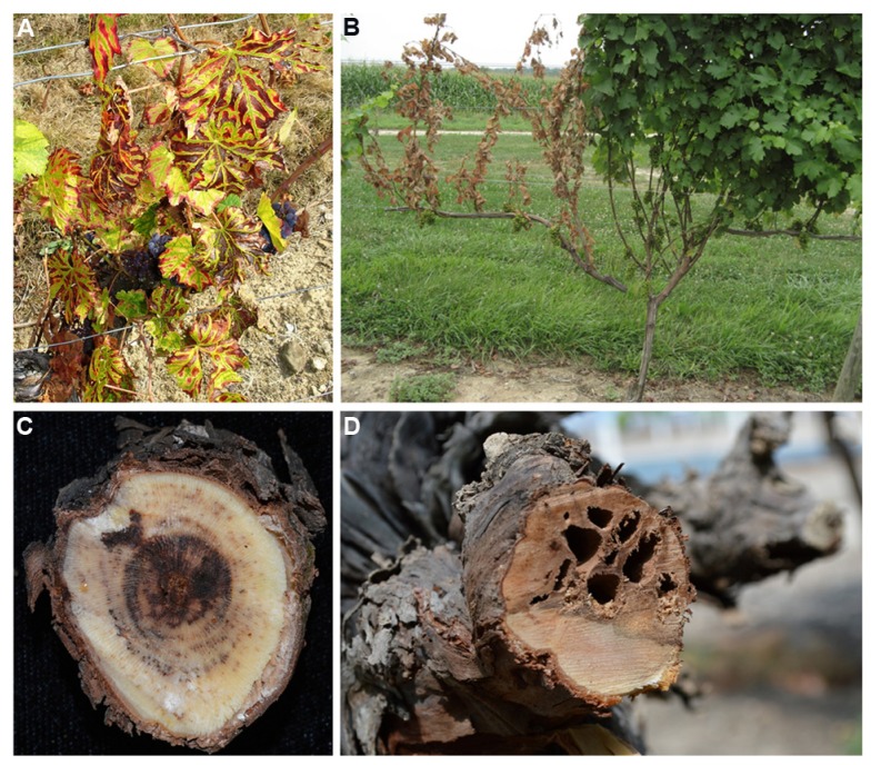
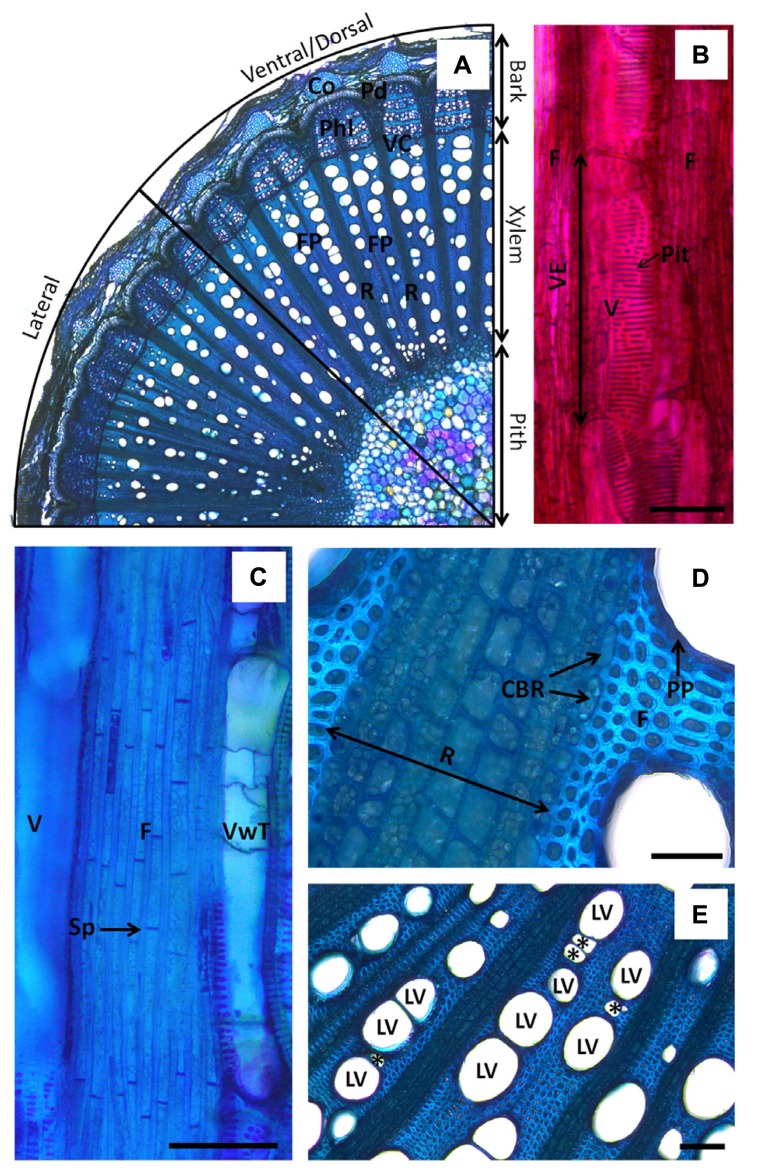
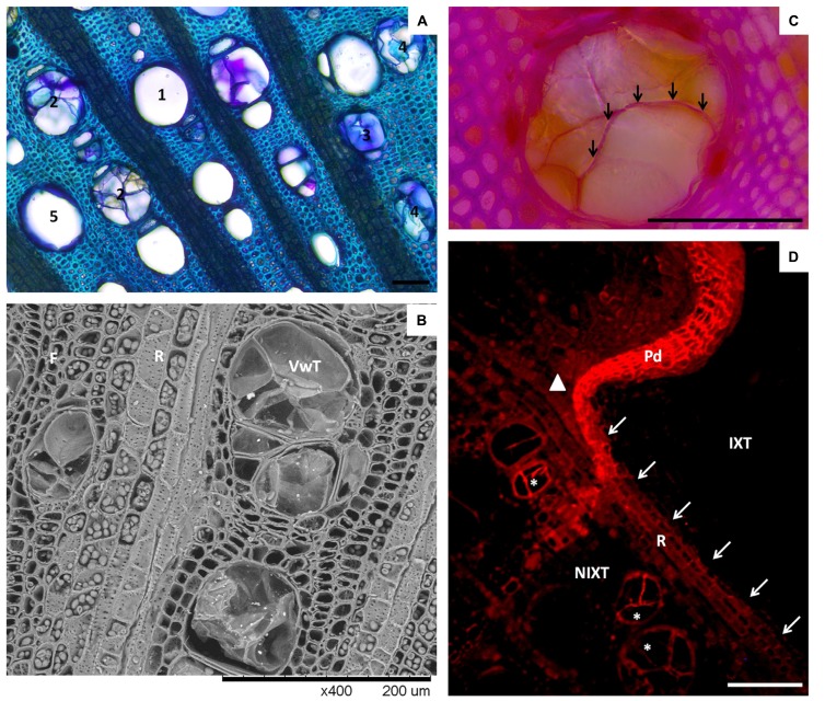
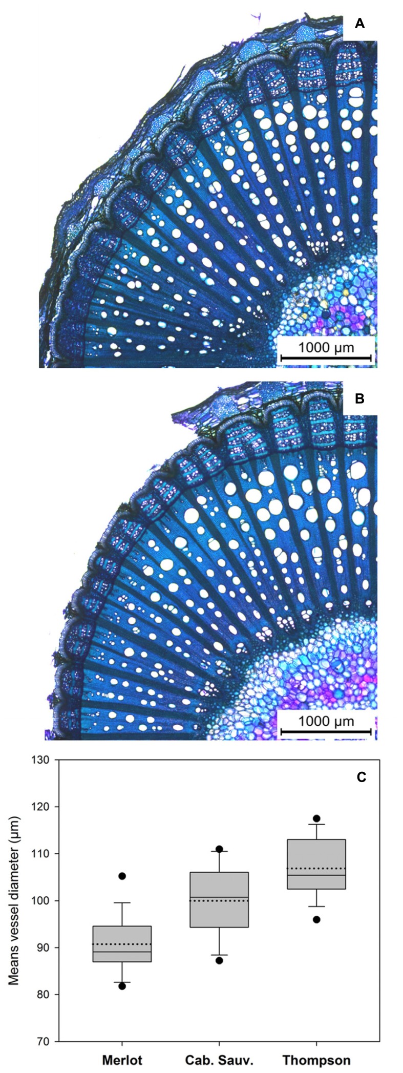
This review illuminates key findings in our understanding of grapevine xylem resistance to fungal vascular wilt diseases. Grapevine (Vitis spp.) vascular diseases such as esca, botryosphaeria dieback, and eutypa dieback, are caused by a set of taxonomically unrelated ascomycete fungi. Fungal colonization of the vascular system leads to a decline of the plant host because of a loss of the xylem function and subsequent decrease in hydraulic conductivity. Fungal vascular pathogens use different colonization strategies to invade and kill their host. Vitis vinifera cultivars display different levels of tolerance toward vascular diseases caused by fungi, but the plant defense mechanisms underlying those observations have not been completely elucidated. In this review, we establish a parallel between two vascular diseases, grapevine esca disease and Dutch elm disease, and argue that the former should be viewed as a vascular wilt disease. Plant genotypes exhibit differences in xylem morphology and resistance to fungal pathogens causing vascular wilt diseases. We provide evidence that the susceptibility of three commercial V. vinifera cultivars to esca disease is correlated to large vessel diameter. Additionally, we explore how xylem morphological traits related to water transport are influenced by abiotic factors, and how these might impact host tolerance of vascular wilt fungi. Finally, we explore the utility of this concept for predicting which V. vinifera cultivars are most vulnerable of fungal vascular wilt diseases and propose new strategies for disease management.

Keywords: Phaeomoniella chlamydospora; Vascular wilt; compartmentalization; esca; grapevine trunk diseases; xylem morphology.


Figures

FIGURE 1
FIGURE 1
Symptoms expressed in grapevine affected with fungal vascular diseases. (A) Vines showing tiger-stripe leaf symptoms associated with the chronic form of esca. (B) Symptoms of apoplexy on a young grapevine; note the foliar symptoms between the apparently healthy (right) and apoplectic (left) cordon. (C) Cross section of a grapevine wood spur infected with esca; note the necrotic spots in vessels organized in rings and the brown necrosis in the center of the wood. (D) Cross section of a grapevine cordon infected with eutypa dieback; note the wedge-shaped canker.
FIGURE 2
FIGURE 2
Anatomy of Vitis vinifera xylem. (A) Micrograph showing organization of stem tissues in cross-section (toluidine blue O). Note the segmentation of xylem in fascicular portions (FP) where large vessels (LV) are packed. Fascicular portions are separated by large rays (R). Note the change in vessel diameters in the lateral and ventral/dorsal sector of the stem. (B) Micrograph showing a large vessel in longitudinal section (safranin O). Vessel element (VE) is shown. Note the great area occupied by scalariform pits in the vessel cell wall. (C) Micrograph showing septate fibers in longitudinal section (toluidine blue O). (D) Micrograph showing close view of ray parenchyma in stem cross-section (toluidine blue O). Note the shape of cells bordering the ray (CBR) compared to ray cells and fibers. Change in staining of the wall due to differential lignification is also observed according to the side in contact with ray cells or in contact with fibers. Note the presence of a single-seriate layer of flat cells forming the paratracheal parenchyma around vessels. (E) Micrograph of stem cross-section showing LV in contact with each other or connected by vessel relays (*). Note the presence of LV in contact with ray parenchyma. The notations CBR stands for cells bordering ray, Co is for cork, F is for fibers, FP is for fascicular portion, LV is for large vessel, Pd is for periderm, Phl is for phloem, PP is for paratracheal parenchyma, R is for ray parenchyma, Sp is for septation, V is for vessel, VC is for vascular cambium, VwT is for vessel with tyloses. Scale bars = 100 μm.
FIGURE 3
FIGURE 3
Vessel occlusions in Vitis vinifera xylem. (A) Micrograph showing different types of occlusion in cross-section of grapevine xylem (bright field, toluidine O). (1) stands for open vessel (no occlusion), (2) stands for vessels occluded by tyloses, (3) stands for vessels occluded by gels, (4) stands for vessels occluded by both tyloses and gels, (5) stands for open vessels harboring a layer of gels coating their walls. (B) SEM micrograph of a stem cross-section showing tyloses in LV. Note the abundance of intercellular junctions occurring in the wall of ray parenchyma cells (R). (C) Close view of an occluded vessel showing the occurrence of lignin in a tylosis wall (arrows), demonstrated by the deep purple color developed in reaction to phloroglucinol/HCl staining. (D) Epifluorescent micrograph showing suberin location in a stem cross-section 2 months after mechanical injury (sudan IV, Ex 590–650 nm/Em > 667 nm). Note the accumulation of suberin in ray (arrows) separating injured (IXT) and non-injured (NIXT) xylem tissues, in the continuity of the periderm (Pd) formed in response to injury. Also, note the presence of a clear signal from the wall of tylosis in some vessels (*). The position of the vascular cambium at the time of injury is indicated by a solid triangle. Scale bars = 100 μm.
FIGURE 4
FIGURE 4
Morphological and physiological features of the xylem of one year-old Vitis vinifera stems cvs. Merlot and Cabernet Sauvignon. (A,B) Micrograph of cross-section of stem of V. vinifera cvs. Merlot (A) and Cabernet Sauvignon (B). (C) Box plot representing means of equivalent circle diameters of LV measured in Merlot, Cabernet Sauvignon, and Thompson Seedless cultivars (n = 18): the median and mean are represented by solid and dotted lines, respectively. Top and bottom lines of the box correspond to the 25th and 75th percentiles of the data, respectively. Error bars represent the 10th and 90th percentiles. Circles represent outliers.

References

    1. Agrios G. N. (2005). Plant Pathology Fifth Edition. San Diego, USA: Elsevier-Academic press
    1. Agustí-Brisach C., Gramaje D., García-Jiménez J., Armengol J. (2013). Detection of black-foot and Petri disease pathogens in soils of grapevine nurseries and vineyards using bait plants. Plant Soil 364 5–13 10.1007/s11104-012-1333-1 - DOI
    1. Allen C. D., Macalady A. K., Chenchouni H., Bachelet D., McDowell N., Vennetier M., et al. (2010). A global overview of drought and heat-induced tree mortality reveals emerging climate change risks for forests. For. Ecol. Manage. 259 660–684 10.1016/j.foreco.2009.09.001 - DOI
    1. Alsina M. M., De Herralde F., Aranda X., Save R., Biel C. (2007). Water relations and vulnerability to embolism are not related: experiments with eight grapevine cultivars. Vitis 46 1–6
    1. Andolfi A., Cimmino A., Evidente A., Iannaccone A., Capparelli R., Mugnai L., et al. (2009). A new flow cytometry technique to identify Phaeomoniella chlamydospora exopolysaccharides and study mechanisms of esca grapevine foliar symptoms. Plant Dis. 93 680–684 10.1094/PDIS-93-7-0680 - DOI - PubMed