. 2014:2014:724670.
doi: 10.1155/2014/724670. Epub 2014 May 25.

Lycopene Inhibits Urotensin-II-Induced Cardiomyocyte Hypertrophy in Neonatal Rat Cardiomyocytes

Affiliations

Lycopene Inhibits Urotensin-II-Induced Cardiomyocyte Hypertrophy in Neonatal Rat Cardiomyocytes

Hung-Hsing Chao et al. Evid Based Complement Alternat Med. 2014.

Abstract

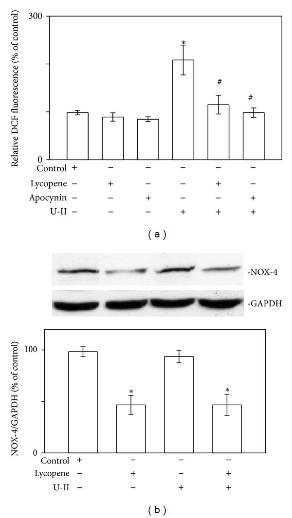
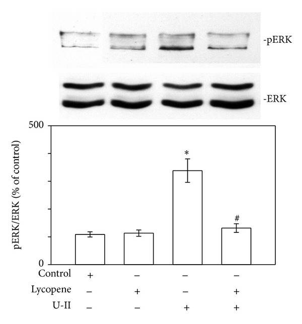
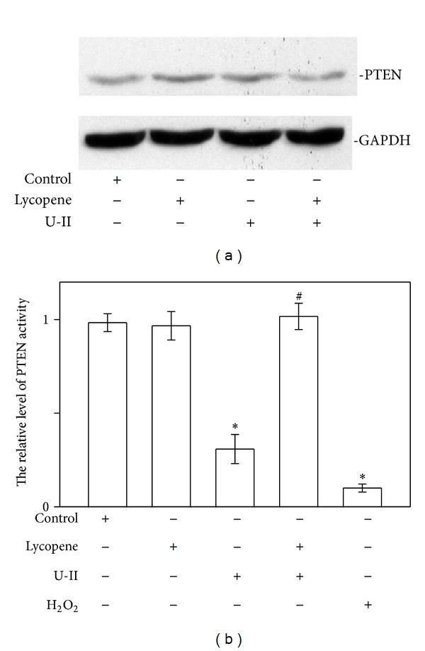
This study investigated how lycopene affected urotensin-II- (U-II-) induced cardiomyocyte hypertrophy and the possible implicated mechanisms. Neonatal rat cardiomyocytes were exposed to U-II (1 nM) either exclusively or following 6 h of lycopene pretreatment (1-10 μ M). The lycopene (3-10 μ M) pretreatment significantly inhibited the U-II-induced cardiomyocyte hypertrophy, decreased the production of U-II-induced reactive oxygen species (ROS), and reduced the level of NAD(P)H oxidase-4 expression. Lycopene further inhibited the U-II-induced phosphorylation of the redox-sensitive extracellular signal-regulated kinases. Moreover, lycopene treatment prevented the increase in the phosphorylation of serine-threonine kinase Akt and glycogen synthase kinase-3beta (GSK-3 β ) caused by U-II without affecting the protein levels of the phosphatase and tensin homolog deleted on chromosome 10 (PTEN). However, lycopene increased the PTEN activity level, suggesting that lycopene prevents ROS-induced PTEN inactivation. These findings imply that lycopene yields antihypertrophic effects that can prevent the activation of the Akt/GSK-3 β hypertrophic pathway by modulating PTEN inactivation through U-II treatment. Thus, the data indicate that lycopene prevented U-II-induced cardiomyocyte hypertrophy through a mechanism involving the inhibition of redox signaling. These findings provide novel data regarding the molecular mechanisms by which lycopene regulates cardiomyocyte hypertrophy.


Figures

Figure 1
Figure 1
Effect of  lycopene on U-II-induced cardiomyocyte hypertrophy. (a) Effect of  lycopene on U-II-induced morphologic changes in cardiomyocytes. Cultured cardiomyocytes were exposed to vehicle control (control: top left), to lycopene alone at 3 μM (lycopene: top right), or to U-II at 1 nM for 24 h in the absence (U-II: bottom left) or presence of lycopene (U-II + lycopene: bottom right) and then immunostained with an anti-α-actinin antibody (red), and the nucleus was stained with DAPI (blue). The figure shows a representative stain prepared based on 3 independent experiments. Scale bar shows 20 μm. (b) Relative cardiomyocyte size. The cultured cardiomyocytes were exposed to vehicle control or U-II at 1 nM or 24 h in the absence or presence of lycopene (1, 3, and 10 μM). The surface areas of the cardiomyocytes were measured using NIH image software in 60 randomly chosen cells from 3 dishes. The data are presented as mean ± SEM. (c) Measuring protein synthesis by using [3H]-leucine incorporation. The results are shown as mean ± SEM (n = 6). *P < 0.05 versus control; # P < 0.05 versus U-II alone.
Figure 2
Figure 2
Effects of lycopene on U-II-induced ROS production in cardiomyocytes. (a) ROS levels after adding lycopene or the NAD(P)H oxidase-4 (NOX-4) inhibitor apocynin in cells exposed to U-II for 2 min. The ROS levels of cells pretreated for 6 h with lycopene (3 μM) or 30 min with apocynin (0.3 mM) and exposed to U-II (1 nM) for 2 min. (b) Upper panels: representative western blot analysis of NOX-4 in cells pretreated for 6 h with lycopene (3 μM) then exposed to U-II. Lower panels: the indicated values represent the ratio of NOX-4 to GAPDH. The results are shown as mean ± SEM (n = 6).*P < 0.05 versus control.
Figure 3
Figure 3
Effects of lycopene on U-II-induced ERK phosphorylation in cardiomyocytes. Upper panels: representative western blot analyses of ERK in cells pretreated for 6 h with lycopene (3 μM) then exposed to U-II (1 nM) for 30 min. Lower panels: the indicated values represent the ratio of phosphorylated protein to total protein. The results are shown as mean ± SEM (n = 6). *P < 0.05 versus control; # P < 0.05 versus U-II alone.
Figure 4
Figure 4
Lycopene treatment prevents Akt/GSK-3β phosphorylation in U-II-stimulated neonatal rat cardiomyocytes. Protein extracts from cardiomyocytes were subjected to immunoblot analysis. (a) Effects of lycopene on U-II-induced Akt phosphorylation in cardiomyocytes. Upper panels: representative immunoblots using total and antiphospho-Akt (Ser473) antibodies. Lower panels: the indicated values represent the ratio of phosphorylated protein to total protein. (b) Effects of lycopene on U-II-induced GSK-3β phosphorylation in cardiomyocytes. Upper panels: representative immunoblots using total and antiphospho-GSK-3β (Ser9) antibodies. Lower panels: the indicated values represent the ratio of phosphorylated protein to total protein. Neonatal rat cardiomyocytes were nonstimulated (control) or stimulated with U-II (1 nM for 2 min). When indicated, lycopene (3 μM) was added 6 h before U-II stimulation. The results are shown as mean ± SEM (n = 6). *P < 0.05 versus control; # P < 0.05 versus U-II alone.
Figure 5
Figure 5
Lycopene treatment prevents PTEN oxidation in U-II-stimulated neonatal rat cardiomyocytes. (a) Lycopene does not affect PTEN protein levels in cardiomyocytes. Neonatal rat cardiomyocytes were exposed to U-II (1 nM for 1 h) in the presence or absence of lycopene (3 μM) added 6 h prior to the stimuli. Representative immunoblots using anti-PTEN antibodies or anti-GAPDH antibodies are shown. The blot data are representative of 3 experiments. (b) Effect of lycopene on PTEN activity. Cardiomyocytes were treated with U-II (1 nM) in the absence or presence of lycopene (3 μM) as indicated. The oxidation of the endogenous PTEN in the cardiomyocytes exposed to 0.5 mM H2O2. The level of PTEN activity in each sample was detected using a PTEN Malachite Green Assay Kit. The relative level of PTEN activity is shown as mean ± SEM (n = 6). *P < 0.05 versus control; # P < 0.05 versus U-II alone.

References

    1. Swynghedauw B. Remodeling of the heart in chronic pressure overload. Basic Research in Cardiology. 1991;86:99–105. - PubMed
    1. Wang Y. Signal transduction in cardiac hypertrophy—dissecting compensatory versus pathological pathways utilizing a transgenic approach. Current Opinion in Pharmacology. 2001;1(2):134–140. - PubMed
    1. Ames RS, Sarau HM, Chambers JK, et al. Human urotensin-II is a potent vasoconstrictor and agonist for the orphan receptor GPR14. Nature. 1999;401(6750):p. 282, p. 286. - PubMed
    1. Ross B, McKendy K, Giaid A. Role of urotensin II in health and disease. American Journal of Physiology, Regulatory Integrative and Comparative Physiology. 2010;298(5):R1156–R1172. - PubMed
    1. Liu J-C, Chen C-H, Chen J-J, Cheng T-H. Urotensin II induces rat cardiomyocyte hypertrophy via the transient oxidization of Src homology 2-containing tyrosine phosphatase and transactivation of epidermal growth factor receptor. Molecular Pharmacology. 2009;76(6):1186–1195. - PubMed

LinkOut - more resources