Uncommon locations and presentations of hydatid cyst
- PMID: 24971224
- PMCID: PMC4071749
- DOI: 10.4103/2141-9248.133476
Uncommon locations and presentations of hydatid cyst
Abstract
Background: Hydatid disease (HD) is an ancient disease and even was known to Hippocrates. This disease involves all human parts and most common affected organs are liver and lungs. Incidence of unusual site is about 8-10%. The clinical picture depends upon the involved organs, its effects on adjacent structures, complications due to secondary infection, rupture, and anaphylaxis caused by hydatid cysts.
Aim: The aim of this study was to find out incidence of unusual location of hydatid cyst in the human body.
Materials and methods: A retrospective study of HD was carried in a medical college between July 2007 and June 2012. A total 79 cases of HD were treated during this period. Information on clinical presentation and management were reviewed, and results presented as summary statistics.
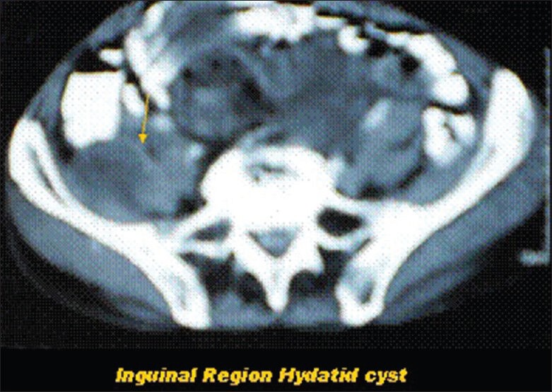
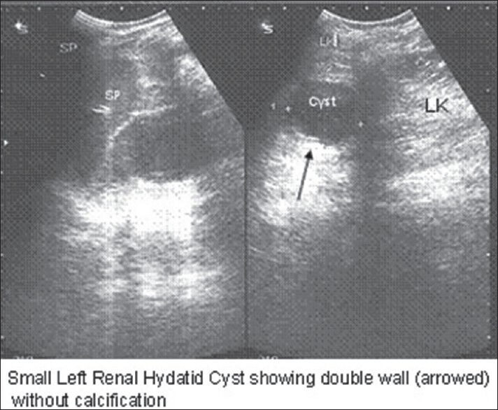
Results: Sixty one cases were of liver HD, and 11 were with hydatid lung disease. Fifty cases were with right lobe involvement, and rest 11 were with both lobe involvement. Out of 11 lung hydatid only one case was with bilateral lung involvement. Only eight cases of HD of uncommon locations and presentations were encountered during this period. First case presented with left hypochondriac mass as splenic HD, second with pelvic HD along with obstructive uropathy, third with non-functioning right kidney with bilateral psoas muscles HD, fourth with HD involving mesentery, fifth with pelvic pain due to right ovary HD, sixth with simultaneous involvement of the liver and right subdiaphragmatic region, seventh with HD of right inguinal region, and eighth with hydatid cyst of the left kidney. Even though, there was no mortality found in these patients, there was high morbidity.
Conclusion: We conclude that Echinococcus granulosus can affect any organ in the body from head to toe, and a high suspicion of this disease is justified in endemic regions. Moreover, medical treatment should be given in the pre-operative period as well as in the post-operative period for 4-6 weeks.
Keywords: Hydatid disease; Ovary; Psoas muscle; Renal; Spleen; Unusual locations.
Conflict of interest statement
Figures


References
-
- McManus DP, Zhang W, Li J, Bartley PB. Echinococcosis. Lancet. 2003;362:1295–304. - PubMed
-
- Kouskos E, Chatziantoniou J, Chrissafis I, Anitsakis C, Zamtrakis S. Uncommon locations of hydatid cysts. Singapore Med J. 2007;48:e119–21. - PubMed
-
- Czermak BV, Unsinn KM, Gotwald T, Niehoff AA, Freund MC, Waldenberger P, et al. Echinococcus granulosus revisited: Radiologic patterns seen in pediatric and adult patients. AJR Am J Roentgenol. 2001;177:1051–6. - PubMed
-
- Pedrosa I, Saíz A, Arrazola J, Ferreirós J, Pedrosa CS. Hydatid disease: Radiologic and pathologic features and complications. Radiographics. 2000;20:795–817. - PubMed
LinkOut - more resources
Full Text Sources
Other Literature Sources