. 2014:2014:124063.
doi: 10.1155/2014/124063. Epub 2014 May 29.

Cardioprotective effects of osteopontin-1 during development of murine ischemic cardiomyopathy

Affiliations

Cardioprotective effects of osteopontin-1 during development of murine ischemic cardiomyopathy

Georg D Duerr et al. Biomed Res Int. 2014.

Abstract

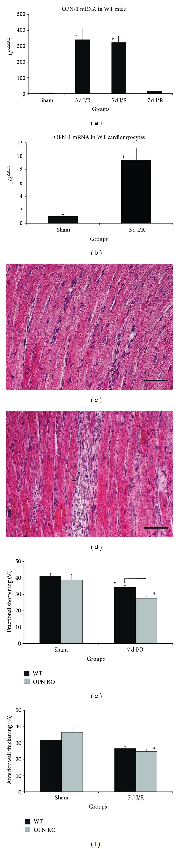
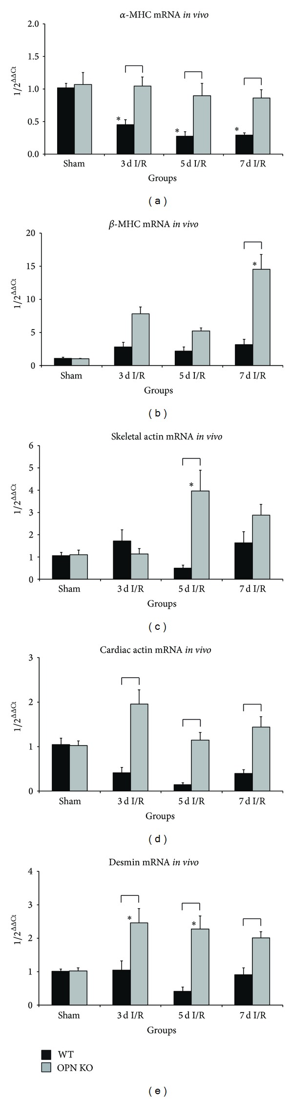
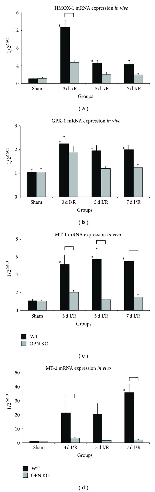
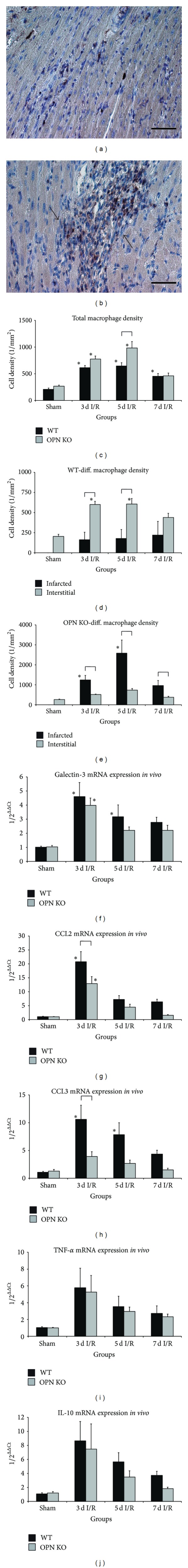
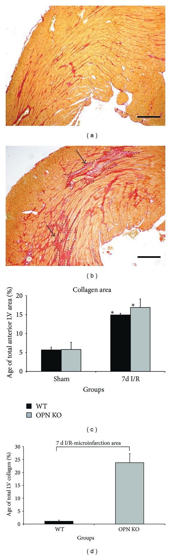
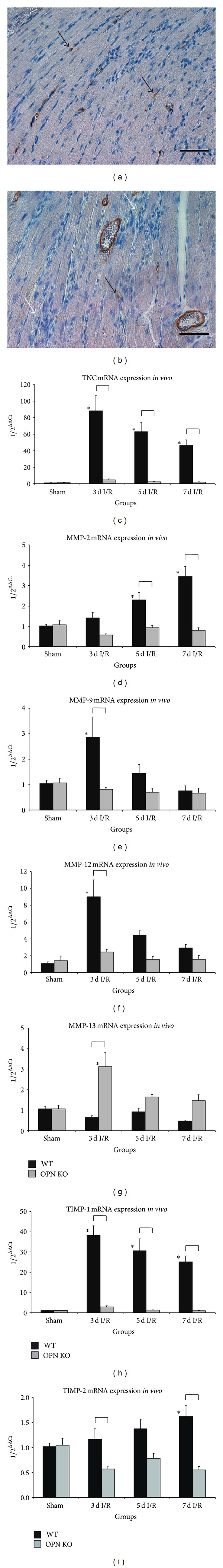
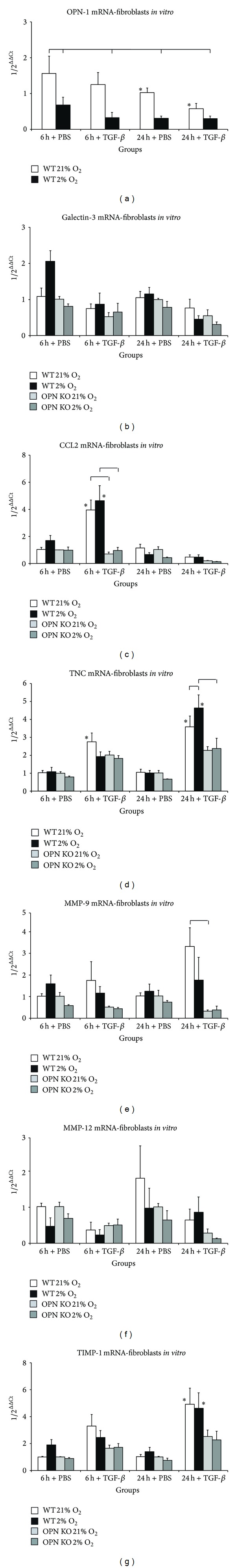
Repetitive brief ischemia and reperfusion (I/R) is associated with ventricular dysfunction in pathogenesis of murine ischemic cardiomyopathy and human hibernating myocardium. We investigated the role of matricellular protein osteopontin-1 (OPN) in murine model of repetitive I/R. One 15-min LAD-occlusion followed by reperfusion was performed daily over 3, 5, and 7 consecutive days in C57/Bl6 wildtype- (WT-) and OPN(-/-)-mice (n = 8/group). After echocardiography hearts were processed for histological and mRNA-studies. Cardiac fibroblasts were isolated, cultured, and stimulated with TGF- β 1. WT-mice showed an early, strong, and cardiomyocyte-specific osteopontin-expression leading to interstitial macrophage infiltration and consecutive fibrosis after 7 days I/R in absence of myocardial infarction. In contrast, OPN(-/-)-mice showed small, nontransmural infarctions after 3 days I/R associated with significantly worse ventricular dysfunction. OPN(-/-)-mice had different expression of myocardial contractile elements and antioxidative mediators and a lower expression of chemokines during I/R. OPN(-/-)-mice showed predominant collagen deposition in macrophage-rich small infarctions. We found lower induction of tenascin-C, MMP-9, MMP-12, and TIMP-1, whereas MMP-13-expression was higher in OPN(-/-)-mice. Cultured OPN(-/-)-myofibroblasts confirmed these findings. In conclusion, osteopontin seems to modulate expression of contractile elements, antioxidative mediators, and inflammatory response and subsequently remodel in order to protect cardiomyocytes in murine ischemic cardiomyopathy.


Figures

Figure 1
Figure 1
Osteopontin-deficiency leads to cardiomyocyte loss and poor left ventricular function. (a) Osteopontin mRNA-expression in whole WT-mouse hearts during repetitive I/R and (b) in Langendorff-isolated adult WT-cardiomyocytes after 3 days I/R compared to sham. (c) Representative HE-stained section of WT-heart after 7 days I/R shows interstitial cellular infiltration in contrast to (d) irreversible loss of cardiomyocytes in OPN/-mice (arrow). (e) Fractional shortening was impaired in both strains, but loss of left ventricular contractility was significantly reduced in OPN/-mice after 7 d I/R compared to WT. (f) Anterior wall thickening was significantly reduced in OPN/-mice after 7 days I/R. n = 8–11/group. Scale bars represent 50 μm. RT-qPCR using Taqman and mRNA-expression is related to controls and GAPDH using comparative ΔΔCt-method. Bracket indicates P ≤ 0.05 between genotypes; ∗ indicates P ≤ 0.05 versus respective shams.
Figure 2
Figure 2
Ischemic OPN/-hearts show different expression contractile elements. mRNA-expression of (a) α-myosin heavy chain (α-MHC), (b) β-MHC, (c) skeletal actin, (d) cardiac actin, and (e) desmin in OPN/-mice compared to WT-mice during repetitive I/R. n = 8-9/group. RT-qPCR using Taqman and mRNA-expression is related to controls and GAPDH using comparative ΔΔCt-method. Bracket indicates P ≤ 0.05 between genotypes; ∗ indicates P ≤ 0.05 versus respective shams.
Figure 3
Figure 3
Ischemic OPN/-hearts show different expression of antioxidative mediators. mRNA-expression of antioxidative mediators (a) heme oxygenase- (HMOX-) 1 and (b) glutathione peroxidase- (GPX-) 1. mRNA-expression of zinc-storage proteins (c) metallothionein (MT)-1 and (d) MT-2. n = 8-9/group. RT-qPCR using Taqman and mRNA-expression is related to controls and GAPDH using comparative ΔΔCt-method. Bracket indicates P ≤ 0.05 between genotypes; ∗ indicates P ≤ 0.05 versus respective shams.
Figure 4
Figure 4
Cellular and molecular changes in response to I/R. (a) Representative section of left ventricle after 7 days I/R in a WT-heart shows low interstitial macrophage accumulation (F4/80). (b) High accumulation of macrophages in areas of small nontransmural infarctions in OPN/-hearts (arrows). (c) Cell density of F4/80 positive macrophages in WT- compared to OPN/-hearts during I/R. (d) Differential macrophage cell count in WT-hearts revealed only few cells within areas of small infarctions (infarcted) in comparison to interstitial space. (e) In contrast, significantly more cells invaded areas of small infarctions in OPN/-mice than interstitial space. mRNA-induction of the galectin-3 (f), chemokines (g) CCL2 and (h) CCL3, and cytokines (i) TNF-α and (j) IL-10 in OPN/-hearts compared to WT-hearts during repetitive I/R. n = 8–11/group. Scale bars represent 50 μm. RT-qPCR using Taqman and mRNA-expression is related to controls and GAPDH using comparative ΔΔCt-method. Bracket indicates P ≤ 0.05 between genotypes; ∗ indicates P ≤ 0.05 versus respective shams.
Figure 5
Figure 5
Collagen deposition in areas of small, nontransmural infarctions in OPN/-hearts during repetitive I/R. (a) Representative section of left ventricle in a WT-heart after 7 days I/R shows interstitial fibrosis in picrosirius red staining. (b) In contrast, OPN/-mice reveal relatively loose collagen deposition in areas of cardiomyocyte loss-small, nontransmural infarctions (arrows and dotted lines show confluent areas of patchy replacement fibrosis), but less interstitial collagen in the ischemic area. (c) Planimetrical analysis of collagen stained area between the genotypes after 7 days I/R. (d) Differential analysis of collagen deposition in small, nontransmural infarctions. n = 8–11/group. Scale bars represent 200 μm. Bracket indicates P ≤ 0.05 between genotypes; ∗ indicates P ≤ 0.05 versus respective shams.
Figure 6
Figure 6
Attenuated remodeling in ischemic OPN/-hearts. (a) Representative section of left ventricle after 7 days I/R in a WT-heart shows numerous α-smac positive myofibroblasts (black arrows) in the ischemic myocardium. (b) At the same time point, α-smac positive signals (black arrow) were almost absent in small, nontransmural infarctions (white arrows) of OPN/-hearts. mRNA-induction of (c) tenascin-C (TNC), (d) MMP-2, (e) MMP-9, (f) MMP-12, (g) MMP-13, (h) TIMP-1, and (i) TIMP-2 in WT- and OPN/-hearts during repetitive I/R. n = 8–11/group. Scale bars represent 50 μm. RT-qPCR using Taqman and mRNA-expression is related to controls and GAPDH using comparative ΔΔCt-method. Bracket indicates P ≤ 0.05 between genotypes; ∗ indicates P ≤ 0.05 versus respective shams.
Figure 7
Figure 7
Expression of remodeling markers in myofibroblasts in vitro. Cultured myofibroblasts from WT- and OPN/-hearts were stimulated with human TGF-β1 or PBS as control under normoxia (21% O2) or hypoxia (2% O2). mRNA-expression of (a) OPN-1 in WT-cells only. mRNA-expression of (b) galectin-3, (c) CCL2, (d) tenascin-C (TNC), (e) MMP-9, (f) MMP-12, and (g) TIMP-1. n = 5–7/group. RT-qPCR using Taqman and mRNA-expression is related to controls and 18 S (in vitro) using comparative ΔΔCt-method. Bracket indicates P ≤ 0.05 between genotypes; ∗ indicates P ≤ 0.05 versus 6 h + PBS normoxia group (control).

Similar articles

Cited by

References

    1. Kloner RA, Bolli R, Marban E, Reinlib L, Braunwald E. Medical and cellular implications of stunning, hibernation, and preconditioning: an NHLBI workshop. Circulation. 1998;97(18):1848–1867. - PubMed
    1. Frangogiannis NG, Shimoni S, Chang S, et al. Active interstitial remodeling: an important process in the hibernating human myocardium. Journal of the American College of Cardiology. 2002;39(9):1468–1474. - PubMed
    1. Frangogiannis NG, Shimoni S, Chang SM, et al. Evidence for an active inflammatory process in the hibernating human myocardium. American Journal of Pathology. 2002;160(4):1425–1433. - PMC - PubMed
    1. Dewald O, Frangogiannis NG, Zoerlein M, et al. Development of murine ischemic cardiomyopathy is associated with a transient inflammatory reaction and depends on reactive oxygen species. Proceedings of the National Academy of Sciences of the United States of America. 2003;100(5):2700–2705. - PMC - PubMed
    1. Frangogiannis NG, Dewald O, Xia Y, et al. Critical role of monocyte chemoattractant protein-1/CC chemokine ligand 2 in the pathogenesis of ischemic cardiomyopathy. Circulation. 2007;115(5):584–592. - PubMed

Publication types

MeSH terms