. 2014 Jun 27;9(6):e99801.
doi: 10.1371/journal.pone.0099801. eCollection 2014.

Coupling genetic and chemical microbiome profiling reveals heterogeneity of archaeome and bacteriome in subsurface biofilms that are dominated by the same archaeal species

Affiliations

Coupling genetic and chemical microbiome profiling reveals heterogeneity of archaeome and bacteriome in subsurface biofilms that are dominated by the same archaeal species

Alexander J Probst et al. PLoS One. .

Abstract

Earth harbors an enormous portion of subsurface microbial life, whose microbiome flux across geographical locations remains mainly unexplored due to difficult access to samples. Here, we investigated the microbiome relatedness of subsurface biofilms of two sulfidic springs in southeast Germany that have similar physical and chemical parameters and are fed by one deep groundwater current. Due to their unique hydrogeological setting these springs provide accessible windows to subsurface biofilms dominated by the same uncultivated archaeal species, called SM1 Euryarchaeon. Comparative analysis of infrared imaging spectra demonstrated great variations in archaeal membrane composition between biofilms of the two springs, suggesting different SM1 euryarchaeal strains of the same species at both aquifer outlets. This strain variation was supported by ultrastructural and metagenomic analyses of the archaeal biofilms, which included intergenic spacer region sequencing of the rRNA gene operon. At 16S rRNA gene level, PhyloChip G3 DNA microarray detected similar biofilm communities for archaea, but site-specific communities for bacteria. Both biofilms showed an enrichment of different deltaproteobacterial operational taxonomic units, whose families were, however, congruent as were their lipid spectra. Consequently, the function of the major proportion of the bacteriome appeared to be conserved across the geographic locations studied, which was confirmed by dsrB-directed quantitative PCR. Consequently, microbiome differences of these subsurface biofilms exist at subtle nuances for archaea (strain level variation) and at higher taxonomic levels for predominant bacteria without a substantial perturbation in bacteriome function. The results of this communication provide deep insight into the dynamics of subsurface microbial life and warrant its future investigation with regard to metabolic and genomic analyses.


Conflict of interest statement

Competing Interests: TZD and AJP are paid employees of Second Genome, Inc. (www.secondgenome.com), patent on PhyloChip G3 pending. This does not alter adherence to PLOS ONE policies on sharing data and materials. Second Genome is a gastrointestinal therapeutics company with a pipeline of intestinal microbiome modulators that impact metabolic diseases and IBD. The publication of archaeal microbiome dynamics in sulfidic groundwater does not influence the value of any of Second Genome's therapeutic assets.

Figures

Figure 1
Figure 1. The conversion of biofilm to string-of-pearls community in the spring water originating from the subsurface.
A: Biofilm. B: Intermediate transition state. C: String-of-pearls community. Row 1: Schematic drawings. Orange: SM1 euryarchaeal cocci, Green: Filamentous, sulfide-oxidizing bacteria. Row 2: Photographs and scanning electron micrograph (2B) of different stages. Row 3: FISH images of different stages (for MSI samples please see ; Archaea orange (CY3), Bacteria green (RG)). A: SM-BF, showing high dominance of Archaea. B: Attachment of archaea to filamentous bacteria. C: String-of-pearls communities with large archaeal colony and bacterial mantle. Arrows point to archaeal microcolonies, manteled by filamentous bacteria. It is proposed that attachment of SM1 Euryarchaeota to filamentous bacteria (B) mediates the transition from biofilm (A) to the string-of-pearls community (C). Scale bars: A3: 10 µm, B2: 1 µm B3: 10 mm, C3: 25 µm.
Figure 2
Figure 2. Abundance of Archaea and Bacteria in samples and the overall community relationship.
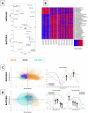
Small panels present binary images of infrared data collected for three sample types, SM-BF (Sippenauer Moor, biofilm), SM-SOPC (Sippenauer Moor, string-of-pearls community) and MSI-BF (Mühlbacher Schwefelquelle, Isling, biofilm). Infrared maps show the distribution of Archaea and Bacteria in the samples. One pixel corresponds to 2 µm. Large panel: Hierarchical clustering based weighted UniFrac of abundance values of eOTUs (Bacteria and Archaea). Two different clusters separating the samples based on hydrogeology were observed.
Figure 3
Figure 3. Detailed community profiling using PhyloChip G3 and SR-FTIR.
A: Ordination analysis of PhyloChip G3 data based on weighted UniFrac measure of eOTU abundances followed by non-metric multidimensional scaling (NMDS). Stress for NMDS of archaeal eOTUs (#37): 0.0088. Stress for NMDS of bacterial eOTUs (#1300): 0.0223. B: Heatmap displaying significantly different families found between the two biofilm types, MSI-BF and SM-BF by PhyloChip G3 assay. Significance is based on aggregated HybScores of eOTUs on family level followed by a Welch-test. For false discovery detection please see Fig. S6. C: Ordination analysis of SR-FTIR data based on a linear discriminant analysis and principal component analysis (PCA-LDA) in the spectral region of 2800–3100 cm−1 on the archaea spectra extracted from the maps from the three different locations. On the right there is the plot of PCA-LDA loadings. PCA-LDA1 explains for the 93.4% of the variance, PCA-LDA2 for 5.3% and PCA-LDA3 for 0.9%. Arrows point to the infrared signals used to explain the difference between the samples: 2975 cm−1, 2965 cm−1, 2924 cm−1 and 2850 cm−1. D: PCA-LDA in the spectral regions of 900–1280 cm−1 and 2800–3100 cm−1 on SR-FTIR spectra of the bacteria “pixels” from the chemical maps of the samples at the three different locations. On the right there is a plot of PCA-LDA loadings in the two spectral region of interest. PCA-LDA1 explains for the 54.5% of the variance, PCA-LDA2 for 28.6% and PCA-LDA3 for 7.3%. Arrows point to the main infrared signals used to explain the difference between the samples: 2958 cm−1, 2925 cm−1, 2870 cm−1 and 2850 cm−1, in the second panel 1250 cm−1, 1110 cm−1, 1080 cm−1 and 1045 cm−1.
Figure 4
Figure 4. Scanning and transmission electron micrographs of biofilms, cells and hami.
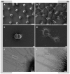
Left panels: MSI, right panel: SM. A: Scanning electron micrograph of MSI biofilm, showing SM1 euryarchaeal cells with defined distances and cell-cell connections. Bar: 1 µm. B: Scanning electron micrograph of SM biofilm, showing SM1 euryarchaeal cells with defined distances and fine-structured cell-cell connections. In-between: Bacterial filamentous and rod-shaped cells. Bar: 1 µm. C: Scanning electron micrograph of dividing SM1 euryarchaeal cell (MSI) with cell surface appendages. Bar: 200 nm. D: Scanning electron micrograph of dividing SM1 euryarchaeal cell (SM) with cell surface appendages. Bar: 200 nm. E: Transmission electron micrograph of cell surface appendages (hami) of SM1 euryarchaeal cells from the MSI biofilm. The hami carry the nano-grappling hooks, but besides that appear bare (square), without prickles (Moissl et al 2005). Bar: 100 nm. F: Transmission electron micrograph of cell surface appendages and matrix of SM1 euryarchaeal cells from the SM biofilm. The hami reveal the typical ultrastructure, with nano-grappling hooks and barbwire-like prickle region (square, Moissl et al 2005). Bar: 100 nm.
Figure 5
Figure 5. Scanning electron micrograph of filamentous bacterium and surrounded and cocooned by the SM1 euryarchaeal cells (SM-BF).
Bar: 1 µm.

References

    1. Whitman WB, Coleman DC, Wiebe WJ (1998) Prokaryotes: the unseen majority. Proc Natl Acad Sci U S A 95: 6578–6583. - PMC - PubMed
    1. Kallmeyer J, Pockalny R, Adhikari RR, Smith DC, D'Hondt S (2012) Global distribution of microbial abundance and biomass in subseafloor sediment. Proc Natl Acad Sci U S A 109: 16213–16216. - PMC - PubMed
    1. Ulrich GA, Martino D, Burger K, Routh J, Grossman EL, et al. (1998) Sulfur Cycling in the Terrestrial Subsurface: Commensal Interactions, Spatial Scales, and Microbial Heterogeneity. Microb Ecol 36: 141–151. - PubMed
    1. Wrighton KC, Thomas BC, Sharon I, Miller CS, Castelle CJ, et al. (2012) Fermentation, hydrogen, and sulfur metabolism in multiple uncultivated bacterial phyla. Science 337: 1661–1665. - PubMed
    1. Castelle CJ, Hug LA, Wrighton KC, Thomas BC, Williams KH, et al. (2013) Extraordinary phylogenetic diversity and metabolic versatility in aquifer sediment. Nat Commun 4: 2120. - PMC - PubMed

Publication types

LinkOut - more resources