. 2014 May-Jun;5(3):313-25.
doi: 10.4161/gmic.28932. Epub 2014 Jun 27.

Maternally acquired genotoxic Escherichia coli alters offspring's intestinal homeostasis

Affiliations

Maternally acquired genotoxic Escherichia coli alters offspring's intestinal homeostasis

Delphine Payros et al. Gut Microbes. 2014 May-Jun.

Abstract

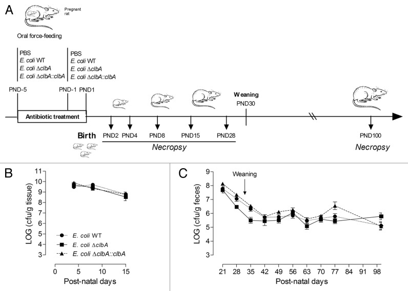
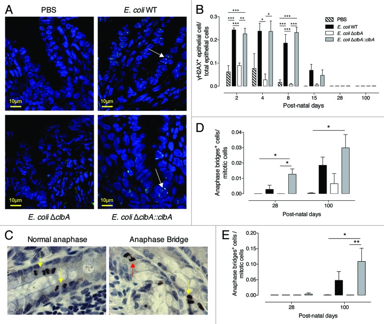
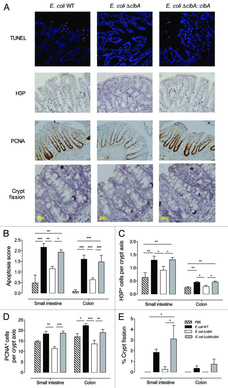
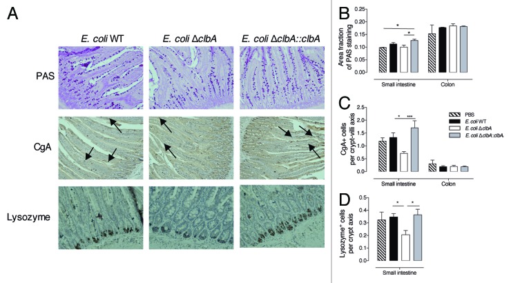
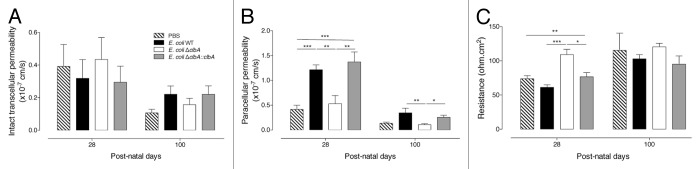
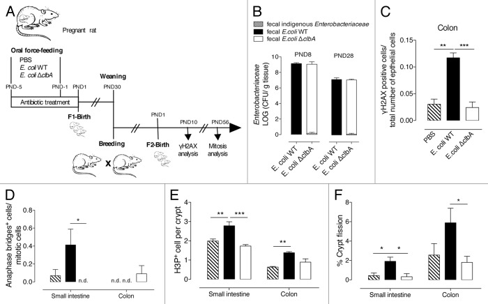
The neonatal gut is rapidly colonized by a newly dominant group of commensal Escherichia coli strains among which a large proportion produces a genotoxin called colibactin. In order to analyze the short- and long-term effects resulting from such evolution, we developed a rat model mimicking the natural transmission of E. coli from mothers to neonates. Genotoxic and non-genotoxic E. coli strains were equally transmitted to the offspring and stably colonized the gut across generations. DNA damage was only detected in neonates colonized with genotoxic E. coli strains. Signs of genotoxic stress such as anaphase bridges, higher occurrence of crypt fission and accelerated renewal of the mature epithelium were detected at adulthood. In addition, we observed alterations of secretory cell populations and gut epithelial barrier. Our findings illustrate how critical is the genotype of E. coli strains acquired at birth for gut homeostasis at adulthood.

Keywords: Escherichia coli; colibactin; epithelial differentiation; epithelial proliferation; genotoxicity; gut; intestinal barrier; neonate.


Figures

None
Figure 1. Commensal E. coli strains are transmitted from mother to the offspring and persistently colonized the gut at adulthood. (A) Experimental design of a rat model mimicking long-lasting colonization of the gut by commensal E. coli strains from birth to adulthood. Pregnant rats were treated with streptomycin (5 g/L) in drinking water and were inoculated twice with 109 CFU by intragastric gavage before parturition with E. coli WT, ΔclbA or ΔclbA::clbA strains or with PBS. Newborns were sacrificed at Post-Natal Days (PND) PND2, PND4, PND8, PND15, PND28 and PND100. (B) Evaluation of gut colonization in cecum/colon homogenate of neonates at early time-points (PND4, PND8 and PND15). (C) Evaluation of gut colonization in feces homogenate at later time-points (PND21, PND28, PND35 to PND100). Weaning is highlighted with a black arrow. Groups of 3–4 rats (PND4), 6–7 (PND8), 6–8 (PND15) and 15–20 (PND21-PND100) were used. Mean values ± SEM are shown.
None
Figure 2. Gut colonization with commensal E. coli strains producing colibactin exacerbates DNA double-strand breaks in gut epithelial cells of neonates and induces signs of increased chromosomal instability at adulthood. Immunofluorescence analysis of intestinal and colonic epithelium of neonates and adults (PND2-PND100) colonized since birth by commensal E. coli WT, E. coli ΔclbA or E. coli ΔclbA::clbA strains or treated with PBS. (A) Representative colon frozen sections at PND4. DNA was stained in blue and γH2AX foci in green. Scaled bars = 10 µm. White arrows show γH2AX foci in nucleus. (B) Quantification of γH2AX-positive epithelial cells. Groups of 3–6 rats were analyzed. Mean value ± SEM are shown. Two-way ANOVA with Bonferroni Multiple Comparison test, * P ≤ 0.05, ** P ≤ 0.01 and *** P ≤ 0.001. Immunohistochemestry analysis of intestinal and colonic epithelium of rats at PND28 and PND100. (C) Representative colon sections at PND100. Phospho-H3 was stained in black and allows detection of normal anaphases (left, yellow arrows) and abnormal mitosis with anaphase bridge (right, red arrow). Magnification used for photomicrographs = x20. Yellow arrows show normal anaphase figures and red arrow shows anormal mitotic cell with anaphase bridge. (D-E) Quantification of mitotic cells with anaphase bridge in small intestine (D) and colon (E). Groups of 5–10 rats were analyzed. Mean ± SEM are shown. Two-way ANOVA with Bonferroni Multiple Comparison test, * P ≤ 0.05 and ** P ≤ 0.01.
None
Figure 3. Early gut colonization with commensal E. coli strains producing colibactin increases the intestinal epithelial cells apoptosis, proliferation and crypt fission at adulthood. Histological and immunofluorescence analysis of the intestinal and colonic epithelium of adult animals early colonized by commensal E. coli WT, ΔclbA or ΔclbA::clbA strains or treated with PBS. (A) Representative colon frozen sections at PND100. DNA was stained in blue and apoptotic cells were stained in red with anti-TdT antibody (Tunel). Scale bars = 10 µm. Representative colon sections at PND100. Phospho-H3 was stained in black. PCNA was stained in black. Representative colon sections at PND100 and stained with hematoxylin-eosin. (B) Quantification of intestinal apoptotic score (see Methods section). (C) Quantification of H3P+ cells per crypt. (D) Quantification of PCNA+ cells per crypt. (E) Quantification of crypt fission. Black arrows show the fission of crypt in the colon. Magnification used for photomicrographs = x20. Groups of 5–10 rats were used, Mean values ± SEM are shown. One-way ANOVA, Bonferroni Multiple Comparison test, * P ≤ 0.05, ** P ≤ 0.01 and *** P ≤ 0.001.
None
Figure 4. Early gut colonization with commensal E. coli strains producing colibactin modulates small intestine epithelial cell differentiation at adulthood (PND100). Histological analysis of the intestinal epithelium of adult animals early colonized by commensal E. coli WT, ΔclbA or ΔclbA::clbA strains or treated with PBS. (A) Representative small intestine sections stained with Periodic-Acid Shiff (PAS) method showing goblet cells in magenta. Enteroendocrine cells (black arrows) were stained in brown with anti-chromogranin A (CgA) antibody. Paneth cells were stained in brown with anti-Lysozyme (Lyz) antibody. (B) Quantification of PAS staining area on total epithelial surface. (C) Quantification of CgA+ cells along the crypt-villus axis. (D) Quantification of Lyz+ cells along the crypt-villus axis in the small intestine. Magnification used for photomicrographs = x20. Groups of 5–10 rats were used. Mean values ± SEM are shown. One-way ANOVA with Bonferroni Multiple Comparison test, * P ≤ 0.05, ** P ≤ 0.01.
None
Figure 5. Early gut colonization with commensal E. coli strains producing colibactin alters intestinal permeability at adulthood (PND28 and PND100). Duodenal permeability was analyzed in adult animals early colonized by commensal E. coli WT, ΔclbA or ΔclbA::clbA strains or treated with PBS. (A) Transcellular permeability was assessed measuring mucosal to serosal flux of HRP. (B) Paracellular permeability was assessed measuring mucosal to serosal flux of FITC-dextran. (C) Electric transepithelial resistance was recorder during the permeability analysis. Groups of at least 10 rats were used. Mean values ± SEM are shown. One-way ANOVA with Bonferroni Multiple Comparison test, * P ≤ 0.05, ** P ≤ 0.01, *** P ≤ 0.001.
None
Figure 6. Alterations in intestinal physiology, induced by early gut colonization with commensal E. coli producing colibactin, are transmitted across generations. (A) Experimental design of a rat model mimicking transgenerational colonization of the gut with commensal E. coli strains. Adult rats, early gut colonized with commensal E. coli WT, ΔclbA strains or treated with PBS (F1), are breed and give rise to a second generation (F2) that is subsequently analyzed. (B) Evaluation of gut colonization in cecum/colon homogenate of neonates at PND8 and feces homogenate of adults at PND28. (C) Quantification of colonic γH2AX-positive epithelial cells. (D) Quantification of mitotic cells with anaphase bridge in small intestine and colon. (E) Quantification of H3P+ cells per crypt in small intestine and colon. (F) Quantification of crypt fission in small intestine and colon. Groups of 6 rats were used. Mean values ± SEM are shown. One-way ANOVA with Bonferroni Multiple Comparison test, * P ≤ 0.05, ** P ≤ 0.01, *** P ≤ 0.001.

Similar articles

Cited by

References

    1. Rautava S, Luoto R, Salminen S, Isolauri E. Microbial contact during pregnancy, intestinal colonization and human disease. Nat Rev Gastroenterol Hepatol. 2012;9:565–76. doi: 10.1038/nrgastro.2012.144. - DOI - PubMed
    1. Sommer F, Bäckhed F. The gut microbiota--masters of host development and physiology. Nat Rev Microbiol. 2013;11:227–38. doi: 10.1038/nrmicro2974. - DOI - PubMed
    1. Adlerberth I, Wold AE. Establishment of the gut microbiota in Western infants. Acta Paediatr. 2009;98:229–38. doi: 10.1111/j.1651-2227.2008.01060.x. - DOI - PubMed
    1. Costello EK, Stagaman K, Dethlefsen L, Bohannan BJ, Relman DA. The application of ecological theory toward an understanding of the human microbiome. Science. 2012;336:1255–62. doi: 10.1126/science.1224203. - DOI - PMC - PubMed
    1. Dominguez-Bello MG, Costello EK, Contreras M, Magris M, Hidalgo G, Fierer N, Knight R. Delivery mode shapes the acquisition and structure of the initial microbiota across multiple body habitats in newborns. Proc Natl Acad Sci U S A. 2010;107:11971–5. doi: 10.1073/pnas.1002601107. - DOI - PMC - PubMed

Publication types