Comparative Study
. 2014 Sep 1;279(2):220-9.
doi: 10.1016/j.taap.2014.06.010. Epub 2014 Jun 24.

Comparative toxicity and efficacy of engineered anthrax lethal toxin variants with broad anti-tumor activities

Affiliations
Comparative Study

Comparative toxicity and efficacy of engineered anthrax lethal toxin variants with broad anti-tumor activities

Diane E Peters et al. Toxicol Appl Pharmacol. .

Abstract

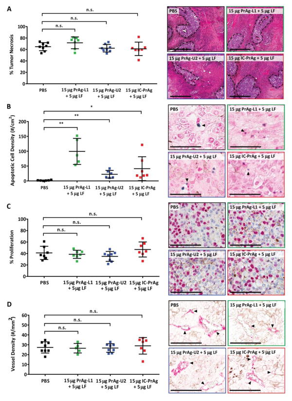
We have previously designed and characterized versions of anthrax lethal toxin that are selectively cytotoxic in the tumor microenvironment and which display broad and potent anti-tumor activities in vivo. Here, we have performed the first direct comparison of the safety and efficacy of three engineered anthrax lethal toxin variants requiring activation by either matrix-metalloproteinases (MMPs), urokinase plasminogen activator (uPA) or co-localized MMP/uPA activities. C57BL/6J mice were challenged with six doses of engineered toxins via intraperitoneal (I.P.) or intravenous (I.V.) dose routes to determine the maximum tolerated dose for six administrations (MTD6) and dose-limiting toxicities. Efficacy was evaluated using the B16-BL6 syngraft model of melanoma; mice bearing established tumors were treated with six I.P. doses of toxin and tumor measurements and immunohistochemistry, paired with terminal blood work, were used to elaborate upon the anti-tumor mechanism and relative efficacy of each variant. We found that MMP-, uPA- and dual MMP/uPA-activated anthrax lethal toxins exhibited the same dose-limiting toxicity; dose-dependent GI toxicity. In terms of efficacy, all three toxins significantly reduced primary B16-BL6 tumor burden, ranging from 32% to 87% reduction, and they also delayed disease progression as evidenced by dose-dependent normalization of blood work values. While target organ toxicity and effective doses were similar amongst the variants, the dual MMP/uPA-activated anthrax lethal toxin exhibited the highest I.P. MTD6 and was 1.5-3-fold better tolerated than the single MMP- and uPA-activated toxins. Overall, we demonstrate that this dual MMP/uPA-activated anthrax lethal toxin can be administered safely and is highly effective in a preclinical model of melanoma. This modified bacterial cytotoxin is thus a promising candidate for further clinical development and evaluation for use in treating human cancers.

Keywords: Bacterial cytotoxin; Cancer; Melanoma; Prodrug; Protease.


Figures

Figure 1
Figure 1. GI toxicity is dose-limiting when C57BL/6J mice are treated with six I.P. doses of MMP-, uPA- or dual MMP/uPA-activated anthrax lethal toxins
Representative H&E sections of small intestine depicting the dose-dependent progression of GI toxicity observed when PrAg-L1 + LF (B,F,J,N) PrAg-U2 + LF (C,G,K,O) or IC-PrAg + LF (D,H,L,P) are administered intraperitoneally. At the MTD6 for each toxin, no GI pathology was present at the gross or microscopic level in 29/30 experimental mice (B–D). At this dose, note the similarity in appearance to control-treated mice receiving 6 I.P. doses of either PBS (A), uncleavable anthrax PrAg paired with LF, 100 μg PrAg-U7 + 33 μg LF (E), or MMP-activated PrAg paired with enzymatically-inactive cytotoxin, 100 μg PrAg-L1 + 33 μg LF-E687A (I). As doses were increased above the MTD6, GI toxicity initially presented as mild small intestinal dilation (F–H) which progressed in severity to involve GI inflammation, regions of villous necrosis, denuded and ulcerated GI epithelium and/or grossly visible GI hemorrhage (J–L, arrowheads depict necrotic villi). (M–P) Higher magnification images showing inflammation in the lamina propria of toxin-treated mice, but not controls. (P) * indicates inflammatory cells that have invaded into the lumen of the small intestine. Scale bars are 300 μm A–L; 100 μm M–P.
Figure 2
Figure 2. I.P. or I.V. treatment with MMP-activated PrAg-L1 + LF caused reduced GI motility
3 doses of PBS (black) or PrAg-L1 + LF (green) were administered either intraperitoneally (A) or intravenously (B). At both the I.P. and I.V. MTD6s, no physical evidence of GI abnormalities was anticipated, while at doses of 2XMTD6 GI histopathology was expected. P-values were determined using a Student’s T-test, two-tailed.
Figure 3
Figure 3. Engineered anthrax lethal toxins reduce B16-BL6 tumor burden and improve survival in a dose-dependent manner
(A–C) Tumor growth and (D–F) survival of C57BL/6J mice bearing B16-BL6 melanoma syngrafts that were treated with six I.P. doses of either PBS (black lines) or engineered anthrax lethal toxins, MMP-activated PrAg-L1 + LF (green), uPA-activated PrAg-U2 + LF (blue) or dual MMP/uPA-activated IC-PrAg + LF (red), at time points indicated by grey arrowheads. All cohorts were treated in parallel and the PBS control groups depicted in panels A–C and D–F are the same. Note that the anti-tumor effect was independent of the proteolytic-activation site; PrAg-L1 + LF, PrAg-U2 + LF and IC-PrAg + LF exhibited indistinguishable anti-tumor effects when administered at protein equivalent doses (A–C). Tumor volume data are expressed as mean tumor weight ± standard error of the mean; *, P<0.05, **, P<0.01, Student’s two-tailed t-test. (D–F) Kaplan-Meier survival curves were compared via Log-Rank test; *, P<0.05, **, P<0.01. (G) Table summarizing anti-tumor and survival effects associated with engineered toxin administration. Abbreviations: n = mouse number, [d.X] = trial day on which maximum % tumor reduction was observed. P-value for % tumor reduction on day 14 was calculated in comparison to the control PBS-treated tumor cohort using a two-tailed Student’s t-test; *, P<0.05, **, P<0.01. P-value for % survival was calculated by comparing Kaplan-Meier survival curves of experimental cohorts with that of the PBS-treated cohort using the Log-Rank test, two-tailed;*, P<0.05, **, P<0.01.
Figure 4
Figure 4. Treatment of B16-BL6 melanoma-bearing mice with a dual MMP/uPA-activated anthrax lethal toxin leads to normalization of blood work values
B16-BL6 melanoma caused significant changes in complete blood count and blood chemistry values relative to tumor-free mice receiving otherwise identical treatments (A). Administration of IC-PrAg + LF led to dose dependent normalization of absolute red blood cell count (B) and alkaline phosphatase levels (C). One toxin-dependent change in blood work values was observed; Treatment with IC-PrAg + LF led to a dose-dependent elevation in absolute platelet count (D). (A) Student’s t-test, two-tailed;*, P<0.05;**, P<0.01;***, P<0.001. (B–D) Bars indicate medians, dashed line indicates disease-free median.
Figure 5
Figure 5. Mechanism of tumoricidal activity of engineered anthrax toxins
Quantification of necrosis (A), apoptosis (B), proliferation (C) and vessel density (D) in B16-BL6 tumors harvested from mice treated with 6 I.P. doses of engineered anthrax lethal toxins at a concentration of 15 μg engineered PrAg + 5 μg LF. In all panels: PBS (black), MMP-activated PrAg-L1 + LF (green), uPA-activated PrAg-U2 + LF (blue) and dual MMP/uPA-activated IC-PrAg + LF (red). (A–D) Student’s two-tailed t-test; **, P<0.01. (A, right) H&E stain. Regions of viable cells are outlined with black dashed lines, scale bars = 500 μm. (B, right) TUNEL stain. Arrowheads highlight examples of TdT positive apoptotic cells stained with TACS blue label, scale bars = 100 μm. (C, right) Ki67 positive nuclei are stained pink with Vulcan FastRed, scale bars = 100 μm. (D, right) Arrowheads highlight CD31 positive blood vessels stained with Vuclan FastRed, scale bars = 100 μm. In all cases representative images are shown.

Similar articles

Cited by

References

    1. Andreasen PA, Kjoller L, Christensen L, Duffy MJ. The urokinase-type plasminogen activator system in cancer metastasis: a review. Int J Cancer. 1997;72:1–22. - PubMed
    1. Bugge TH. Proteolysis in carcinogenesis. In: Ensley JF, Gutkind JS, Jacob JR, Lippman SM, editors. Head and Neck Cancer. Academic Press; San Diego: 2003. pp. 137–49.
    1. Duffy MJ, Mullooly M, O’Donovan N, Sukor S, Crown J, Pierce A, McGowan PM. The ADAMs family of proteases: new biomarkers and therapeutic targets for cancer? Clin Proteomics. 2011;8:9. - PMC - PubMed
    1. Kontos CK, Scorilas A. Kallikrein-related peptidases (KLKs): a gene family of novel cancer biomarkers. Clin Chem Lab Med. 2012;50:1877–91. - PubMed
    1. Roy R, Yang J, Moses MA. Matrix metalloproteinases as novel biomarkers and potential therapeutic targets in human cancer. J Clin Oncol. 2009;27:5287–97. - PMC - PubMed

Publication types

MeSH terms