Review
. 2014 Sep 1;393(1):109-23.
doi: 10.1016/j.ydbio.2014.06.014. Epub 2014 Jun 24.

Symmetry breakage in the vertebrate embryo: when does it happen and how does it work?

Affiliations
Review

Symmetry breakage in the vertebrate embryo: when does it happen and how does it work?

Martin Blum et al. Dev Biol. .

Abstract

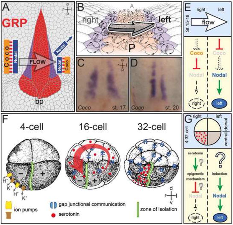
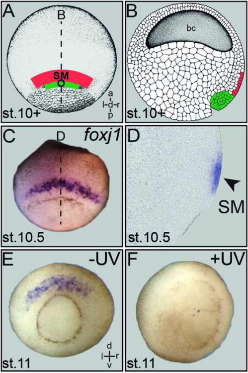
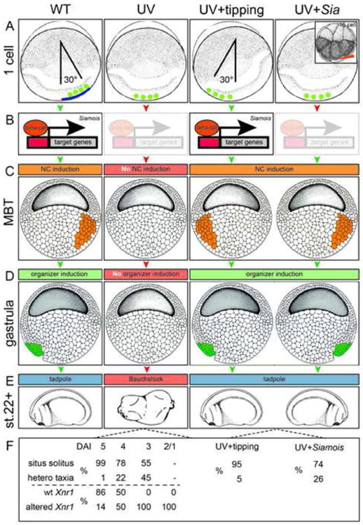
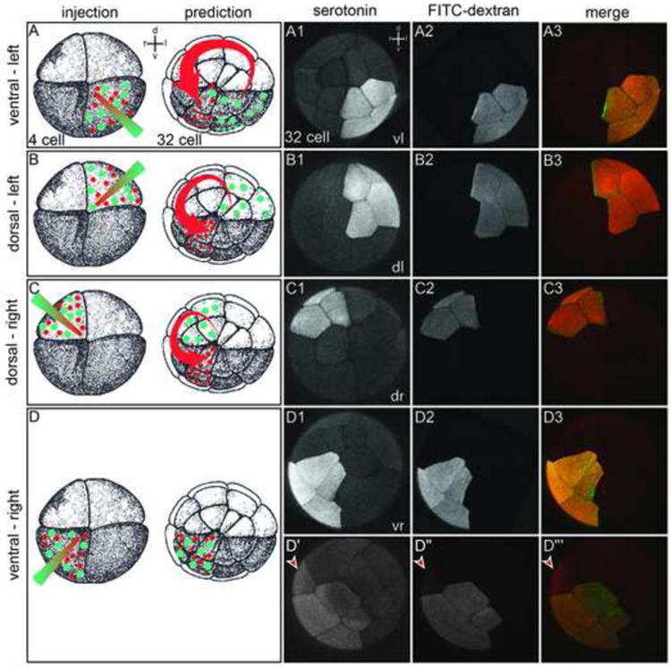
Asymmetric development of the vertebrate embryo has fascinated embryologists for over a century. Much has been learned since the asymmetric Nodal signaling cascade in the left lateral plate mesoderm was detected, and began to be unraveled over the past decade or two. When and how symmetry is initially broken, however, has remained a matter of debate. Two essentially mutually exclusive models prevail. Cilia-driven leftward flow of extracellular fluids occurs in mammalian, fish and amphibian embryos. A great deal of experimental evidence indicates that this flow is indeed required for symmetry breaking. An alternative model has argued, however, that flow simply acts as an amplification step for early asymmetric cues generated by ion flux during the first cleavage divisions. In this review we critically evaluate the experimental basis of both models. Although a number of open questions persist, the available evidence is best compatible with flow-based symmetry breakage as the archetypical mode of symmetry breakage.

Keywords: Cilia; Ion-flux model; Leftward flow; Left–right asymmetry; Symmetry breakage.


Figures

Fig. 1
Fig. 1. Prevailing models of symmetry breakage in the frog Xenopus
(A-E). Leftward flow. (A) Schematic representation of a stage 17 archenteron roof in ventral perspective. Flow occurs from the right to the left side of the ciliated gastrocoel roof plate (GRP; red). Nodal and Coco are co-expressed at the lateral GRP margins on both sides (purple). Flow represses Coco, activating Nodal by release of repression. bp, blastopore. (B) GRP at higher magnification. Polarized and flow-producing cilia at the GRP center are bordered by Nodal/Coco-positive cells (purple) which harbor unpolarized, sensory cilia. (C, D) Coco expression during (C) and following (D) leftward flow. Note the decrease in signal intensity on the left at post-flow stage 20 (D). (E) Schematic depiction of events on the left and right side leading up to asymmetric Nodal cascade induction in the left lateral plate mesoderm (LPM). (F, G). Ion-flux. (F) Asymmetrically expressed ion pumps create a voltage gradient in the 4-cell embryo which initiates the electrogenic transfer of serotonin through gap junctional communication to the ventral-right lineage at the 32-cell stage. Serotonin accumulates in this lineage because the ventral midline is devoid of GJC. (F) Schematic depiction of events on the left and right side leading up to asymmetric Nodal cascade induction in the left LPM. Question marks indicate unproven interactions and mechanisms.
Fig. 2
Fig. 2
Structural and functional relationship of Spemann's organizer and superficial mesoderm. (A) Schematic depiction of superficial mesoderm (SM; red) and organizer (O; green) in whole-mount stage 10+ gastrula embryo shown in dorsal view. (B) Arrangement of organizer and SM in a sagittal section. (C) SM foxj1 expression in a whole-mount gastrula embryo. (D) Sagittal section (plane indicated by dashed line in C) demonstrates foxj1 mRNA in the SM (arrowhead). (E, F) Loss of SM foxj1 expression (E) in UV-ventralized gastrula embryo (F), demonstrating the dependence of SM specification on organizer function.
Fig. 3
Fig. 3
Laterality in UV-treated and rescued embryos. In wildtype (WT) embryos, cortical rotation (blue line) relocates determinants (green circles) to the future dorsal pole (A). Wnt/β-catenin dependent transcription of siamois (B) induces the Nieuwkoop Center (NC; C; orange), following the onset of zygotic transcription at the mid-blastula transition (MBT). The NC induces the formation of the gastrula organizer and SM (D, green), resulting in tadpoles with normal laterality specification (E). UV-irradiation of the zygote inhibits cortical rotation and prevents relocation of determinants (green) to the future dorsal pole. Lack of NC and organizer induction results in ventralized embryos (“Bauchstück” or belly piece of dorso-anterior index: DAI=0) without discernible organs or organ situs. (F) Depending on the degree of UV-induced ventralization, graded responses were observed, both with respect to organ situs (Danos and Yost, 1995) as well as left-asymmetric Xnr1 expression in the LPM (Lohr et al., 1997). Manual tipping of UV-irradiated specimens relocates determinants, induces NC, Siamois, organizer and SM function and thus rescues laterality, as does Siamois mRNA injection into vegetal blastomeres at random positions of the 16-cell embryo (Vandenberg and Levin, 2010b).
Fig. 4
Fig. 4
Serotonin fails to relocalize according to the ion-flux model's prediction. Embryos were selected that displayed regular and symmetric cleavage patterns relative to the pigmentation differences established by the sperm entry point. (A-D) Experimental design (injection) and prediction. A mixture of serotonin and fixable fluorescein dextran (FITC) was microinjected into ventral-left, dorsal-left, dorsal-right, or ventral-right quadrants of 4-cell embryos, respectively. According to the ion-flux model (prediction), serotonin (red) should translocate counterclockwise from the site of injection and accumulate in the ventral right quadrant, while the fluorescein-dextran (green) should remain in situ. (A1-D3) Embryos were fixed at the 32-cell stage and examined via whole mount confocal microscopy to detect potential redistribution of serotonin (A1-D1) relative to the fluorescein dextran (A2-D2). Enrichment of serotonin in the ventral-right lineage was not observed (merged images; A3-D3). (D′-D‴) Displacement of serotonin into uninjected lineage is frequently accompanied by lineage label (cf. Table 3). (D′-D‴) In the individual case of a ventral-right injection shown here, serotonin has leaked across the second cleavage plane into the dorsal-right quadrant (D″, arrowhead). The same blastomere also received small amounts of fluorescent dextran (D‴, arrowhead), revealing the existence of a persistent cytoplasmic bridge between the dorsal and ventral lineages (cf. Fig. 5).
Fig. 5
Fig. 5
Cytoplasmic bridges maintain continuity between subsets of sister blastomeres. Shown is the blastocoel-facing surface of four sister blastomeres of an embryo fixed at the onset of the 5th cleavage (16->32 cell stage), with two cleavage furrows indicated. An older, nearly-complete furrow (arrows), produces sister blastomeres 1 and 2. The nearly-closed cytoplasmic bridge remains in the form of a midbody suspended between 1 and 2 across the blastocoel. With the next cleavage cycle, a later cleavage furrow (arrowheads), separates 1′ from 1″ and 2′ from 2″. It is evident that 1″ and 2″ will become topologically isolated from each other, while cytoplasmic continuity can persist between 1′ and 2′ as long as the cytoplasmic bridge remains.
Fig. 6
Fig. 6
Hypothesis: “Early” determinants are required for SM specification through serotonin accumulation in epithelial cells. (A) Schematic depiction of serotonin accumulation in the epithelial layer of the blastocoel roof before the onset of gastrulation. Transversal sections of 32- and 128-cell embryos (plane indicated in light blue) and sagittal (animal-vegetal) section of stage 9 blastula embryo. bc, blastocoel; d, dorsal; v, ventral. Redrawn from (Beyer et al., 2012a; Vandenberg and Levin, 2010b). (B) Apical enrichment of serotonin through Rab11/Kif16b/microtubule (MT) mediated exocytosis, re-uptake through SERT and re-packaging into vesicles through V-MAT. (C) Epithelialization of the outer layer of the blastula. For details see text.

References

    1. Adams DS, Robinson KR, Fukumoto T, Yuan S, Albertson RC, Yelick P, Kuo L, McSweeney M, Levin M. Early, H+-V-ATPase-dependent proton flux is necessary for consistent left-right patterning of non-mammalian vertebrates. Development. 2006;133:1657–1671. - PMC - PubMed
    1. Aizawa H. Habenula and the asymmetric development of the vertebrate brain. Anat Sci Int. 2013;88:1–9. - PubMed
    1. Amack JD, Yost HJ. The T box transcription factor no tail in ciliated cells controls zebrafish left-right asymmetry. Curr Biol. 2004;14:685–690. - PubMed
    1. Aw S, Adams DS, Qiu D, Levin M. H,K-ATPase protein localization and Kir4.1 function reveal concordance of three axes during early determination of left-right asymmetry. Mech Dev. 2008;125:353–372. - PMC - PubMed
    1. Aw S, Koster JC, Pearson W, Nichols CG, Shi NQ, Carneiro K, Levin M. The ATP-sensitive K(+)-channel (K(ATP)) controls early left-right patterning in Xenopus and chick embryos. Dev Biol. 2010;346:39–53. - PMC - PubMed

Publication types

MeSH terms

LinkOut - more resources