. 2014 Jun 27;15(1):72.
doi: 10.1186/1465-9921-15-72.

Impaired macrophage phagocytosis of bacteria in severe asthma

Affiliations

Impaired macrophage phagocytosis of bacteria in severe asthma

Zhike Liang et al. Respir Res. .

Abstract

Background: Bacteria are frequently cultured from sputum samples of severe asthma patients suggesting a defect in bacterial clearance from the airway. We measured the capacity of macrophages from patients with asthma to phagocytose bacteria.

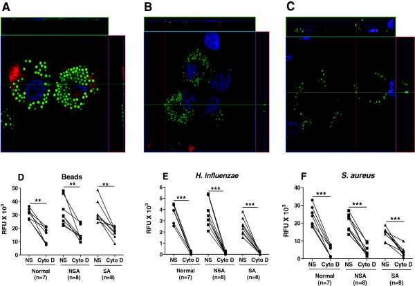
Methods: Phagocytosis of fluorescently-labelled polystyrene beads, Haemophilus influenzae or Staphylococcus aureus by broncholaveolar lavage alveolar macrophages (AM) and by monocyte-derived macrophages (MDM) from non-asthmatics, mild-moderate and severe asthmatic patients was assessed using fluorimetry.

Results: There were no differences in phagocytosis of polystyrene beads by AMs or MDMs from any of the subject groups. There was reduced phagocytosis of Haemophilus influenzae and Staphylococcus aureus in MDMs from patients with severe asthma compared to non-severe asthma (p < 0.05 and p < 0.01, respectively) and healthy subjects (p < 0.01and p < 0.001, respectively). Phagocytosis of Haemophilus influenzae and Staphylococcus aureus by AM was also reduced in severe asthma compared to normal subjects (p < 0.05). Dexamethasone and formoterol did not suppress phagocytosis of bacteria by MDMs from any of the groups.

Conclusions: Persistence of bacteria in the lower airways may result partly from a reduced phagocytic capacity of macrophages for bacteria. This may contribute to increased exacerbations, airway colonization and persistence of inflammation.


Figures

Figure 1
Figure 1
Phagocytosis of fluorescently-labelled beads (Panel A), H. influenzae (Panel B) or S. aureus (Panel C) by monocyte-derived macrophages (MDM). MDM were obtained from normal subjects (●, n = 14) and patients with non-severe asthma (NSA; ■, n = 14) or severe asthma (SA; ▲, n = 14). Phagocytosis was reported as relative fluorescence unit (RFU). Data are presented as dot plots and medians. A Kruskal-Wallis test followed by Dunnett's post-hoc test was used for multi-group comparisons, where *p < 0.05 and **p < 0.01.
Figure 2
Figure 2
Phagocytosis of beads, H. influenzae or S. aureus by alveolar macrophages. Alveolar macrophages from normal subjects (●, n = 7) and patients with non-severe asthma (NSA; ■, n = 6) or severe asthma (SA; ▲, n = 8) were obtained by bronchoalveolar lavage. Alveolar macrophages were exposed to fluorescently-labeled beads (Panel A) or H. influenzae or S. aureus(Panels B and C) for 4 h and phagocytosis reported as relative fluorescence units (RFU). Data are presented as dot-plots and medians. A Kruskal-Wallis test by Dunnett's post-hoc test was used for multi-group comparisons where *p < 0.05. Direct comparison between normal subjects and SA for S.aureus (C) was performed using a Mann-Whitney t-test where $p < 0.01.
Figure 3
Figure 3
Relationship between phagocytosis and lung function parameters. MDM were derived from normal subjects (●, n = 14), non-severe (■, n = 14) and severe asthmatics (▲, n = 14) and exposed to either fluorescently-labeled H. influenzae(Panel A) or S. aureus(Panel B). Alveolar macrophages from normal subjects (●, n = 7) and patients with non-severe asthma (NSA; ■, n = 6) or severe asthma (SA; ▲, n = 8) were exposed to either fluorescently-labeled H. influenzae(Panel C) or S. aureus(Panel D). Correlations were determined using Spearman rank correlation coefficient.
Figure 4
Figure 4
Confocal microscopy of fluorescently-labeled beads or bacteria by MDM and effect of cytochalasin D. Panels A-C: MDM were derived from a patient with non-severe asthma and exposed to either fluorescently-labelled beads (Panel A) or S. aureus for 4 h (green) (Panel B). Panel C shows lack of phagocytosis of S.aureus in the presence of cytochalasin D. Cell cytoplasm was stained with Cell Tracker Red and the nuclei with DAPI (blue). Panels D-F: MDMs were derived from normal subjects (●), non-severe asthmatics (NSA; ■) or severe asthmatics (SA; ▲) and pretreated with cytochalasin D (cyto D; 5 μg/ml) prior to incubation with beads (Panel D), H. i influenzae(Panel E) or S. aureus(Panel F) for 4 h. Phagocytosis was measured by fluorimetry and reported as relative fluorescence units (RFU). NS = not stimulated. A paired t-test was used to determine the effect of Cytochalasin D on phagocytosis. **p < 0.01, ***p < 0.001.
Figure 5
Figure 5
Effect of dexamethasone on phagocytosis by MDM. MDM from normal subjects (●, n = 14), non-severe asthmatics (■, n = 14) and severe asthmatics (▲, n = 14) were pre-treated with dexamethasone for 1 h prior to exposure to fluorescently-labelled beads (Panel A), H. influenzae(Panel B) or S.aureus(Panel C) for 4 h. Phagocytosis was measured by fluorimetry and presented as mean ± SEM. A Mann-Whitney test was used to compare the effect of given concentration of dexamethasone with that of untreated samples. **p < 0.01 and ***p < 0.001 compared to untreated cells.
Figure 6
Figure 6
Effect of formoterol alone or in combination with dexamethasone on phagocytosis. MDM were derived from from normal subjects (●, n = 5), non-severe asthmatics (■, n = 6) and severe asthmatics (▲, n = 5) were pre-treated with formoterol in the absence or presence of dexamethasone for 1 h prior to exposure to H. influenzae(Panels A and C) or S. aureus(Panels B and D) for 4 h. Phagocytosis was measured by fluorimetry and data presented as mean ± SEM. Differences between the effects of dexamethasone or formoterol or dexamethasone and formoterol treatment were analysed using Wilcoxon paired t-test, where *p < 0.05 and **p < 0.01 compared to untreated cells. D = dexamethasone, F = formoterol.

Similar articles

Cited by

References

    1. Chung KF, Godard P, Adelroth E, Ayres J, Barnes N, Barnes P, Bel E, Burney P, Chanez P, Connett G, Corrigan C, de Blic J, Fabbri L, Holgate ST, Ind P, Joos G, Kerstjens H, Leuenberger P, Lofdahl C-G, McKenzie S, Magnussen H, Postman, Saetta D, Salmeron S, Silverman M, Sterk P. Difficult/therapy-resistant asthma: the need for an integrated approach to define clinical phenotypes, evaluate risk factors, understand pathophysiology and find novel therapies. ERS Task Force on Difficult/Therapy-Resistant Asthma. European Respiratory Society. Eur Respir J. 1999;13:1198–1208. - PubMed
    1. Moore WC, Bleecker ER, Curran-Everett D, Erzurum SC, Ameredes BT, Bacharier L, Calhoun WJ, Castro M, Chung KF, Clark MP, Dweik RA, Fitzpatrick AM, Gaston B, Hew M, Hussain I, Jarjour NN, Israel E, Levy BD, Murphy JR, Peters SP, Teague WG, Meyers DA, Busse WW, Wenzel SE. Characterization of the severe asthma phenotype by the National Heart, Lung, and Blood Institute’s Severe Asthma Research Program. J Allergy Clin Immunol. 2007;119:405–413. - PMC - PubMed
    1. Bisgaard H, Hermansen MN, Buchvald F, Loland L, Halkjaer LB, Bønnelykke K, Brasholt M, Heltberg A, Vissing NH, Thorsen SV, Stage M, Pipper CB. Childhood asthma after bacterial colonization of the airway in neonates. N Eng J Med. 2007;357:1487–1495. - PubMed
    1. Martin RJ, Kraft M, CHU HW, Berns EA, Cassell GH. A link between chronic asthma and chronic infection. J Allergy Clin Immunol. 2001;107:595–601. - PubMed
    1. Jackson DJ, Johnston SL. The role of viruses in acute exacerbations of asthma. J Allergy Clin Immunol. 2010;125:1178–1187. - PMC - PubMed

Publication types