. 2014 Nov;70(2):1459-67.
doi: 10.1007/s12013-014-0083-5.

Mechanisms behind the inhibition of lung adenocarcinoma cell by shikonin

Affiliations

Mechanisms behind the inhibition of lung adenocarcinoma cell by shikonin

Wenjing Lan et al. Cell Biochem Biophys. 2014 Nov.

Abstract

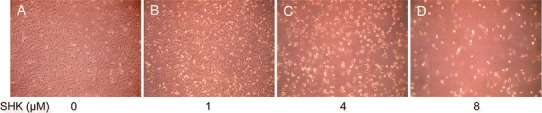
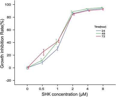
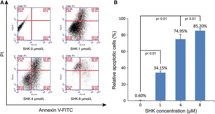
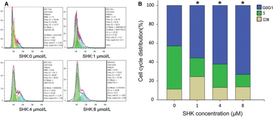
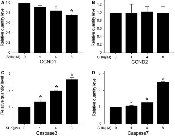
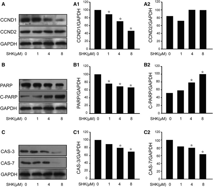
Shikonin, a natural naphthoquinone isolated from a traditional Chinese medicinal herb, can exert inhibitory effect on tumor cell growth. However, little has been known concerning the effect of shikonin on lung adenocarcinoma cell and underlying mechanisms. In the present study, we investigated the effect of shikonin on the proliferation, cell cycle arrest, and apoptosis in human lung adenocarcinoma cells. We found that shikonin significantly suppressed the proliferation of lung adenocarcinoma cells compared with control in dose- and time-dependent manner (P < 0.05). In the meantime, our results showed that shikonin markedly increased the proportion of A549 cells at stage G1 as well as induced apoptosis in A549 cells. Furthermore, suppressed CCND1 and elevated caspase3 and caspase7 expression levels at mRNA were found in this study, indicating that shikonin may inhibit the growth of lung adenocarcinoma cell by changing cell cycle and promoting cell apoptosis through the regulation of CCND1, caspase3, and caspase7. Although more studies are needed, this study suggests that shikonin has the potential to be used as an anti-cancer agent in the treatment of lung adenocarcinoma.


Figures

Fig. 1
Fig. 1
The effect of shikonin on the morphology of A549 cells. a Control group; b Cells were treated with shikonin at the concentration of 1.0 μM for 24 h. The number of A549 decreased and the morphology of A549 obviously changed; c Cells were treated with shikonin at the concentration of 4 μM for 24 h. The number of A549 obviously decreased and the morphology of A549 is different from that of the control with weak adherence; d Cells were treated with shikonin at the concentration of 8 μM for 24 h. Many cells died, and cell debris were found in substrate
Fig. 2
Fig. 2
Shikonin inhibited A549 cells proliferation in a dose-dependent and time-dependent manner. SHK shikonin
Fig. 3
Fig. 3
The effect of shikonin on the apoptosis of A549 cells. a Representative images of the flow cytometry analysis. Cells in the lower right quadrant (Q2-LR) indicate the percentage of Annexin-positive, early apoptotic cells. Cells in the lower left quadrant (Q2-LL) indicate the percentage of Annexin-negative/PI-negative, viable cells. Cells in the upper right quadrant (Q2-UR) indicate the percentage of Annexin-positive/PI-positive, late apoptotic cells. Cells in the upper left quadrant (Q2-UL) indicate the percentage of PI-positive, necrotic cells; b Graphic representation of apoptotic levels (early plus late apoptosis) after shikonin treatment at different concentrations for 24 h. Data were expressed as mean ± SEM
Fig. 4
Fig. 4
The effect of shikonin on cell cycle of A549 cells. a Flow cytometric analysis on A549 cell line to determine the stage of cells after incubation with shikonin at 0, 1.0, 4.0, and 8.0 μM. b The percentages of cells in each phase of cell cycle are shown. With the increase of shikonin concentrations, the proportion of G0/G1 stage increased as shown in the blue column. *P < 0.01 vs control
Fig. 5
Fig. 5
Expression levels of apoptotic genes and cycle-related genes by RT-PCR. a The levels of CCND1 markedly decreased when exposed to various concentrations (0, 1, 4, and 8 μM) of shikonin. b The levels of CCND2 did not change with the increase of shikonin concentration. c The level of caspase3 markedly increased with the increase of shikonin. d The level of caspase7 markedly increased with the increase of shikonin. *P < 0.05 vs control
Fig. 6
Fig. 6
Expression levels of apoptotic genes and cycle-related genes by Western blotting. a The levels of CCND1 and CCND2. a1 The expression level of CCND1 reduced with the increase of shikonin. a2 The level of CCND2 remained stable whatever was the concentration of shikonin. b The levels of PARP and cleaved PARP. b1 The level of PARP was markedly declined with the increase of shikonin concentrations. b2 The level of cleaved PARP increased. c The levels of caspase3 c1 and caspase7 c2. The expression levels of caspase3 and caspase7 were decreased with the increase of shikonin concentrations. *P < 0.05 vs control

Similar articles

Cited by

References

    1. Chen Y, Peng W, Lu Y, et al. MiR-200a enhances the migrations of A549 and SK-MES-1 cells by regulating the expression of TSPAN1. Journal of Biosciences. 2013;38:523–532. doi: 10.1007/s12038-013-9351-6. - DOI - PubMed
    1. Jemal A, Bray F, Center MM, et al. Global cancer statistics. CA: A Cancer Journal for Clinicians. 2011;61:69–90. - PubMed
    1. Jemal A, Siegel R, Ward E, et al. Cancer statistics. CA: A Cancer Journal for Clinicians. 2009;59:225–249. - PubMed
    1. Wingo PA, Cardinez CJ, Landis SH. Long-term trends in cancer mortality in the United States, 1930–1998. Cancer. 2003;97:3133–3275. doi: 10.1002/cncr.11380. - DOI - PubMed
    1. Lynch TJ, Bell DW, Sordella R, et al. Activating mutations in the epidermal growth factor receptor underlying responsiveness of non-small cell lung cancer to gefitinib. New England Journal of Medicine. 2004;350:2129–2139. doi: 10.1056/NEJMoa040938. - DOI - PubMed

Publication types

MeSH terms