. 2014 Aug;6(8):1075-89.
doi: 10.15252/emmm.201403864.

RGS5 promotes arterial growth during arteriogenesis

Affiliations

RGS5 promotes arterial growth during arteriogenesis

Caroline Arnold et al. EMBO Mol Med. 2014 Aug.

Abstract

Arteriogenesis-the growth of collateral arterioles-partially compensates for the progressive occlusion of large conductance arteries as it may occur as a consequence of coronary, cerebral or peripheral artery disease. Despite being clinically highly relevant, mechanisms driving this process remain elusive. In this context, our study revealed that abundance of regulator of G-protein signalling 5 (RGS5) is increased in vascular smooth muscle cells (SMCs) of remodelling collateral arterioles. RGS5 terminates G-protein-coupled signalling cascades which control contractile responses of SMCs. Consequently, overexpression of RGS5 blunted Gαq/11-mediated mobilization of intracellular calcium, thereby facilitating Gα12/13-mediated RhoA signalling which is crucial for arteriogenesis. Knockdown of RGS5 evoked opposite effects and thus strongly impaired collateral growth as evidenced by a blockade of RhoA activation, SMC proliferation and the inability of these cells to acquire an activated phenotype in RGS5-deficient mice after the onset of arteriogenesis. Collectively, these findings establish RGS5 as a novel determinant of arteriogenesis which shifts G-protein signalling from Gαq/11-mediated calcium-dependent contraction towards Gα12/13-mediated Rho kinase-dependent SMC activation.

Keywords: G‐protein; RGS5; arteriogenesis; remodelling; vascular smooth muscle cells.


Figures

Figure 1
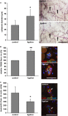
Figure 1. RGS5 protein abundance is increased in remodelling collateral arterioles
A   Arteriogenic remodelling of collateral arterioles in the mouse hindlimb was analysed 7 days post ligation of the femoral artery. Growth of the remodelling arterioles (arrows) is significantly increased during this period (*P < 0.05 versus control, n = 5; scale bar: 1 mm). B   Quantification of RGS5-specific immunofluorescence intensity (red fluorescence staining) in the SMCs of these arterioles reveals a significant rise in RGS5 abundance over this period (**P < 0.05 versus control, n = 5, analysing up to 3 collaterals per animal; scale bar: 50 μm). C   Under these conditions, the fluorescence intensity of myocardin (red), detected in the collateral media, was significantly decreased (*P < 0.05 versus control, n = 5; arrows: endothelial cell; CD31 staining: green; scale bar: 50 μm).
Figure 2
Figure 2. Nitric oxide and cyclic stretch—critical determinants of arteriogenesis—increase RGS5 protein levels
A, B   Cultured human umbilical artery SMCs were exposed to the NO donor NONOate (100 μM) or the cell-permeable cyclic GMP analog 8pCPT-cGMP (100 μM) for up to 96 h. Subsequent changes in RGS5 mRNA levels were quantified by real-time PCR analysis (*P < 0.05 versus 0 h; ***P < 0.001 versus 0 h, n = 3; expression of the housekeeping gene RPL32 was utilized as internal standard). C   Treatment (72 h) of isolated femoral artery segments with NONOate also reveals a significant rise in RGS5-specific immunofluorescence (red fluorescence staining) in the medial SMCs (*P < 0.05 versus 0 h, n = 3, nuclei were counterstained with DAPI (blue fluorescence staining), scale bar: 50 μm). D   Immunofluorescence analyses of control cells cultured under static and stretch-stimulated conditions (0.5 Hz, 0 to 15%) HUASMCs showed an increase in RGS5-specific (red) fluorescence (*P < 0.05 versus control, n = 3; RGS5 abundance in the cells was quantified by determining the fluorescence intensity in at least five microscopic fields of view per condition. Control levels were set to 100%; scale bar: 100 μm).
Figure 3
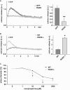
Figure 3. GPCR agonist-induced mobilization of intracellular calcium and arterial constriction is enhanced in RGS5-deficient SMCs
A   Cultured human umbilical artery SMCs were transduced with an adenoviral control (GFP) or RGS5 expression vector (RGS5) and then loaded with the calcium-sensing fluorophore Rhod-4 AM. Sphingosine-1-phosphate (S1P, 10 μM) elicits a rapid but transient rise in intracellular calcium in GFP-expressing cells which is virtually abrogated in cells overexpressing RGS5 (**P < 0.01 versus GFP-expressing cells, n = 4; calcium transients were quantified by determining the area under the curve). B   Cultured arterial SMCs derived from wild-type (WT) or RGS5-deficient mice (RGS5−/−) were loaded with the calcium-sensing fluorophore Rhod-4 AM. S1P moderately increased the intracellular calcium concentration in control SMCs. This effect was significantly reinforced in RGS5-deficient SMCs (*P < 0.05 versus control, n = 4). C   Similarly, mesenteric artery segments of RGS5-deficient mice responded with a more pronounced constriction to increasing concentrations of norepinephrine as compared to segments of wild-type (WT) animals (*P < 0.05 versus WT, n = 3).
Figure 4
Figure 4
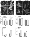
A–G   Cultured human umbilical artery SMCs were transduced with an adenoviral control vector (GFP; A, C, E) or RGS5 expression vector (RGS5; B, D, F). Thereafter, stress fibres (F-actin) were visualized by exposing the cells to TRITC-labelled phalloidin (1:200). RGS5 overexpression alone facilitates stress fibre formation (B and G, #P < 0.05 versus GFP-expressing cells, n = 3). Stimulation with sphingosine-1-phosphate (C and D, S1P, 10 μM) increases stress fibre formation in cells overexpressing RGS5. This effect is abolished by pretreatment of control and RGS5-overexpressing cells with 5 μM of the Rho-kinase inhibitor Y27632 and subsequent S1P stimulation (E, F and G, *P < 0.05 versus RGS5-overexpressing cells, n = 3; cumulative fluorescence intensity of 3–6 whole cells was measured in at least 6 different fields of view, scale bar 20 μm). H   RGS5 overexpression facilitates S1P-stimulated RhoA activation as evidenced by G-LISA-based analyses (*P < 0.05 versus GFP-expressing/S1P-stimulated cells, n = 3). I–M   As compared to SMCs transfected with control siRNA (ctr. siRNA; I and K), knockdown of RGS5 significantly decreases stress fibre formation at baseline (RGS5 siRNA; I, J and M) but stimulation with S1P (K, L and M) does not further affect stress fibre formation upon RGS5 knockdown (M, *P < 0.05 versus ctr. siRNA-transfected cells, n = 3; cumulative fluorescence intensity of 3–6 whole cells was measured in at least 6 different fields of view, scale bar: 20 μm). N   Loss of RGS5 in HUASMCs inhibits S1P-stimulated RhoA activation as evidenced by G-LISA-based analyses (**P < 0.01 versus ctr. siRNA/S1P-treated cells, n = 4).
Figure 5
Figure 5. RGS5 deficiency inhibits growth of collateral arterioles
A   To determine collateral-dependent blood flow recovery, hind foot perfusion was measured before, just after and 7 days after ligation. Blood flow recovery (expressed as flow in percent of non-ligated legs) was significantly attenuated in RGS5-deficient mice as compared to wild-type mice (wild type: ###P < 0.001 versus control, $$P < 0.001 versus ligation 0 days (wild type); RGS5−/−: ###P < 0.001 versus control not significant (n.s.) versus ligation 0 days (RGS5−/−), **P < 0.01 versus ligation 7 days (wild type), n = 6–11; control values were set to 100%). B   Arteriogenic remodelling of collateral arterioles in the mouse hindlimb was analysed 7 days post ligation of the femoral artery. Growth of the remodelling arterioles (arrows) is significantly increased during this period in WT (***P < 0.001 versus control, n = 5) but not in RGS5-deficient mice (B, ###P < 0.001 versus WT ligation, n = 5; scale bar: 1 mm). C   Likewise, wall thickness is not increased during arteriogenesis in RGS5-deficient mice (***P < 0.001 versus control (WT) and not significant (n.s.) versus control (RGS5−/−), n = 5). D   Immunofluorescence detection of active RhoA (Rho-GTP) in cross sections of collateral arterioles revealed a significant increase in RhoA activity during arteriogenesis in WT but not in RGS5-deficient mice (**P < 0.01 versus control (WT), ##P < 0.01 versus ligation (WT) and not significant (n.s.) versus control (RGS5−/−), n = 5, analysing up to three collaterals per animal; scale bar: 25 μm).
Figure 6
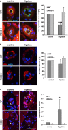
Figure 6. RGS5 deficiency preserves the differentiated SMC phenotype
A, B   Immunofluorescence analyses of cross sections of arterioles undergoing arteriogenesis revealed a significant decline in myocardin (A) and alpha-smooth muscle actin (B) abundance in WT but not in RGS5-deficient mice over this period (*P < 0.05, ***P < 0.001 versus control, n = 5, analysing up to 3 collaterals per animal; asterisk: myoglobin-related red background fluorescence). C   Likewise, PCNA-positive nuclei (C, red fluorescence, arrows) indicating proliferating cells were detected in the SMCs of remodelling collaterals of WT mice but not in RGS5-deficient mice (*P < 0.05 versus control, n = 5; n.d: none detected; arrowheads indicate collateral arterioles; scale bar: 50 μm).
Figure 7
Figure 7. Impact of RGS5 on SMC function
A   Contractile responses of vascular smooth muscle cells (vSMC) were evoked by Gαq/11 signalling which is sparsely inhibited under baseline conditions. B   Loss of RGS5 enhances these responses to some extend and leads to a slight increase in blood pressure. C   The increase in biomechanical stretch and release of nitric oxide (NO) triggers the expression of RGS5 during the onset of arteriogenesis, thereby favouring RhoA-mediated signal responses which promote biomechanical sensitivity of SMCs and the activated SMC phenotype. D   In RGS5-deficient mice, RhoA signalling is not amplified impairing the SMC phenotype switch under these conditions.

References

    1. Adams LD, Geary RL, McManus B, Schwartz SM. A comparison of aorta and vena cava medial message expression by cDNA array analysis identifies a set of 68 consistently differentially expressed genes, all in aortic media. Circ Res. 2000;87:623–631. - PubMed
    1. Althoff TF, Juarez JA, Troidl K, Tang C, Wang S, Wirth A, Takefuji M, Wettschureck N, Offermanns S. Procontractile G protein-mediated signaling pathways antagonistically regulate smooth muscle differentiation in vascular remodeling. J Exp Med. 2012;209:2277–2290. - PMC - PubMed
    1. Banai S, Wolf Y, Golomb G, Pearle A, Waltenberger J, Fishbein I, Schneider A, Gazit A, Perez L, Huber R, et al. PDGF-receptor tyrosine kinase blocker AG1295 selectively attenuates smooth muscle cell growth in vitro and reduces neointimal formation after balloon angioplasty in swine. Circulation. 1998;97:1960–1969. - PubMed
    1. Berger M, Bergers G, Arnold B, Hammerling GJ, Ganss R. Regulator of G-protein signaling-5 induction in pericytes coincides with active vessel remodeling during neovascularization. Blood. 2005;105:1094–1101. - PubMed
    1. Berman DM, Wilkie TM, Gilman AG. GAIP and RGS4 are GTPase-activating proteins for the Gi subfamily of G protein alpha subunits. Cell. 1996;86:445–452. - PubMed

Publication types

MeSH terms

Substances