. 2014 Sep;80(17):5530-41.
doi: 10.1128/AEM.01239-14. Epub 2014 Jun 27.

LitR is a repressor of syp genes and has a temperature-sensitive regulatory effect on biofilm formation and colony morphology in Vibrio (Aliivibrio) salmonicida

Affiliations

LitR is a repressor of syp genes and has a temperature-sensitive regulatory effect on biofilm formation and colony morphology in Vibrio (Aliivibrio) salmonicida

Hilde Hansen et al. Appl Environ Microbiol. 2014 Sep.

Abstract

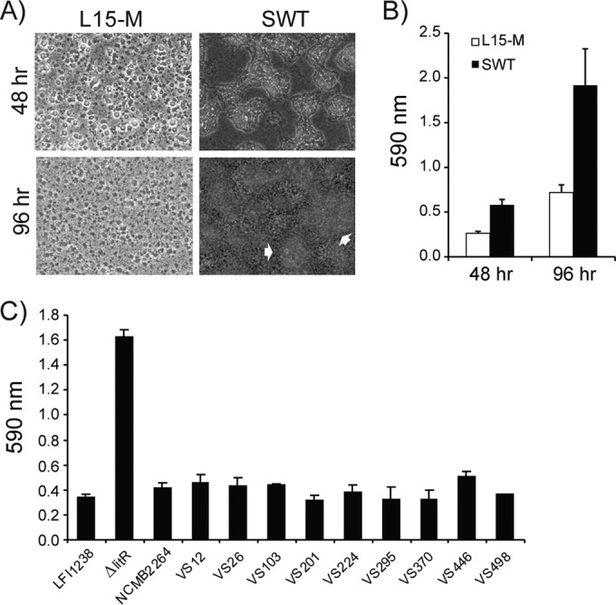
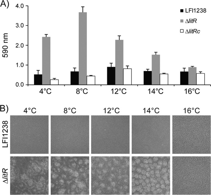
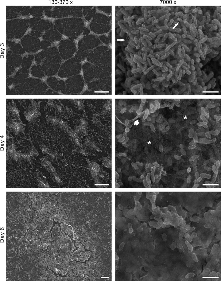
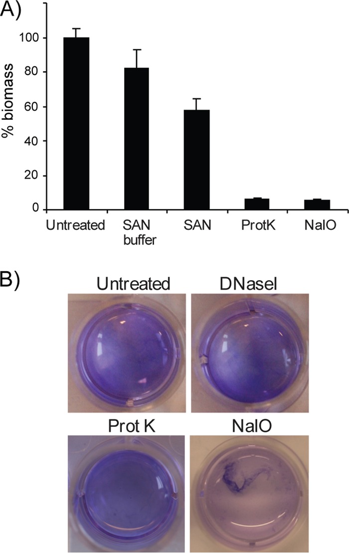
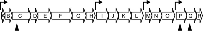
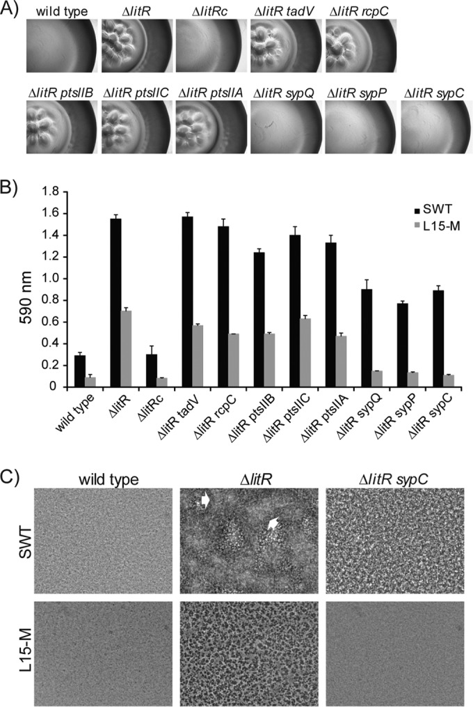
Vibrio (Aliivibrio) salmonicida is the etiological agent of cold water vibriosis, a disease in farmed Atlantic salmon (Salmo salar) that is kept under control due to an effective vaccine. A seawater temperature below 12°C is normally required for disease development. Quorum sensing (QS) is a cell density-regulated communication system that bacteria use to coordinate activities involved in colonization and pathogenesis, and we have previously shown that inactivation of the QS master regulator LitR attenuates the V. salmonicida strain LFI1238 in a fish model. We show here that strain LFI1238 and a panel of naturally occurring V. salmonicida strains are poor biofilm producers. Inactivation of litR in the LFI1238 strain enhances medium- and temperature-dependent adhesion, rugose colony morphology, and biofilm formation. Chemical treatment and electron microscopy of the biofilm identified an extracellular matrix consisting mainly of a fibrous network, proteins, and polysaccharides. Further, by microarray analysis of planktonic and biofilm cells, we identified a number of genes regulated by LitR and, among these, were homologues of the Vibrio fischeri symbiosis polysaccharide (syp) genes. The syp genes were regulated by LitR in both planktonic and biofilm lifestyle analyses. Disruption of syp genes in the V. salmonicida ΔlitR mutant alleviated adhesion, rugose colony morphology, and biofilm formation. Hence, LitR is a repressor of syp transcription that is necessary for expression of the phenotypes examined. The regulatory effect of LitR on colony morphology and biofilm formation is temperature sensitive and weak or absent at temperatures above the bacterium's upper threshold for pathogenicity.


Figures

FIG 1
FIG 1
Biofilm formation of V. salmonicida wild-type strains and the LFI1238-derived ΔlitR mutant. (A and B) Morphology (A) and quantitation (B) of biofilms formed by the ΔlitR mutant in L15-M and SWT after 48 and 96 h of static incubation at 4°C. The morphology was monitored by phase-contrast microscopy (Leica), and the white arrows point to irregular mushroom-like structures in the biofilm. (C) Quantification of the biofilm formed by the different wild-type strains and the ΔlitR mutant in SWT medium after 72 h of incubation at 4°C. The biofilms were stained with crystal violet, and the absorbance was read at 590 nm. The error bars represent the standard deviations of three biological replicates.
FIG 2
FIG 2
Biofilm formation in SWT medium at different temperatures. (A) Quantitation of crystal violet stained biofilms formed by wild-type LFI1238, ΔlitR, and ΔlitRc strains at the indicated temperatures. The biofilms were grown statically in the medium for 72 h before being quantified. The error bars represent the standard deviations of three biological replicates. (B) Morphologies of the biofilms formed by wild-type LFI1238 and the ΔlitR mutant at the indicated temperatures. The morphologies of biofilms were inspected in a Zeiss Primo Vert microscope (10× magnification) and photographed after 72 h of growth.
FIG 3
FIG 3
Colony morphology of wild-type LFI1238 and ΔlitR strains. (A) The colonies were allowed to form on blood agar (BL), SWT, or LB agar plates for 10 days at 4°C before being photographed with a Canon camera. (B) The central and peripheral parts of colonies grown on SWT agar at 4, 8, and 14°C were viewed in a Zeiss Primo Vert microscope at 4× magnification. (C) Thin sections of wild-type and ΔlitR colonies grown on BL and analyzed by transmission electron microscopy. Scale bar, 2 μm.
FIG 4
FIG 4
SEM analyses of ΔlitR biofilms after 3, 4, and 6 days. The biofilms were grown in SWT medium at 4°C before being fixed. The panel to the left shows the biofilms using a low magnification (scale bar, 100 μm for days 3 and 4 and 20 μm for day 6). The panel to the right shows the biofilm at a higher magnification (scale bar, 2 μm). At day 4 a network of fibers (*) is clearly visible, and at day 6 the bacteria are completely encapsulated in the extracellular matrix. The thin arrows point at some flagellum-like structures, and the thick arrow points to a sheet of collapsed extracellular matrix.
FIG 5
FIG 5
Characterization of extracellular matrix using a biofilm inhibition assay and a biofilm detachment assay. (A) For the inhibition assay, the different treatments (SAN, proteinase K, or NaIO4) were added directly to the SWT medium before the biofilms were allowed to form. After 3 days of static incubation at 4°C, the biofilms were stained with crystal violet, and the absorbance was read at 590 nm. The inhibition is shown as the percent biomass relative to the untreated control. (B) For the detachment assay, the biofilm was allowed to form for 3 days at 4°C before the medium (SWT) was removed, and the different treatments (bovine DNase I, proteinase K, or NaIO4) were added. The wells were incubated for 24 h at 37°C before dissolution of the biofilm was visualized by adding a small amount crystal violet into the wells. The error bars represent the standard deviations of three biological replicates.
FIG 6
FIG 6
Genetic organization of the syp locus in V. salmonicida LFI1238. The locus consists of 18 genes (sypA to sypR) and four operons. Putative σ54 binding sites were predicted using Fuzznuc and are indicated by bent arrows. The syp genes that were interrupted by plasmid insertions in the present study are indicated by black arrowheads.
FIG 7
FIG 7
Colony morphology and biofilm formation of V. salmonicida wild-type LFI1238 and the different mutants. (A) The colony morphology of the different strains shows that the disruption of sypC, sypP, and sypQ in the ΔlitR mutant alleviates the rugose appearance. (B) Quantitation of the biofilms formed in L15-M and SWT medium after 3 days of static incubation. The error bars show the standard deviations of three biological replicates. (C) Phase-contrast microscopy (Leica) shows that disruption of syp results in a flat and regular biofilm when formed in SWT medium. The white arrows point to irregular mushroom-like structures in the biofilm. All experiments were performed at 4°C.

Similar articles

Cited by

References

    1. Karatan E, Watnick P. 2009. Signals, regulatory networks, and materials that build and break bacterial biofilms. Microbiol. Mol. Biol. Rev. 73:310–347. 10.1128/MMBR.00041-08 - DOI - PMC - PubMed
    1. Sutherland IW. 2001. The biofilm matrix—an immobilized but dynamic microbial environment. Trends Microbiol. 9:222–227. 10.1016/S0966-842X(01)02012-1 - DOI - PubMed
    1. Whitchurch CB, Tolker-Nielsen T, Ragas PC, Mattick JS. 2002. Extracellular DNA required for bacterial biofilm formation. Science 295:1487. 10.1126/science.295.5559.1487 - DOI - PubMed
    1. Flemming HC, Neu TR, Wozniak DJ. 2007. The EPS matrix: the “house of biofilm cells.” J. Bacteriol. 189:7945–7947. 10.1128/JB.00858-07 - DOI - PMC - PubMed
    1. Harmsen M, Lappann M, Knochel S, Molin S. 2010. Role of extracellular DNA during biofilm formation by Listeria monocytogenes. Appl. Environ. Microbiol. 76:2271–2279. 10.1128/AEM.02361-09 - DOI - PMC - PubMed

Publication types

MeSH terms