. 2014 Sep;141(1):155-65.
doi: 10.1093/toxsci/kfu121. Epub 2014 Jun 27.

Sialic acid rescues repurified lipopolysaccharide-induced acute renal failure via inhibiting TLR4/PKC/gp91-mediated endoplasmic reticulum stress, apoptosis, autophagy, and pyroptosis signaling

Affiliations

Sialic acid rescues repurified lipopolysaccharide-induced acute renal failure via inhibiting TLR4/PKC/gp91-mediated endoplasmic reticulum stress, apoptosis, autophagy, and pyroptosis signaling

Chih-Ching Yang et al. Toxicol Sci. 2014 Sep.

Abstract

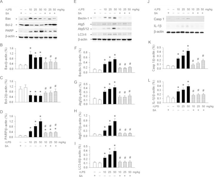
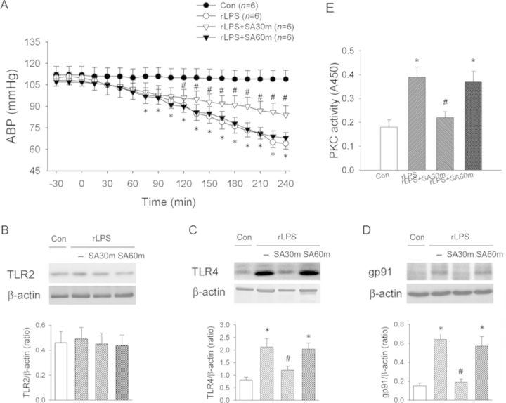
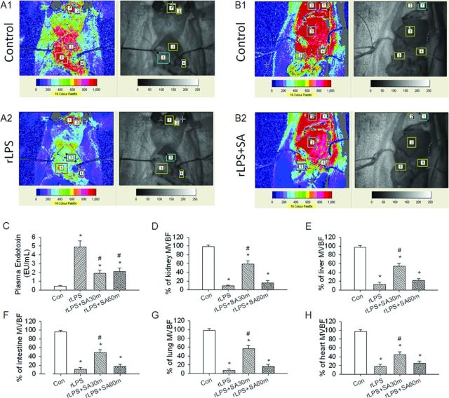
Lipopolysaccharides (LPS) through Toll-like receptor 2 (TLR2) and Toll-like receptor 4 (TLR4) activation induce systemic inflammation where oxidative damage plays a key role in multiple organ failure. Because of the neutralization of LPS toxicity by sialic acid (SA), we determined its effect and mechanisms on repurified LPS (rLPS)-evoked acute renal failure. We assessed the effect of intravenous SA (10 mg/kg body weight) on rLPS-induced renal injury in female Wistar rats by evaluating blood and kidney reactive oxygen species (ROS) responses, renal and systemic hemodynamics, renal function, histopathology, and molecular mechanisms. SA can interact with rLPS through a high binding affinity. rLPS dose- and time-dependently reduced arterial blood pressure, renal microcirculation and blood flow, and increased vascular resistance in the rats. rLPS enhanced monocyte/macrophage (ED-1) infiltration and ROS production and impaired kidneys by triggering p-IRE1α/p-JNK/CHOP/GRP78/ATF4-mediated endoplasmic reticulum (ER) stress, Bax/PARP-mediated apoptosis, Beclin-1/Atg5-Atg12/LC3-II-mediated autophagy, and caspase 1/IL-1β-mediated pyroptosis in the kidneys. SA treatment at 30 min, but not 60 min after rLPS stimulation, gp91 siRNA and protein kinase C-α (PKC) inhibitor efficiently rescued rLPS-induced acute renal failure via inhibition of TLR4/PKC/NADPH oxidase gp91-mediated ER stress, apoptosis, autophagy and pyroptosis in renal proximal tubular cells, and rat kidneys. In response to rLPS or IFNγ, the enhanced Atg5, FADD, LC3-II, and PARP expression can be inhibited by Atg5 siRNA. Albumin (10 mg/kg body weight) did not rescue rLPS-induced injury. In conclusion, early treatment (within 30 min) of SA attenuates rLPS-induced renal failure via the reduction in LPS toxicity and subsequently inhibiting rLPS-activated TLR4/PKC/gp91/ER stress/apoptosis/autophagy/pyroptosis signaling.

Keywords: apoptosis; autophagy; lipopolysaccharide; pyroptosis; reactive oxygen species; sialic acid; toll-like receptors.


Figures

FIG. 1.
FIG. 1.
The procedures for measurement of kinetic interaction between SA and rLPS. (A) The procedures for immobilization of SA on an amine coupling kit activated CM5 chip. (B) The original tracing of SA interacts with rLPS at the level of RU = 165 during 100–210 s. (C) Representative sensograms of kinetic experiments between the increased rLPS concentration and immobilized SA. Using the single-cycle kinetics approach, samples are injected one after the other in the same cycle with no intervening regeneration steps. Here, a dilution series of rLPS was prepared at concentrations of 1, 2, 3, and 4 μg/ml and sequentially injected over a sensor surface prepared with SA (MW = 309.27) immobilized at 165 RU. Red line contrasts the measured data from the simulated fits (black line).
FIG. 2.
FIG. 2.
Effect of SA on 50 mg of LPS or rLPS-induced changes of arterial blood pressure, renal blood flow, and vascular resistance in the rats. (A) Typical laser speckle imaging perfusions are displayed on a 16-level color palette in four rats receiving saline (control), rLPS treatment, rLPS plus SA treatment (LPS + SA), or rLPS plus albumin treatment (LPS + Alb), respectively. The mean changes of mean arterial blood pressure (B), total renal blood flow (C), and renal vascular resistance (D) was continuously recorded during 6 h of rLPS treatment. *p < 0.05 versus the matched time-course point of the control group without rLPS and SA treatment. #p < 0.05 versus the matched time-course point of the rLPS group.
FIG. 3.
FIG. 3.
Effect of SA on LPS or rLPS-induced oxidative stress in the kidney and blood and renal dysfunction in the rats. The level of kidney ROS (A), blood ROS (B), BUN (C), and creatinine (D) was significantly and dose-dependently elevated after 6 h of rLPS (10, 25, and 50 mg) treatment. Thirty minutes after LPS or rLPS treatment, SA at 10 mg significantly decreased the oxidative stress markers and ameliorated renal dysfunction. All the parameters were similar between 50 mg of rLPS or LPS treatment. *p < 0.05 versus the control group without rLPS and SA treatment. #p < 0.05 versus respective dosage of rLPS or LPS treatment.
FIG. 4.
FIG. 4.
Effect of SA treatment on rLPS-induced renal injury. rLPS induced marked histologic changes (B), ED-1 infiltration (E), Beclin-1-autophagy (H), TUNEL-apoptosis formation (K), and caspase 1-pyroptosis (N) in the damaged kidneys when compared with respective control sections (A, D, G, J, and M). Intravenous SA at 10 mg significantly improved rLPS-induced pathologic parameters (C, F, I, L, and O). These markers of oxidative injury are indicated by green arrows. The scale bar is 50 μm. The statistic data is shown in P–T. *p < 0.05 versus the control group without rLPS and SA treatment. #p < 0.05 versus rLPS treatment.
FIG. 5.
FIG. 5.
Effect of SA treatment on rLPS induced apoptosis-, autophagy-, and pyroptosis-related proteins expression in the rat kidneys. Original data of apoptosis-related Bax, Bcl-2, and PARP and autophagy-related Beclin-1, Atg5/12, and LC3-II proteins was demonstrated in (A), (E), and (J), respectively. The statistic data was indicated in (B–D), (F–I), and (K–L), respectively. *p < 0.05 versus the control group without rLPS and SA treatment. #p < 0.05 versus rLPS treatment.
FIG. 6.
FIG. 6.
Effect of gp91 siRNA or SA treatment on rLPS induced TLR4, gp91, apoptosis-, autophagy-, and pyroptosis-related proteins expression (A), O2−• production (H), percentage of cell death (I), and MDC positive autophagic cells (J) in the renal proximal tubular cells. Original data of TLR4, gp91, PARP, and autophagy-related Beclin-1 and pyroptosis-related caspase 1 (casp 1) and IL-1β protein expressions is demonstrated in (A). The statistic data are indicated in (B–G), respectively. The temporal response of Atg5, LC3-II, and PARP to rLPS is indicated in (K). The effect of siRNA Atg5 on rLPS- or IFNγ-stimulated LC3-II and PARP is demonstrated in (L). *p < 0.05 versus the control group without rLPS and SA treatment. #p < 0.05 versus respective rLPS treatment.
FIG. 7.
FIG. 7.
Effect of SA, NADPH oxidase inhibitor, PKC inhibitor, and TLR4 inhibitor on rLPS-induced ER stress, FADD, apoptosis-, autophagy-, and pyroptosis-related proteins expression in the kidneys. Original data and statistic data of p-IRE1a/IRE1a (A) p-JNK/JNK (B), ATF4 (C), CHOP (D), caspase 12 (E), GRP78 (F), caspase 1 (G), Atg5 (H), FADD (I), caspase 3 (J) expression are demonstrated, respectively. Con, control; S, sialic acid; L, repurified lipopolysaccharide; LS, repurified lipopolysaccharide and sialic acid treatment; LP, repurified lipopolysaccharide and PKC-α inhibitor; LD, repurified lipopolysaccharide plus NADPH oxidase inhibitor DPI; LT, repurified lipopolysaccharide plus TLR4 inhibitor. *p < 0.05 versus Con group. #p < 0.05 versus rLPS treatment.
FIG. 8.
FIG. 8.
Time effect of intravenous SA treatment on arterial blood pressure (A), TLR2 (B), TLR4 (C), NADPH oxidase gp91 subunit expression (D), and PKC activity (E) in the rLPS kidneys with or without SA treatment. Con, control rats without rLPS and SA treatment; rLPS, the rats with iv 50 mg/kg rLPS; rLPS + SA30m, 30 min after rLPS plus iv 10 mg SA; rLPS+SA60m, 60 min after rLPS plus iv 10 mg SA. *p < 0.05 versus the Con group without LPS and SA treatment. #p < 0.05 rLPS30m versus rLPS group.
FIG. 9.
FIG. 9.
Systemic effect of SA on rLPS-induced microvascular blood flow in multiple organs. rLPS induced the reduction of microvascular blood flow in the kidney, liver, small intestine, lung, and heart. After 240 min of rLPS, the decreased percentage of microcirculation of four groups is indicated in (F). Con, control rats without rLPS and SA treatment; rLPS, the rats with iv 50 mg/kg rLPS; rLPS + SA30m, 30 min after rLPS plus iv 10 mg SA; rLPS + SA60m, 60 min after rLPS plus iv 10 mg SA. *p < 0.05 versus the Con group without rLPS and SA treatment. #p < 0.05 rLPS30m versus rLPS group.

References

    1. Angus D. C., Linde-Zwirble W. T., Lidicker J., Clermont G., Carcillo J., Pinsky M. R. Epidemiology of severe sepsis in the United States: Analysis of incidence, outcome, and associated costs of care. Crit. Care Med. 2001;29:1303–1310. - PubMed
    1. Azuma Y., Higurashi K., Matsumoto K. Immobilized alpha2,6-linked sialic acid suppresses caspase-3 activation during anti-IgM antibody-induced apoptosis in Ramos cells. Biochim. Biophys. Acta. 2007;1770:279–285. - PubMed
    1. Bannan J., Visvanathan K., Zabriskie J. B. Structure and function of streptococcal and staphylococcal superantigens in septic shock. Infect. Dis. Clin. North Am. 1999;13:387–396. - PubMed
    1. Bhattacharyya J., Biswas S., Datta A. G. Mode of action of endotoxin: Role of free radicals and antioxidants. Curr. Med. Chem. 2004;11:359–368. - PubMed
    1. Bortoluci K. R., Medzhitov R. Control of infection by pyroptosis and autophagy: Role of TLR and NLR. Cell. Mol. Life Sci. 2010;67:1643–1651. - PMC - PubMed

Publication types

MeSH terms