. 2014 Aug 15;289(33):22835-22849.
doi: 10.1074/jbc.M114.563064. Epub 2014 Jun 27.

Platelet-derived growth factor primes cancer-associated fibroblasts for apoptosis

Affiliations

Platelet-derived growth factor primes cancer-associated fibroblasts for apoptosis

Sumera I Ilyas et al. J Biol Chem. .

Abstract

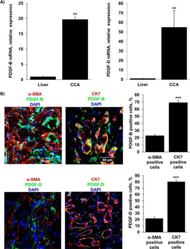
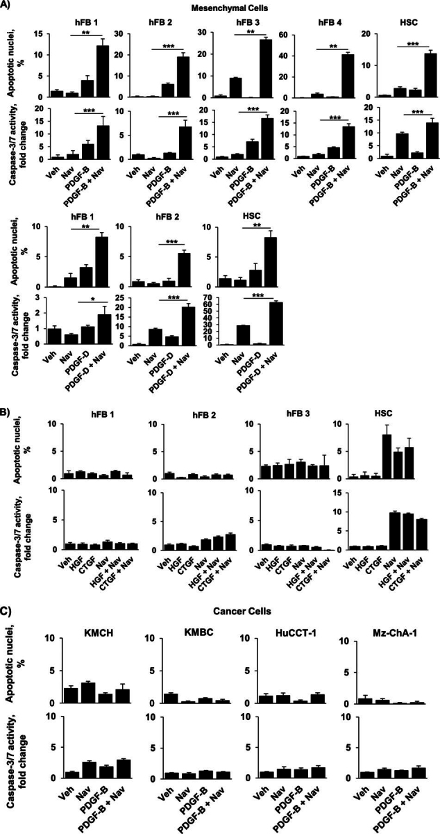
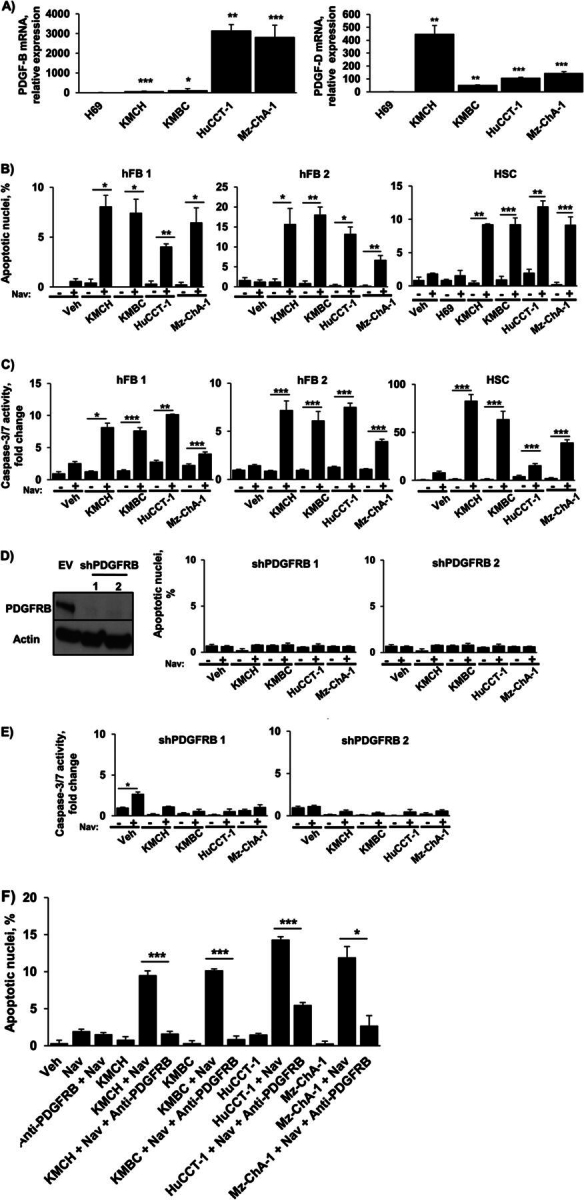
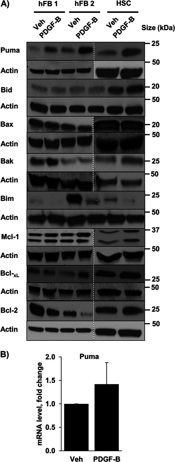
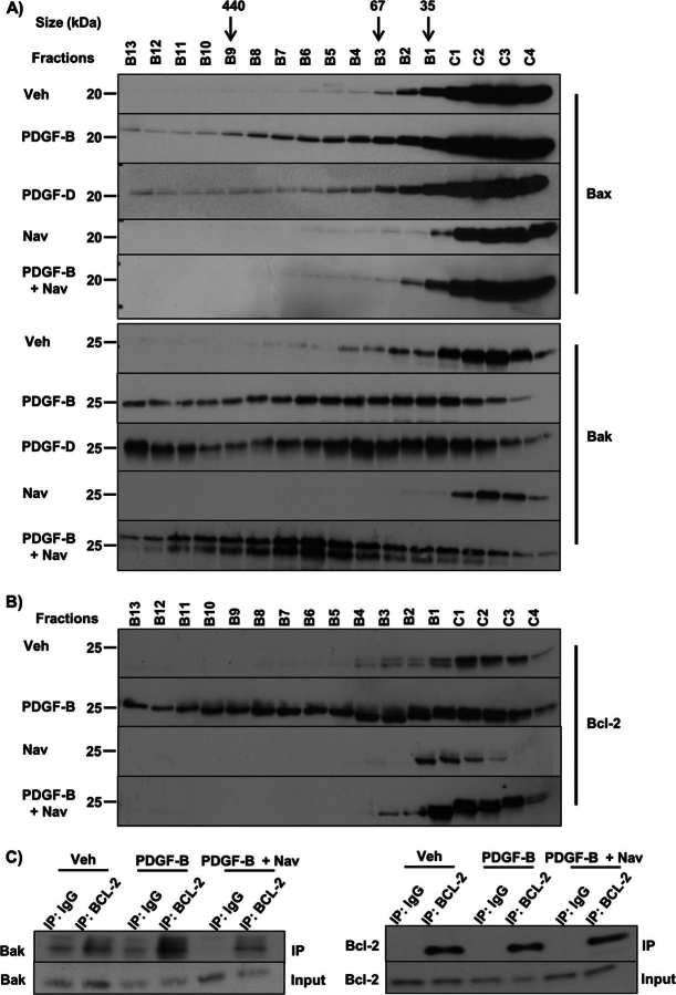
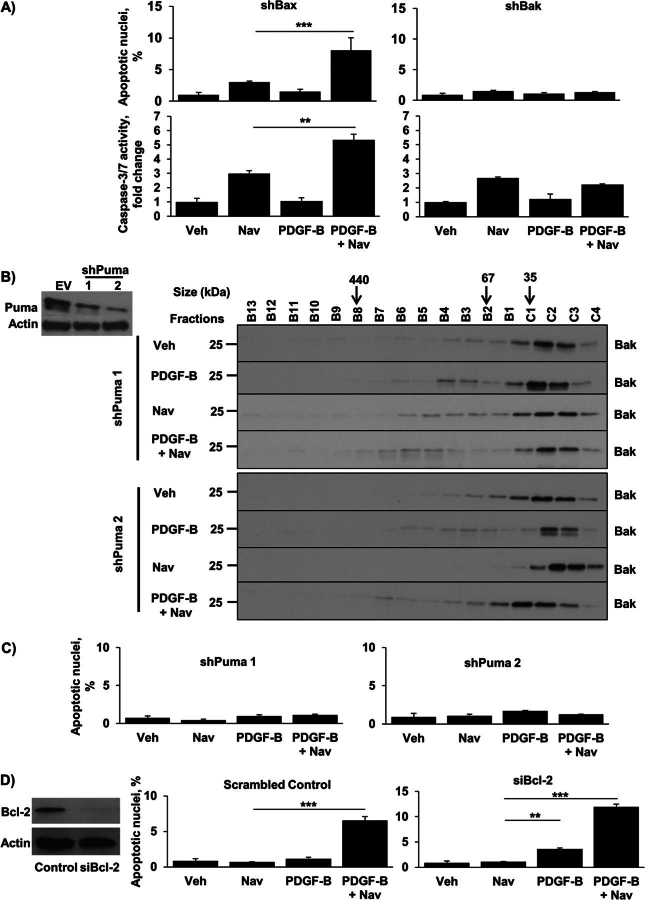
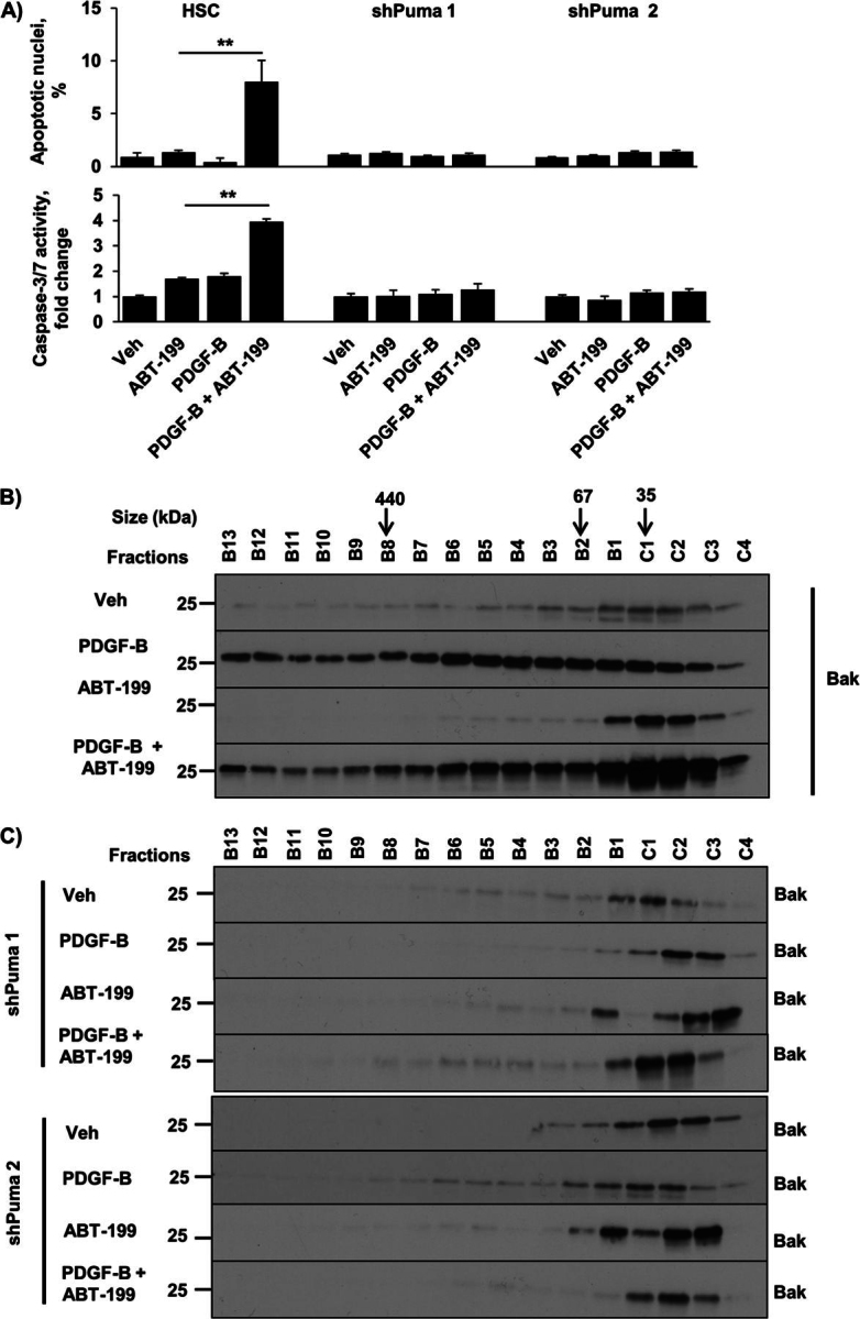
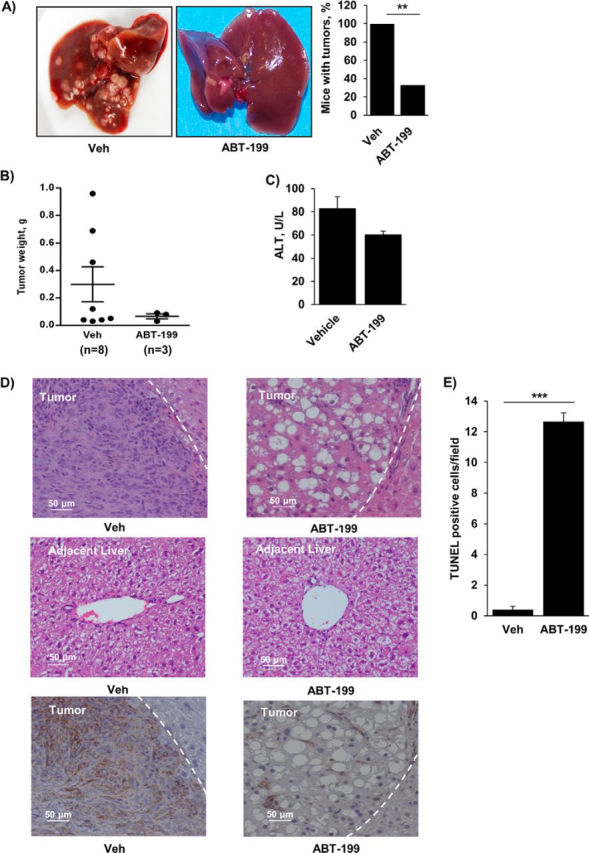
Desmoplastic malignancies such as cholangiocarcinoma (CCA) are characterized by a dense stroma containing an abundance of myofibroblasts termed cancer-associated fibroblasts (CAF). The CAF phenotype represents an "activated state" in which cells are primed for cell death triggered by BH3 mimetics. Accordingly, this primed state may be therapeutically exploited. To elucidate the mechanisms underlying this poorly understood apoptotic priming, we examined the role of platelet-derived growth factor (PDGF) in CAF priming for cell death given its prominent role in CAF activation. PDGF isomers PDGF-B and PDGF-D are abundantly expressed in CCA cells derived from human specimens. Either isomer sensitizes myofibroblasts to cell death triggered by BH3 mimetics. Similar apoptotic sensitization was observed with co-culture of myofibroblasts and CCA cells. Profiling of Bcl-2 proteins expressed by PDGF-primed myofibroblasts demonstrated an increase in cellular levels of Puma. PDGF-mediated increases in cellular Puma levels induced proapoptotic changes in Bak, which resulted in its binding to Bcl-2. Short hairpin RNA-mediated down-regulation of Puma conferred resistance to PDGF-mediated apoptotic priming. Conversely, the BH3 mimetic navitoclax disrupted Bcl-2/Bak heterodimers, allowing Bak to execute the cell death program. Treatment with a Bcl-2-specific BH3 mimetic, ABT-199, reduced tumor formation and tumor burden in a murine model of cholangiocarcinoma. Collectively, these findings indicate that apoptotic priming of CAF by PDGF occurs via Puma-mediated Bak activation, which can be converted to active full-blown apoptosis by navitoclax or ABT-199 for therapeutic benefit.

Keywords: Apoptosis; B-cell Lymphoma 2 (Bcl-2); BH3 Mimetics; Cancer Biology; Cholangiocarcinoma; Myofibroblast; PDGF; Tumor Microenvironment.


Figures

FIGURE 1.
FIGURE 1.
PDGF-B and PDGF-D are increased in CCA cells in human resected specimens. A, expression of PDGF-B and PDGF-D was quantified in human resected CCA specimens and the corresponding normal livers by quantitative real-time-PCR. Mean ± S.E. are depicted for n = 12; **, p < 0.01. B, immunofluorescence was used to detect PDGF-B and PDGF-D expression in CCA cells (CK7-positive) compared with CAF (α-SMA-positive) (left panel). The number of PDGF-B or PDGF-D positive CCA cells and CAF was quantified in five high power fields and expressed as a percentage of total (right panel). Means ± S.E. are depicted for n = 5. ***, p < 0.001. Original magnification, 63×.
FIGURE 2.
FIGURE 2.
PDGF-B and PDGF-D sensitize hFB and HSC, but not CCA cells, to navitoclax-induced apoptosis. A, HSC and four different types of quiescent hFB (hFB 1–4) were treated with vehicle, navitoclax for 48 h, and PDGF-B for 24 h or PDGF-B for 24 h followed by the addition of navitoclax for 48 h. Similarly, HSC cells and two hFB cell types were treated with vehicle (Veh), navitoclax (Nav), PDGF-D, or PDGF-D for 24 h followed by the addition of navitoclax for 48 h. Apoptosis was quantified morphologically using DAPI staining plus fluorescence microscopy (upper panels) and biochemically by measuring caspase-3/7 activity (lower panels). Mean ± S.E. are depicted for n = 3. **. p < 0.01; ***, p < 0.001. B, hFB and HSC were treated with vehicle, hepatocyte growth factor (HGF), connective tissue growth factor (CTGF), and navitoclax for 24 h followed by the addition of navitoclax for 48 h or connective tissue growth factor for 24 h followed by the addition of navitoclax for 48 h. Apoptosis was quantified using DAPI plus fluorescence microscopy (upper panels) and by measuring caspase-3/7 activity (lower panels). Mean ± S.E. are depicted for n = 3. C, four different human CCA cell lines (KMCH, KMBC, HuCCT-1, Mz-ChA-1) were treated as described in A. Apoptosis was quantified using DAPI plus fluorescence microscopy (upper panels) and by measuring caspase-3/7 activity (lower panels). Mean ± S.E. are depicted for n = 3.
FIGURE 3.
FIGURE 3.
Co-culturing hFB or HSC with CCA cell lines significantly increases navitoclax-induced apoptosis. A, expression of PDGF-B and PDGF-D was quantified in the human CCA cell lines KMCH, KMBC, HuCCT-1, and Mz-ChA-1 and the human cholangiocyte cell line H69 by quantitative real-time-PCR. Mean ± S.E. are depicted for n = 3. *, p < 0.05; **, p < 0.01; ***, p < 0.001 versus H69. B, two hFB cell lines and HSC were co-cultured with each of the four CCA cell lines. HSC were also co-cultured with H69 cells. After 24 h, navitoclax (Nav) or vehicle (Veh) was added for an additional 48 h. Apoptosis was assessed by DAPI staining and fluorescence microscopy. Means ± S.E. are depicted for n = 3. *, p < 0.05; **, p < 0.01; ***, p < 0.001. C, conditioned media from the human CCA cell lines KMCH, KMBC, HuCCT-1, and Mz-ChA-1 was added to quiescent hFB and HSC for 24 h. Navitoclax or vehicle was subsequently added for 48 h. Apoptosis was assessed by fluorometric measurement of caspase-3/7 activity. Means ± S.E. are depicted for n = 3. *, p < 0.05; **, p < 0.01; ***, p < 0.001. D, HSC cells with shRNA-targeted knockdown of the PDGF receptor, PDGFRB (Western blotting for PDGFRB in empty vector (EV) and shPDGFRB HSC cells; left panel) were co-cultured with four human CCA cell lines (KMCH, KMBC, HuCCT-1, Mz-ChA-1). After 24 h, navitoclax or vehicle was added for an additional 48 h. Apoptosis was assessed by DAPI staining and fluorescence microscopy (right panel). Means ± S.E. are depicted for n = 3. E, HSC cells with shRNA-targeted knockdown of the PDGF receptor, PDGFRB, were treated with conditioned media from four human CCA cell lines (KMCH, KMBC, HuCCT-1, Mz-ChA-1). After 24 h, navitoclax or vehicle was added for an additional 48 h. Apoptosis was assessed by fluorometric measurement of caspase-3/7 activity. Means ± S.E. are depicted for n = 3. * p < 0.05. F, HSC cells were co-cultured with four human CCA cell lines (KMCH, KMBC, HuCCT-1, Mz-ChA-1). Antibody neutralizing PDGFRB was added to the respective groups. After 24 h, navitoclax or vehicle was added for an additional 48 h. Apoptosis was assessed by DAPI staining and fluorescence microscopy (right panel). Mean ± S.E. are depicted for n = 3. *, p < 0.05; ***, p < 0.001.
FIGURE 4.
FIGURE 4.
PDGF increases cellular Puma levels. A, whole cell lysates were prepared from quiescent hFB and HSC treated with vehicle (Veh) or PDGF-B for 24 h. Cell lysates were subject to immunoblot analysis of the Bcl-2 family of proteins. Actin was used as a loading control. Except where indicated by dashed lines, all lanes were adjacent on the membranes. B, expression of Puma was assessed by quantitative real-time-PCR in HSC. Means ± S.E. are depicted for n = 3.
FIGURE 5.
FIGURE 5.
Navitoclax interferes with PDGF-facilitated binding of Bak and Bcl-2 triggering Bak oligomerization. A, HSC cells were treated with vehicle (Veh), PDGF-B, PDGF-D, navitoclax (Nav), and PDGF-B for 24 h followed by the addition of navitoclax for 24 h or PDGF-D for 24 h followed by the addition of navitoclax for 24 h. Subsequently, whole cell lysates were subjected to FPLC and fractions underwent immunoblot analysis for Bax and Bak. B, HSC cells were treated with vehicle, PDGF-B, navitoclax, or PDGF-B for 24 h followed by the addition of navitoclax for 24 h. Subsequently, whole cell lysates were subjected to FPLC, and fractions underwent immunoblot analysis for Bcl-2. C, immunoprecipitation (IP) of Bcl-2 from cell lysates of HSC cells treated as described in Fig. 5B and subsequent immunoblot analysis for Bak.
FIGURE 6.
FIGURE 6.
Knockdown of Puma or Bak, but not Bax, confers resistance to PDGF-mediated sensitization to navitoclax-induced apoptosis. A, Bak and Bax were knocked down in HSC cells by shRNA technique, and cells were treated as described in Fig. 2A. Apoptosis was quantified using DAPI staining plus fluorescence microscopy (upper panels) and by measuring caspase-3/7 activity (lower panels). Means ± S.E. are depicted for n = 3. **, p < 0.01; ***, p < 0.001. Veh, vehicle; Nav, navitoclax. B, Puma was knocked down in HSC cells by shRNA technique (Western blotting for Puma in empty vector (EV) and shPuma HSC cells; left panel). shPuma HSC cells (clone 1 and 2) were treated as described in Fig. 5B and subjected to FPLC, and fractions subsequently underwent immunoblot analysis for Bak (right panel). C, shPuma cells (clones 1 and 2) were treated as described in Fig. 2A. Apoptosis was quantified using DAPI staining plus fluorescence microscopy. Means ± S.E. are depicted for n = 3. D, cells transfected with a specific siRNA for Bcl-2 (Western blotting for Bcl-2; left panel) were treated as described in Fig. 5B. Cells transfected with scramble siRNA were used as a control. Apoptosis was quantified using DAPI staining plus fluorescence microscopy. Means ± S.E. are depicted for n = 3.
FIGURE 7.
FIGURE 7.
ABT-199 induces apoptosis in sensitized myofibroblasts in a Bak- and Puma-dependent manner. A, HSC and shPuma (clones 1 and 2) cells were treated as described in Fig. 2A. Apoptosis was quantified using DAPI staining plus fluorescence microscopy (top panel) and by measuring caspase-3/7 activity (bottom panel). Veh, vehicle. Means ± S.E. are depicted for n = 3. **, p < 0.01. B, HSC cells were treated with vehicle, PDGF-B for 24 h, ABT-199 for 24 h, and PDGF-B for 24 h followed by the addition of ABT-199 for 24 h. Subsequently, whole cell lysates were subjected to FPLC, and fractions underwent immunoblot analysis for Bak. C, shPuma HSC cells were treated as described in B. Subsequently, whole cell lysates were subjected to FPLC, and fractions underwent immunoblot analysis for Bak.
FIGURE 8.
FIGURE 8.
ABT-199 reduces tumor burden in a murine model of cholangiocarcinoma. A, mice having undergone biliary oncogene transduction, systemic IL-33 administration, and subsequent treatment with vehicle (Veh) or ABT-199 (50 mg/kg/day) were euthanized and examined for the presence of tumors and tumor burden. The mean is depicted for n = 8 (vehicle) and n = 9 (ABT-199). **, p < 0.01. B, tumors, if present, were carefully excised from the liver and weighed. C, serum alanine aminotransferase values were measured by standard techniques in samples from mice treated with vehicle and ABT-199. D, representative photomicrograph of hematoxylin and eosin-stained tumor sections and adjacent liver are shown (upper and middle panels, respectively). Characteristic tumor areas are to the left of the dashed line. Immunohistochemistry was used to detect α-SMA expression in tumors (lower panels). Original magnification. 20×. E, apoptotic cells were quantified by counting TUNEL-positive nuclei in 5 random microscopic fields (63×) using a confocal microscope. Means ± S.E. are depicted. ***, p < 0.001.

Similar articles

Cited by

References

    1. Greaves M., Maley C.C. Clonal evolution in cancer. Nature. 2012;481:306–313. - PMC - PubMed
    1. Almendro V., Marusyk A., Polyak K. Cellular heterogeneity and molecular evolution in cancer. Annu. Rev. Pathol. 2013;8:277–302. - PubMed
    1. Mertens J.C., Fingas C.D., Christensen J.D., Smoot R.L., Bronk S.F., Werneburg N.W., Gustafson M.P., Dietz A.B., Roberts L.R., Sirica A.E., Gores G.J. Therapeutic effects of deleting cancer-associated fibroblasts in cholangiocarcinoma. Cancer Res. 2013;73:897–907. - PMC - PubMed
    1. Hanahan D., Weinberg R.A. Hallmarks of cancer: the next generation. Cell. 2011;144:646–674. - PubMed
    1. Sonnenschein C., Soto A.M. The death of the cancer cell. Cancer Res. 2011;71:4334–4337. - PMC - PubMed

Publication types

MeSH terms