. 2014 Aug 15;289(33):23004-23019.
doi: 10.1074/jbc.M113.495754. Epub 2014 Jun 27.

The serotonin transporter undergoes constitutive internalization and is primarily sorted to late endosomes and lysosomal degradation

Affiliations

The serotonin transporter undergoes constitutive internalization and is primarily sorted to late endosomes and lysosomal degradation

Troels Rahbek-Clemmensen et al. J Biol Chem. .

Abstract

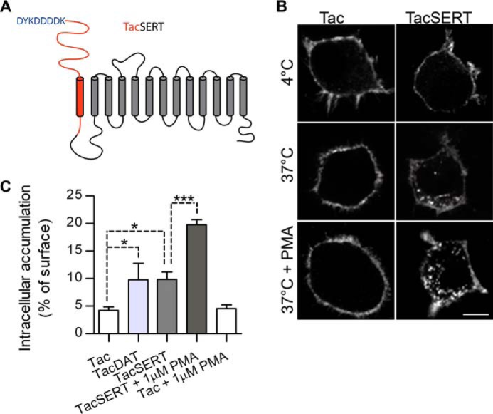
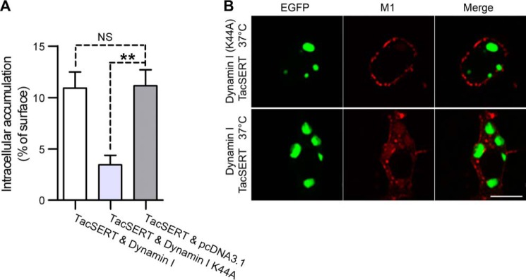
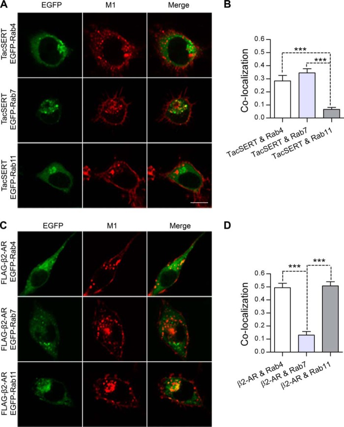
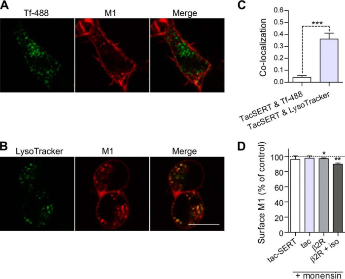
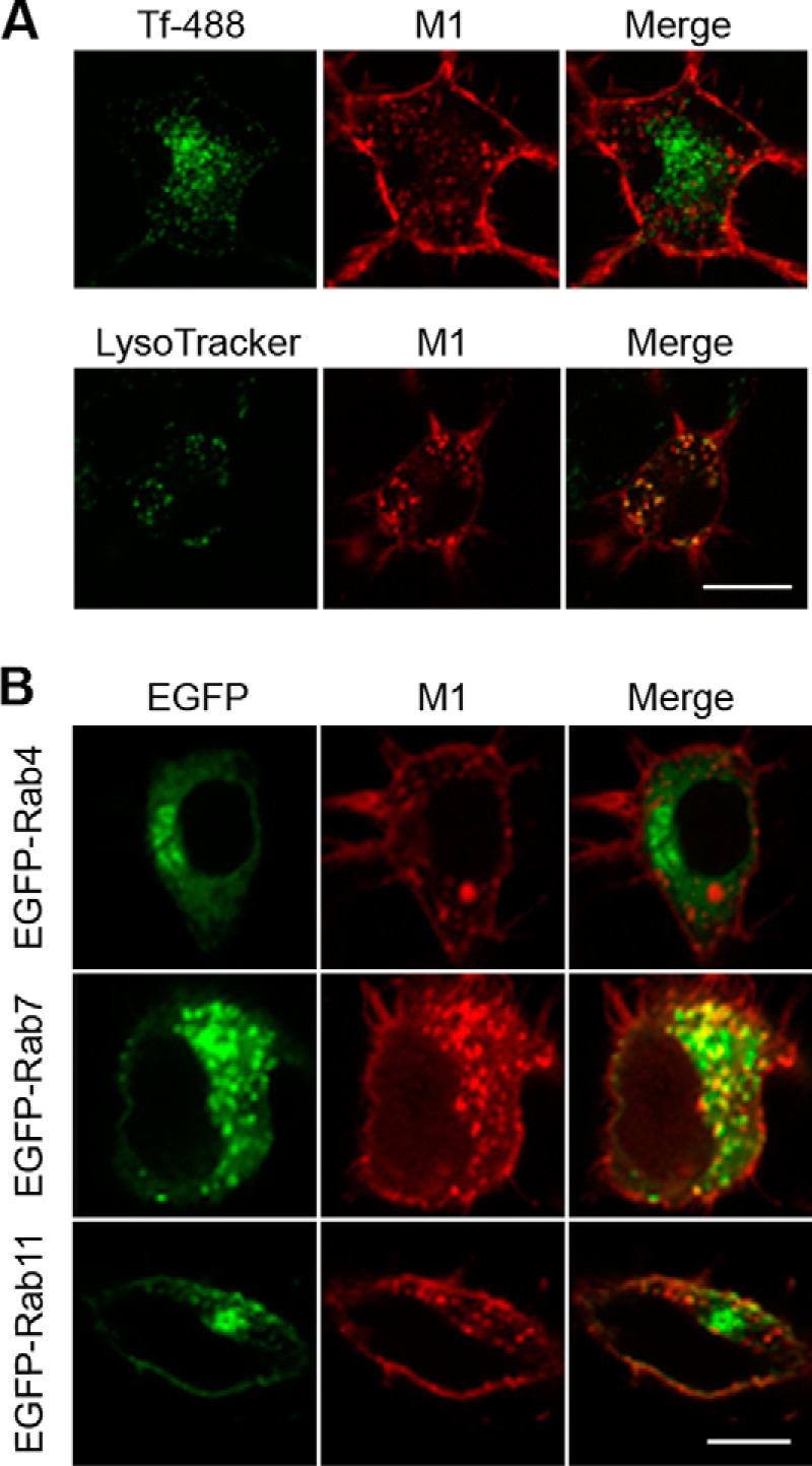
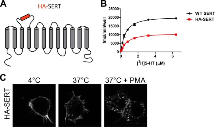
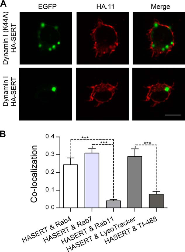
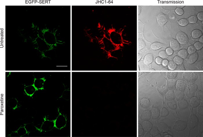
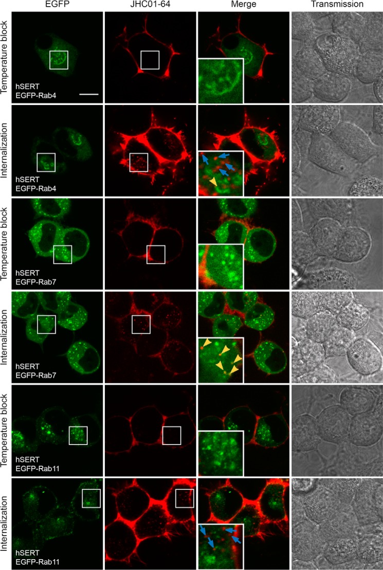
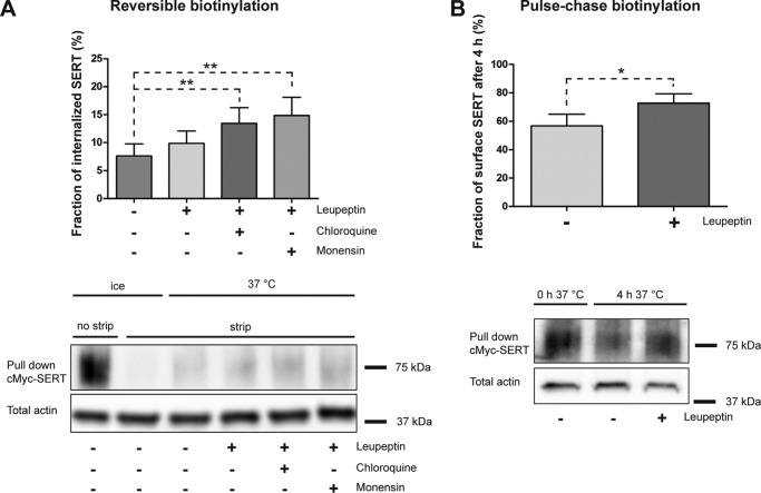
The serotonin transporter (SERT) plays a critical role in regulating serotonin signaling by mediating reuptake of serotonin from the extracellular space. The molecular and cellular mechanisms controlling SERT levels in the membrane remain poorly understood. To study trafficking of the surface resident SERT, two functional epitope-tagged variants were generated. Fusion of a FLAG-tagged one-transmembrane segment protein Tac to the SERT N terminus generated a transporter with an extracellular epitope suited for trafficking studies (TacSERT). Likewise, a construct with an extracellular antibody epitope was generated by introducing an HA (hemagglutinin) tag in the extracellular loop 2 of SERT (HA-SERT). By using TacSERT and HA-SERT in antibody-based internalization assays, we show that SERT undergoes constitutive internalization in a dynamin-dependent manner. Confocal images of constitutively internalized SERT demonstrated that SERT primarily co-localized with the late endosomal/lysosomal marker Rab7, whereas little co-localization was observed with the Rab11, a marker of the "long loop" recycling pathway. This sorting pattern was distinct from that of a prototypical recycling membrane protein, the β2-adrenergic receptor. Furthermore, internalized SERT co-localized with the lysosomal marker LysoTracker and not with transferrin. The sorting pattern was further confirmed by visualizing internalization of SERT using the fluorescent cocaine analog JHC1-64 and by reversible and pulse-chase biotinylation assays showing evidence for lysosomal degradation of the internalized transporter. Finally, we found that SERT internalized in response to stimulation with 12-myristate 13-acetate co-localized primarily with Rab7- and LysoTracker-positive compartments. We conclude that SERT is constitutively internalized and that the internalized transporter is sorted mainly to degradation.

Keywords: Cellular Regulation; Endocytosis; Intracellular Trafficking; Monoamine Transporter; Protein Kinase C (PKC); Protein Sorting; Serotonin Transporter.


Figures

FIGURE 1.
FIGURE 1.
TacSERT is constitutively internalized in CAD cells. A, topology diagram of TacSERT, a chimeric protein of the interleukin 2 receptor α subunit (Tac) fused to the N terminus of SERT. An N-terminal FLAG epitope was added to the extracellular domain of Tac. B, antibody feeding internalization assay. Confocal images of CAD cells expressing FLAG-tagged Tac or TacSERT detected by Alexa568-conjugated M1. After incubation with the antibody at 4 °C, cells were either kept at 4 °C (no trafficking) or at 37 °C for 30 min to allow internalization. When indicated, 1 μm PMA was added during the 37 °C incubation. Scale bar, 10 μm. C, intracellular accumulation detected by ELISA-based internalization assay. Cells transiently transfected with Tac, TacDAT, or TacSERT were labeled with M1 at 4 °C and then either incubated at 4 °C for surface detection or incubated at 37 °C for 1 h to allow internalization. When indicated, 1 μm PMA was added during the 37 °C incubation. To get a measure of internalized protein, the fraction of M1 still present at the surface after the 37 °C incubation was stripped off using acid buffer. Internalization is expressed relative to the initial surface signal (4 °C). Means ± S.E. of n = 3–6, *, p < 0.05; ***, p < 0.001 one-way ANOVA, Dunnett's post-test.
FIGURE 2.
FIGURE 2.
Constitutive internalization of SERT is dynamin-dependent. CAD cells were co-transfected with TacSERT and wild type dynamin I, dominant negative (K44A) dynamin I, or the empty vector pcDNA3.1. A, intracellular accumulation of TacSERT measured in the ELISA-based internalization assay. NS is not significant. Values represent the mean ± S.E. of n = 3, **, p < 0.01; two-way ANOVA. B, immunofluorescence-based internalization assay. Confocal images of cells after 60 min of Alexa568 M1-conjugated antibody internalization. Data are representative of at least three independent experiments. Scale bar,10 μm.
FIGURE 3.
FIGURE 3.
SERT co-localizes with the late endosomal marker Rab7. Confocal microscopy images obtained after 30 min of Alexa568 M1-conjugated antibody internalization, showing putative co-localization between internalized TacSERT (A) or FLAG-tagged β2-AR (C) and EGFP-tagged Rab4, Rab7, or Rab11 co-expressed in CAD cells. For the cells expressing β2-AR, 10 μm isoproterenol was included during the internalization. Scale bar, 10 μm. B and D, quantifications of co-localizations in A and B between internalized TacSERT (B) or internalized β2-AR (D) and the EGFP-tagged Rab4, Rab7, or Rab11 (means ± S.E. ***, p < 0.001, one-way ANOVA, Bonferroni's multiple comparison test). Data were analyzed from 25 to 35 images of each condition from three independent experiments.
FIGURE 4.
FIGURE 4.
SERT co-localizes with the lysosomal marker LysoTracker. Confocal microscopy images of internalized TacSERT and Tf-488 (A) or LysoTracker Green (B) after 30 min of Alexa568 M1-conjugated antibody internalization in the presence of Tf 488 or LysoTracker. Scale bar, 10 μm. C, quantifications of co-localizations in A and C between internalized TacSERT and Tf-488 or LysoTracker Green (means ± S.E. of n = 3, ***, p < 0.001, one-way ANOVA, Bonferroni's multiple comparison test). Data were analyzed from 25 to 35 images of each condition. D, surface ELISA in CAD cells expressing TacSERT, Tac, or β2-AR after 1 h treatment with monensin (25 μm). Isoproterenol (iso) was included when indicated during monensin treatment (means ± S.E. of n = 4–5, *, p < 0.05; **, p < 0.01, paired t test).
FIGURE 5.
FIGURE 5.
Postendocytic sorting of TacSERT after PMA stimulation. Confocal microscopy images of co-localization between internalized TacSERT and Tf-488 or LysoTracker Green (A) or EGFP-tagged Rab proteins (B) after 30 min of Alexa568 M1-conjugated antibody internalization in the presence of 1 μm PMA are shown. Tf-488 or LysoTracker was added to the media during time of internalization. Images are representative of three independent experiments. Scale bar, 10 μm.
FIGURE 6.
FIGURE 6.
HA-tagged SERT expressed at the cell membrane is functional and is internalized constitutively and after PMA treatment. A, topology diagram of HA-SERT with the HA epitope inserted in the EL2. B, kinetic measurements of specific 5-[3H]HT uptake in CAD cells expressing WT SERT (Km, 0.587 μm; Vmax, 21,771 fmol/min/well) or HA-SERT (Km, 0.727 μm; Vmax, 11,430 fmol/min/well). The curve is representative of three independent experiments. C, antibody feeding internalization assay. Confocal images of CAD cells expressing HA-SERT detected by Alexa568-conjugated HA.11. After incubation with the antibody at 18 °C, cells were either kept at 4 °C (no trafficking) or at 37 °C for 30 min to allow internalization. When indicated, 1 μm PMA was added during the 37 °C incubation. Scale bar,10 μm.
FIGURE 7.
FIGURE 7.
HA-SERT is internalized via a dynamin I-dependent pathway and co-localizes with Rab4 and Rab7. A, CAD cells co-transfected with HA-SERT and wild type dynamin I, dominant negative (K44A) dynamin I, or the empty vector pcDNA3.1. B, quantifications of co-localizations between internalized HA-SERT and EGFP-tagged Rab4, Rab7, or Rab11 or LysoTracker or Tf-488 (means ± S.E., ***, p < 0.001, one-way ANOVA, Bonferroni's multiple comparison test). Data were analyzed from 25 to 35 images of each condition from three independent experiments.
FIGURE 8.
FIGURE 8.
JHC1-64 binds specifically to SERT. Confocal microscopy images of co-localization between EGFP-SERT were transiently expressed in CAD and the fluorescent cocaine analog JHC1-64. Images were taken after 10 min of incubation with 10 nm JHC1-64 alone or together with 1 μm of the SERT-specific antagonist paroxetine. Cells treated with paroxetine were preincubated for 10 min before the addition of JHC1-64. Data are representative of three independent experiments. Scale bar, 20 μm.
FIGURE 9.
FIGURE 9.
Internalized JHC1-64 labeled hSERT co-localizes with the late endosomal marker Rab7. Confocal microscopy images of co-localization between hSERT, visualized by JHC1-64, and EGFP-tagged Rab4, Rab7, or Rab11 co-expressed in CAD cells. For the temperature block, images were taken after 30 min at 15 °C with 20 nm JHC1-64 and three washes. For internalization, images were taken after 30 min at 15 °C with 20 nm JHC1-64 and two washes prior to 1 h of internalization in cell media at 37 °C and two washes. Blue arrows indicate JHC1-64-positive vesicles that show no co-localization with EGFP. Yellow arrowheads indicate areas of co-localization between JHC1-64-positive vesicles and EGFP. Data are representative of 3–4 independent experiments. Scale bar, 10 μm.
FIGURE 10.
FIGURE 10.
Blocking proteases and recycling increases the intracellular pool of constitutively internalized c-Myc-SERT. A, reversible biotinylation assay on transiently expressed c-Myc-SERT in CAD cells. c-Myc-SERT was allowed to internalize for 2 h at 37 °C while incubated with no extra treatment, 10 μg/ml leupeptin, 10 μg/ml leupeptin + 200 μm chloroquine, or 10 μg/ml leupeptin + 25 μm monensin, before stripping the surface biotin. Stripping efficiency was 93–96%. Biotinylated protein was analyzed by SDS-PAGE followed by immunoblotting. A representative blot (representative of five independent experiments) is shown in the lower panel, and quantification of the data is shown in the upper panel. The background immunosignal seen in the strip control (ice plus strip) was subtracted from the immunosignal of the internalized samples before normalization. Data are means ± S.E., **, p < 0.01, one-way ANOVA, Bonferroni's multiple comparison test, n = 5. B, pulse-chase biotinylation assay on c-Myc-SERT in HEK293 cells. c-Myc-SERT was biotinylated with a nonreducible biotin and allowed to internalize for 4 h ± 100 μg/ml leupeptin. Remaining biotinylated protein was analyzed by SDS-PAGE followed by immunoblotting. The experiment shown (lower panel) is representative of five independent experiments, and quantification of the data is shown in the upper panel. Quantification of the remaining SERT was normalized to a third sample lysed before internalization. Data are means ± S.E. *, p < 0.05, t test, n = 5.

Similar articles

Cited by

References

    1. Bröer S., Gether U. (2012) The solute carrier 6 family of transporters. Br. J. Pharmacol. 167, 256–278 - PMC - PubMed
    1. Chen N. H., Reith M. E., Quick M. W. (2004) Synaptic uptake and beyond: the sodium- and chloride-dependent neurotransmitter transporter family SLC6. Pflugers Arch. 447, 519–531 - PubMed
    1. Kristensen A. S., Andersen J., Jørgensen T. N., Sørensen L., Eriksen J., Loland C. J., Strømgaard K., Gether U. (2011) SLC6 neurotransmitter transporters: structure, function, and regulation. Pharmacol. Rev. 63, 585–640 - PubMed
    1. Yamashita A., Singh S. K., Kawate T., Jin Y., Gouaux E. (2005) Crystal structure of a bacterial homologue of Na+/Cl−-dependent neurotransmitter transporters. Nature 437, 215–223 - PubMed
    1. Penmatsa A., Wang K. H., Gouaux E. (2013) X-ray structure of dopamine transporter elucidates antidepressant mechanism. Nature 503, 85–90 - PMC - PubMed

Publication types

MeSH terms

LinkOut - more resources