. 2014 Aug 29;289(35):24549-59.
doi: 10.1074/jbc.M114.584037. Epub 2014 Jun 27.

Structural basis of transcription initiation by bacterial RNA polymerase holoenzyme

Affiliations

Structural basis of transcription initiation by bacterial RNA polymerase holoenzyme

Ritwika S Basu et al. J Biol Chem. .

Abstract

The bacterial RNA polymerase (RNAP) holoenzyme containing σ factor initiates transcription at specific promoter sites by de novo RNA priming, the first step of RNA synthesis where RNAP accepts two initiating ribonucleoside triphosphates (iNTPs) and performs the first phosphodiester bond formation. We present the structure of de novo transcription initiation complex that reveals unique contacts of the iNTPs bound at the transcription start site with the template DNA and also with RNAP and demonstrate the importance of these contacts for transcription initiation. To get further insight into the mechanism of RNA priming, we determined the structure of initially transcribing complex of RNAP holoenzyme with 6-mer RNA, obtained by in crystallo transcription approach. The structure highlights RNAP-RNA contacts that stabilize the short RNA transcript in the active site and demonstrates that the RNA 5'-end displaces σ region 3.2 from its position near the active site, which likely plays a key role in σ ejection during the initiation-to-elongation transition. Given the structural conservation of the RNAP active site, the mechanism of de novo RNA priming appears to be conserved in all cellular RNAPs.

Keywords: Promoter; RNA Polymerase; Transcription; Transcription Initiation Factor; X-ray Crystallography.


Figures

FIGURE 1.
FIGURE 1.
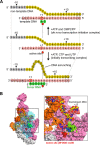
Structure of the transcription initiation complex. A, schematic of the de novo transcription initiation complex (top) and the initially transcribing complex (bottom) obtained from the RNAP-promoter complex. Sequence of the nucleic acid scaffold used for holoenzyme-promoter DNA complex crystallization is shown in the middle (pink, template DNA; yellow, non-template DNA; gray, −10 element). NTPs and 6-mer RNA in the transcription complexes are depicted by green circles. Positions of the RNAP active site (red sphere) as well as template DNA transcription start site (TSS, +1, red circle) are indicated. DNA bases indicated by black dashed circles are disordered in the crystal structures. In a standard format describing RNAP promoter sequence, the transcription start site, which base pairs with the first iNTP and encodes the 5′-end of the RNA chain, is designated +1. DNA positions downstream from the transcription start site increase as positive numbers from +1 and DNA positions upstream of the start site increase as negative numbers counting from −1. B, overall structure of the de novo transcription initiation complex. T. thermophilus RNAP is depicted as a molecular surface model. Each subunit of RNAP is depicted with a unique color (white, α; cyan, β; pink, β′; orange, σ). DNA is depicted as a sphere model with the same color scheme as described in A. Right panel shows a magnified view of the boxed region in the left panel. For clarity, the β subunit was made transparent in the left panel and removed in the right panel. Several key motifs discussed in the text are highlighted (see also supplemental Movie S1).
FIGURE 2.
FIGURE 2.
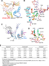
First and second iNTP binding sites of the de novo transcription initiation complex. A, active site structure of the de novo transcription initiation complex. DNA, iNTPs, and amino acid side chains are shown as stick models, and the trigger loop, DFDGD motif, and the σ region 3.2 (σ3.2) are shown as worm models and labeled. The disordered region of the trigger loop is shown as a dashed line. FoFc electron density maps (yellow mesh) showing ATP (first iNTP) and CMPCPP (second iNTP) are superposed on the final model. B, the first iNTP (ATP) binds at the active site through multiple interactions, including base pairing with the +1 DNA base (red dashed lines), base stacking with the −1 purine base (gray dashed lines), water-mediated interactions (yellow dashed lines), and salt bridges with side chains (cyan dashed lines). Amino acid side chains involved in the second iNTP (CMPCPP) binding are also indicated. The transcription start site (TSS) is indicated. C, comparison of the holoenzyme-promoter DNA (Protein Data Bank code 4G7H, magenta), de novo transcription (blue), and transcription elongation complexes (Protein Data Bank code 2O5J, gray and white). RNAP structures were superposed at their active site domains of the β′ subunit. The i and i+1 sites are indicated. Trigger loops (β′ residues 1222–1265) of these structures are shown as tube models. D, ATP and CMPCPP bound in the active site. The aspartate residues of the DFDGD motif of the β′ subunit coordinating the Mg2+ (yellow spheres) are labeled. The distance between the 3′-OH of ATP and the α-phosphate of CMPCPP is 5.4 Å as indicated. E, amino acid residues involved in the first iNTP binding are conserved in all cellular RNAPs.
FIGURE 3.
FIGURE 3.
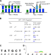
Transcription activities of wild-type and mutant RNAPs on the T7A1 promoter. A, the sequences of the wild-type T7A1 promoter and its variants used in this study. Positions of the −35, −10 promoter elements, and the transcription start site (+1) are indicated. The T7A1 promoter variant has a base substitution at the −1 position (highlighted in yellow); the T7A1 consensus (T7A1cons) promoter contains the consensus −10 element (TATAAT). B, transcription was performed in the presence of low (10 μm all four NTPs with addition of α-[32P]UTP) or high (100 μm ATP, GTP, CTP, and 10 μm UTP) concentrations of NTPs with or without initiating primer CpA (25 μm), corresponding to positions −1/+1 of the promoter. The positions of the run-off (RO) and abortive (ab) RNA products are indicated.
FIGURE 4.
FIGURE 4.
Role of the base stacking interaction between the first iNTP and template DNA in transcription initiation. A, schematic representation of the base stacking interaction between the −1 template DNA purine base and the first iNTP purine base. (see also supplemental Movie S1). B, apparent Km values for the initiating substrates on the wild-type T7A1 promoter (−1C nontemplate sequence) and its variant (−1G non-template sequence) for RNAPs containing wild-type or Δ513–519 σ70 factors. The numbers in blue and boldface type show changes in Km values for the T7A1 promoter variant relative to the wild-type sequence. C, non-template DNA sequence around the transcription start sites of human Pol I, which is adapted from Ref. . The transcription start site is +1. D, sequence conservation of the non-template DNA around transcription start site of human Pol II. The sequence logo was prepared using experimentally determined non-redundant collection of human Pol II promoters, for which the transcription start site (+1) has been determined experimentally. The majority of human Pol II promoters contain −1 pyrimidine and +1 purine bases in the non-template DNA.
FIGURE 5.
FIGURE 5.
The initially transcribing complex of RNAP holoenzyme containing 6-mer RNA. A, FoFc electron density map (yellow mesh) showing 6-mer RNA and a NTP bound at the E site of the initially transcribing complex. The final model of 6-mer RNA and amino acid side chains involved in NTP binding at the E site are shown as stick models and labeled. The DNA is depicted as CPK model. B, active site structure. DNA, RNA, and amino acid side chains contacting the 6-mer RNA are shown as stick models. Disordered regions of the trigger loop (residues 1239–1253) and the σ region 3.2 (σ3.2, residues 321–327) are indicated by dashed lines. The transcription start site (TSS) is indicated. C, comparison of the de novo transcription (blue, template DNA; orange, σ region 3.2) and the 6-mer RNA initially transcribing complexes (pink, template DNA; green, 6-mer RNA). RNAP structures were superposed on the active site domains of the β′ subunit. D, model of an extended RNA transcript placed in the RNA exit channel explains σ release. Structures of the initially transcribing (pink, template DNA; green, 6-mer RNA; orange, σ factor) and the transcription elongation complexes (Protein Data Bank code 2O5J, gray, template DNA; red, RNA; cyan, incoming NTP) were superposed on the active site domains of the β′ subunit. The i and i+1 sites are indicated. E, effects of the E. coli RNAP mutations on abortive synthesis. Transcription was performed on the T7A1cons promoter at high NTP concentrations in the presence of the CpA primer. Positions of the run-off (RO) and abortive products are indicated. The profiles above the gel show distribution of the RNA products for the wild-type, K1065A, and H1237A RNAPs (black, blue, and red, respectively).

References

    1. Vassylyev D. G., Vassylyeva M. N., Zhang J., Palangat M., Artsimovitch I., Landick R. (2007) Structural basis for substrate loading in bacterial RNA polymerase. Nature 448, 163–168 - PubMed
    1. McClure W. R., Cech C. L., Johnston D. E. (1978) A steady state assay for the RNA polymerase initiation reaction. J. Biol. Chem. 253, 8941–8948 - PubMed
    1. Kulbachinskiy A., Mustaev A. (2006) Region 3.2 of the sigma subunit contributes to the binding of the 3′-initiating nucleotide in the RNA polymerase active center and facilitates promoter clearance during initiation. J. Biol. Chem. 281, 18273–18276 - PubMed
    1. Gaal T., Bartlett M. S., Ross W., Turnbough C. L., Jr., Gourse R. L. (1997) Transcription regulation by initiating NTP concentration: rRNA synthesis in bacteria. Science 278, 2092–2097 - PubMed
    1. Turnbough C. L., Jr., Switzer R. L. (2008) Regulation of pyrimidine biosynthetic gene expression in bacteria: repression without repressors. Microbiol. Mol. Biol. Rev. 72, 266–300 - PMC - PubMed

Publication types

Substances

LinkOut - more resources