. 2014 Aug 15;289(33):23065-23074.
doi: 10.1074/jbc.M114.557652. Epub 2014 Jun 27.

β-Agonist-mediated relaxation of airway smooth muscle is protein kinase A-dependent

Affiliations

β-Agonist-mediated relaxation of airway smooth muscle is protein kinase A-dependent

Sarah J Morgan et al. J Biol Chem. .

Abstract

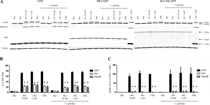
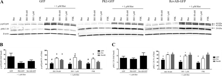
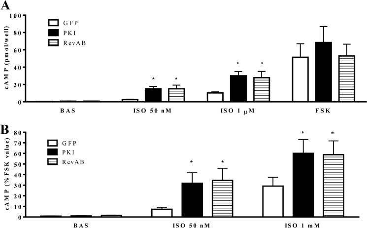
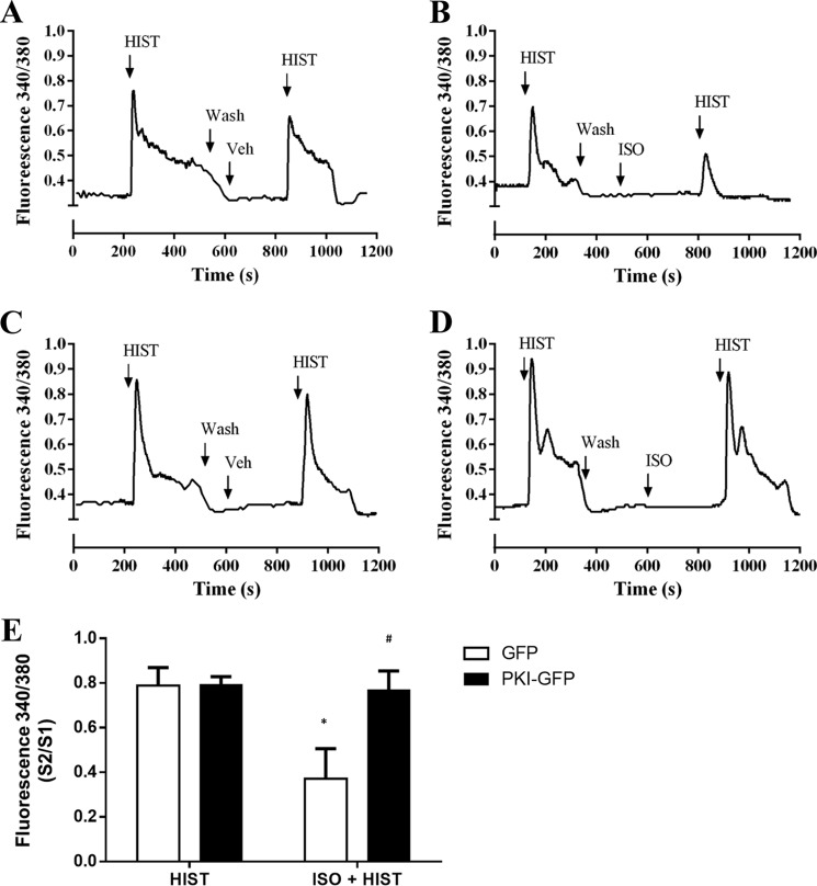
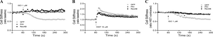
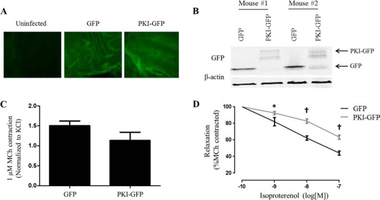
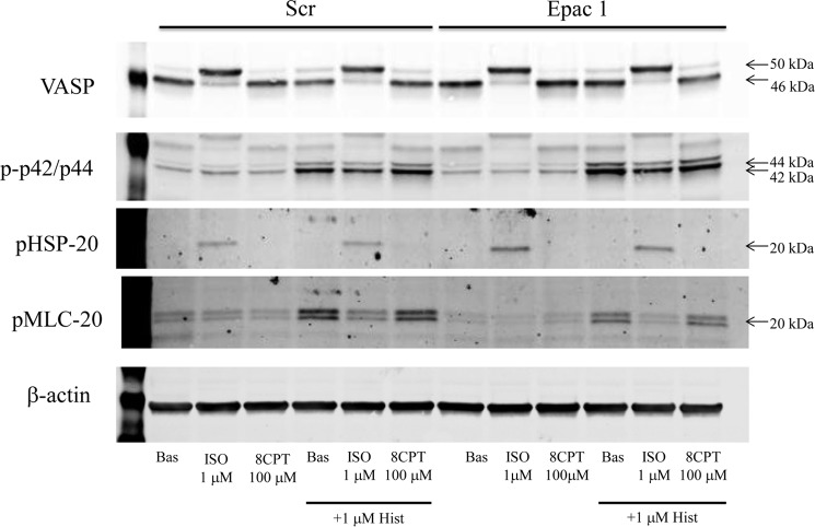
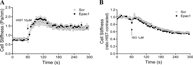
Inhaled β-agonists are effective at reversing bronchoconstriction in asthma, but the mechanism by which they exert this effect is unclear and controversial. PKA is the historically accepted effector, although this assumption is made on the basis of associative and not direct evidence. Recent studies have asserted that exchange protein activated by cAMP (Epac), not PKA, mediates the relaxation of airway smooth muscle (ASM) observed with β-agonist treatment. This study aims to clarify the role of PKA in the prorelaxant effects of β-agonists on ASM. Inhibition of PKA activity via expression of the PKI and RevAB peptides results in increased β-agonist-mediated cAMP release, abolishes the inhibitory effect of isoproterenol on histamine-induced intracellular calcium flux, and significantly attenuates histamine-stimulated MLC-20 phosphorylation. Analyses of ASM cell and tissue contraction demonstrate that PKA inhibition eliminates most, if not all, β-agonist-mediated relaxation of contracted smooth muscle. Conversely, Epac knockdown had no effect on the regulation of contraction or procontractile signaling by isoproterenol. These findings suggest that PKA, not Epac, is the predominant and physiologically relevant effector through which β-agonists exert their relaxant effects.

Keywords: Adrenergic Receptor; Asthma; G Protein-coupled Receptor (GPCR); Protein Kinase A (PKA); Smooth Muscle.


Figures

FIGURE 1.
FIGURE 1.
β-Agonist regulation of VASP and phospho-HSP-20 and the effect of PKA inhibition. GFP-, PKI-GFP-, or RevAB-expressing HASM cells were plated into 12-well plates and stimulated with vehicle (Bas), ISO (50 nm or 1 μm) or FSK (100 μm) ± HIST (1 μm) as described under “Experimental Procedures.” After 5 min of stimulation, cell lysates were harvested, and expression levels of various proteins were assessed by immunoblotting. Representative blots (A) are shown. Band intensities for VASP phosphorylation and phospho-HSP-20 were quantified as described under “Experimental Procedures,” adjusted for quantified β-actin levels and presented in graph form in B and C, respectively. VASP bands present as either a 46- or 50-kDa species, the latter representing VASP in which Ser-157 is phosphorylated by PKA. Expression of GFP in all three lines was confirmed by Western blot using anti-GFP antibody. HSP-20 phosphorylation has also been shown previously to be PKA-dependent. Quantified phospho-HSP20 was normalized to the GFP + FSK value. Data are mean ± S.E. †, p < 0.0001 PKI/RevAB versus GFP.
FIGURE 2.
FIGURE 2.
β-Agonist regulation of phospho-p42/p44 and phospho-MLC-20 levels and the effect of PKA inhibition. Cultures of GFP-, PKI-GFP-, or RevAB-expressing HASM cells were plated into 12-well plates and stimulated with vehicle (Bas), ISO (50 nm or 1 μm), or FSK (100 μm) ± HIST (1 μm) according to the experiments in Fig. 1. Representative blots in A, derived from the same representative experiment depicted in Fig. 1A, depict the regulation of phospho-p42/p44 and phospho-MLC-20 levels by the indicated agonists. Band intensities for phospho-p42/p44 and phospho-MLC-20 were quantified as the ratio of HIST-stimulated to basal (left panel) and as the percent of HIST alone (right panel) and are presented in graph form in B and C, respectively. A, VASP bands present as either a 46- or 50-kDa species, the latter representing VASP in which Ser-157 is phosphorylated by PKA. Expression of GFP in all three lines was confirmed by Western blot analysis using anti-GFP antibody. HSP-20 phosphorylation has also been shown previously to be PKA-dependent. Quantified phospho-HSP20 was normalized to the GFP + FSK value. B and C, phospho-p42/p44 (B) and phospho-MLC-20 (C) levels were quantified as the ratio of HIST-stimulated to basal (left panel) and as the percent of HIST alone (right panel). The HIST alone value (not shown) was set to 100%. Data are mean ± S.E. *, p < 0.05, PKI/RevAB versus GFP; #, p < 0.05 versus HIST alone (n = 5).
FIGURE 3.
FIGURE 3.
Effect of PKI-GFP or RevAB-GFP expression on β-agonist-mediated cAMP accumulation. GFP-, PKI-GFP-, or RevAB-GFP-expressing HASM cells were plated into 24-well plates and stimulated with vehicle (Bas), ISO (50 nm or 1 μm), or FSK (100 μm). Global cAMP accumulation was measured after 10-min stimulation and plotted as raw values (picomoles/well) (A) or percent of the FSK-stimulated value (B). Data are mean ± S.E. *, p < 0.05 PKI/RevAB versus GFP (n = 6).
FIGURE 4.
FIGURE 4.
Effect of PKI-GFP and RevAB-GFP expression on β-agonist inhibition of HIST-stimulated Ca2+ mobilization. HASM cultures expressing GFP or PKI-GFP were loaded with Fura-2/AM. Ca2+ mobilization was assessed in response to stimulation with HIST (10 μm). Cells were then washed and restimulated with HIST ± ISO (1 μm) pretreatment. A—D, representative traces for GFP-expressing (A and B) and PKI-GFP-expressing (C and D) cells assessing the effect of prior vehicle (A and C) or ISO (B and D) treatment. The ratio of second histamine response to the first (S2/S1) was calculated and compared between groups. Data are mean ± S.E. *, p < 0.05 versus HIST alone; #, p < 0.05 versus matched GFP (GFP HIST, n = 82; PKI HIST, n = 93; GFP ISO + HIST, n = 187; PKI ISO + HIST, n = 170).
FIGURE 5.
FIGURE 5.
Effects of PKA inhibition on β-agonist-induced changes in cell stiffness. HASM cultures stably expressing GFP, PKI-GFP, or RevAB-GFP were assessed for changes in cell stiffness in response to HIST and ISO, measured using magnetic twisting cytometry as described under “Experimental Procedures.” A, time-dependent response to ISO (1 μm) from baseline. B, response to HIST (10 μm) from baseline. C, ability of ISO (1 μm) to alter stiffness in cells prestimulated with HIST (10 μm). †, p < 0.0001 PKI/RevAB versus GFP. Differences in mean values for baseline stiffness were less than 20% among the GFP, PKI-GFP, and RevAB-GFP groups. Data points represent mean ± S.E. of ≥ 86 cells/experimental group.
FIGURE 6.
FIGURE 6.
Effect of PKA inhibition on the ability of β-agonist to relax precontracted murine tracheal rings. Tracheal rings were isolated from C57BL6 mice and infected for 48 h with lentivirus encoding GFP or PKI-GFP. A, GFP images of control, GFP-, or PKI-GFP-infected murine tracheal rings. The control ring was maintained in culture for 48 h but not infected with virus. B, representative immunoblot for GFP expression in lysates from GFP- and PKI-GFP-infected murine tracheal rings. C, KCl-normalized contraction of rings stimulated with MCh (1 μm). D, tracheal rings were precontracted with MCh (1 μm), and then changes in contractile force were measured in response to treatment with increasing doses of ISO. *, p < 0.05; †, p < 0.0001 versus GFP (GFP, n = 9; PKI, n = 12).
FIGURE 7.
FIGURE 7.
Effect of Epac knockdown on β-agonist regulation of VASP, phospho-HSP-20, phospho-p42/p44, and phospho-MLC-20 levels. 96 h after transfection of HASM cells with either scrambled (Scr) siRNA or siRNA targeting Epac1, cells plated into 12-well plates were stimulated with vehicle (Bas), ISO (1 μm), 8-pCPT-2′-O-Me-cAMP (8CPT, 100 μm), or FSK (100 μm) ± HIST (1 μm) as described under “Experimental Procedures.” After 5 min of stimulation, cell lysates were harvested, and expression levels of VASP, phospho-HSP-20, phospho-p42/p44 and phospho-MLC-20, and β-actin were assessed by immunoblotting. Blots are representative of three experiments providing similar results.
FIGURE 8.
FIGURE 8.
Effects of Epac knockdown on β-agonist-induced changes in cell stiffness. HASM cells were subjected to Epac1 knockdown as in Fig. 7. Experiments were assessed for changes in cell stiffness in response to HIST and ISO using magnetic twisting cytometry as described under “Experimental Procedures.” Scr, scrambled.

References

    1. Newnham D. M., McDevitt D. G., Lipworth B. J. (1994) Bronchodilator subsensitivity after chronic dosing with eformoterol in patients with asthma. The American Journal of Medicine 97, 29–37 - PubMed
    1. Newnham D. M., Grove A., McDevitt D. G., Lipworth B. J. (1995) Subsensitivity of bronchodilator and systemic β 2 adrenoceptor responses after regular twice daily treatment with eformoterol dry powder in asthmatic patients. Thorax 50, 497–504 - PMC - PubMed
    1. Grove A., Lipworth B. J. (1995) Bronchodilator subsensitivity to salbutamol after twice daily salmeterol in asthmatic patients. Lancet 346, 201–206 - PubMed
    1. Sears M. R. (2002) Adverse effects of b-agonists. J. Allergy Clin. Immunol. 110, S322–S328 - PubMed
    1. Salpeter S. R., Buckley N. S., Ormiston T. M., Salpeter E. E. (2006) Meta-analysis: effect of long-acting b-Agonists on severe asthma exacerbations and asthma-related deaths. Annals Intern. Med. 144, 904–912 - PubMed

Publication types

MeSH terms

Substances

LinkOut - more resources