

Incidental central tear in Descemet membrane endothelial complex during Descemet membrane endothelial keratoplasty
- PMID: 24973345
- PMCID: PMC4078533
- DOI: 10.1136/bcr-2013-202935
Incidental central tear in Descemet membrane endothelial complex during Descemet membrane endothelial keratoplasty
Abstract
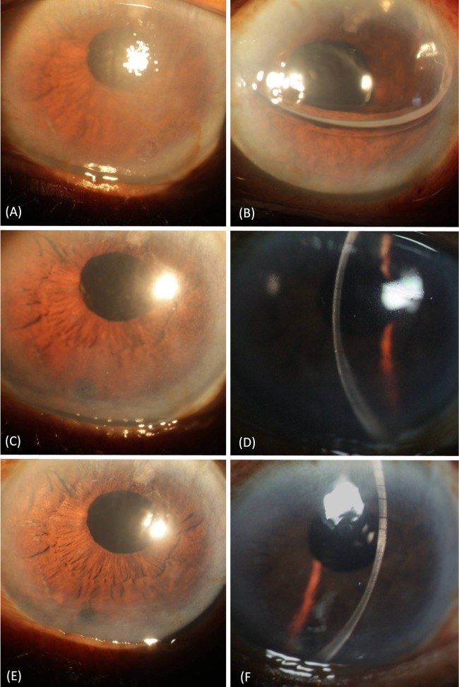
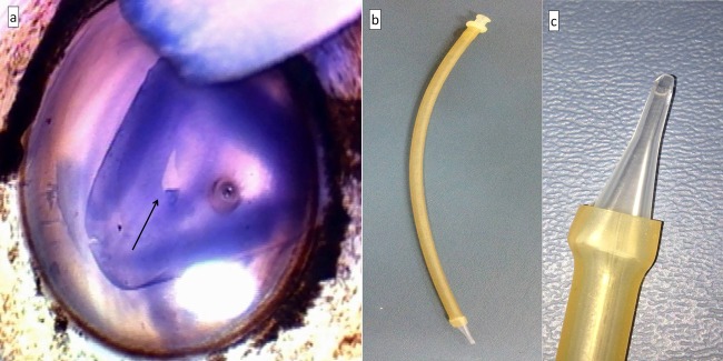
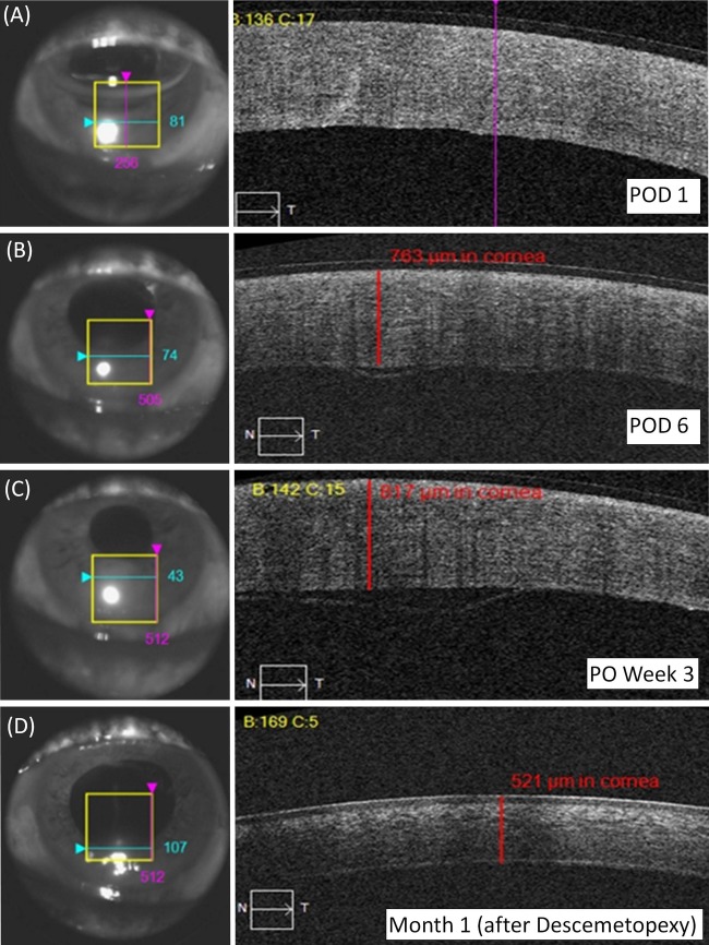
Descemet membrane endothelial keratoplasty (DMEK) was performed in a 70-year-old man diagnosed with pseudophakic bullous keratopathy. During Descemet endothelial complex (DEC) preparation, a central tear was noticed in the DMEK graft. However, the surgery was continued. On sixth postoperative day, a small fluid pocket was observed between the DEC and the posterior host stroma in inferior third of the graft area. It was, however, decided to observe it for spontaneous attachment. At 2 weeks, the inferior DEC detachment had increased with overlying corneal oedema. Descemetopexy with 100% air was performed the same day which reattached the DEC. Subsequently, DEC remained attached and at the last follow-up of 2 months, DEC was well opposed with a clear overlying cornea. The final best-corrected Snellen's visual acuity was 20/60. A small tear in the DEC does not necessitate tissue replacement and a good anatomical and visual outcome can be achieved in such cases.
2014 BMJ Publishing Group Ltd.
Figures

Similar articles
-
Corneal Higher-Order Aberrations in Descemet Membrane Endothelial Keratoplasty versus Ultrathin DSAEK in the Descemet Endothelial Thickness Comparison Trial: A Randomized Clinical Trial.Ophthalmology. 2019 Jul;126(7):946-957. doi: 10.1016/j.ophtha.2019.02.007. Epub 2019 Feb 16. Ophthalmology. 2019. PMID: 30776384 Free PMC article. Clinical Trial.
-
Non-Descemet stripping Descemet membrane endothelial keratoplasty.Cornea. 2013 Dec;32(12):1607-9. doi: 10.1097/ICO.0b013e3182a6d0cb. Cornea. 2013. PMID: 24097182
-
Visual rehabilitation rate after isolated descemet membrane transplantation: descemet membrane endothelial keratoplasty.Arch Ophthalmol. 2009 Mar;127(3):252-5. doi: 10.1001/archophthalmol.2008.619. Arch Ophthalmol. 2009. PMID: 19273786 Clinical Trial.
-
Meta-Analysis of Postoperative Outcome Parameters Comparing Descemet Membrane Endothelial Keratoplasty Versus Descemet Stripping Automated Endothelial Keratoplasty.Cornea. 2017 Dec;36(12):1445-1451. doi: 10.1097/ICO.0000000000001384. Cornea. 2017. PMID: 28957976 Review.
-
[Transplantation of endothelium and Descemet's membrane].Vestn Oftalmol. 2019;135(1):98-103. doi: 10.17116/oftalma201913501198. Vestn Oftalmol. 2019. PMID: 30830081 Review. Russian.
References
-
- Anshu A, Price MO, Tan DTH, et al. Endothelial keratoplasty: a revolution in evolution. Surv Ophthalmol 2012;57:236–52 - PubMed
-
- Tourtas T, Laaser K, Bachmann BO, et al. Descemet membrane endothelial keratoplasty versus descemet stripping automated endothelial keratoplasty. Am J Ophthalmol 2012;153:1082–90 - PubMed
-
- Guerra FP, Anshu A, Price MO, et al. Descemet's membrane endothelial keratoplasty: prospective study of 1-year visual outcomes, graft survival, and endothelial cell loss. Ophthalmology 2011;118:2368–73 - PubMed
-
- Kruse FE, Laaser K, Cursiefen C, et al. A stepwise approach to donor preparation and insertion increases safety and outcome of Descemet membrane endothelial keratoplasty. Cornea 2011;30:580–7 - PubMed
Publication types
MeSH terms
LinkOut - more resources
Full Text Sources
Other Literature Sources
Medical
Research Materials