

Genetic analysis implicates APOE, SNCA and suggests lysosomal dysfunction in the etiology of dementia with Lewy bodies
- PMID: 24973356
- PMCID: PMC4222357
- DOI: 10.1093/hmg/ddu334
Genetic analysis implicates APOE, SNCA and suggests lysosomal dysfunction in the etiology of dementia with Lewy bodies
Abstract
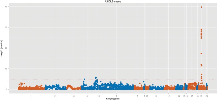
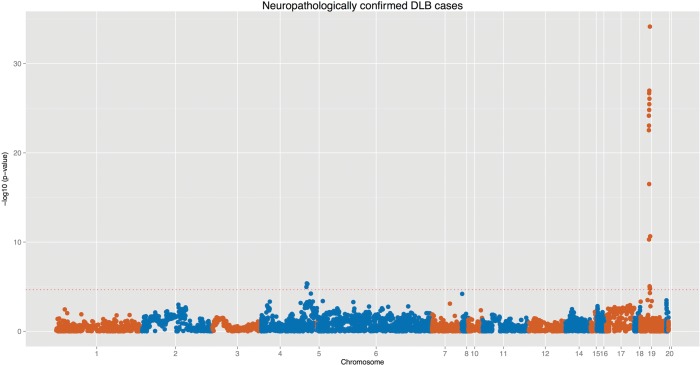
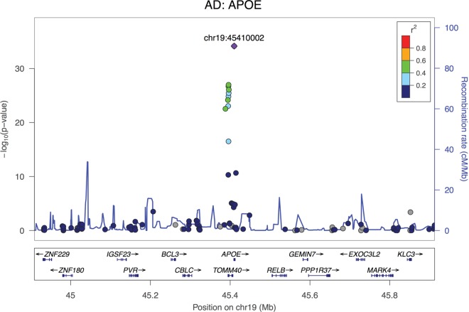
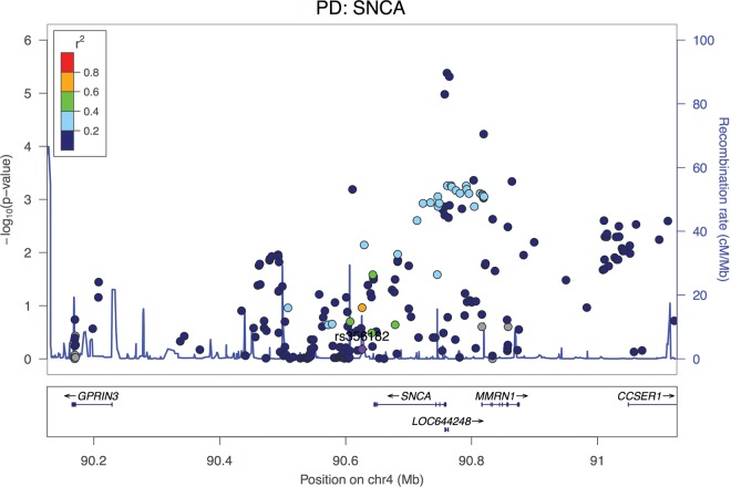
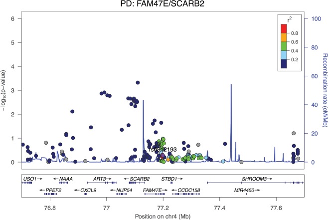
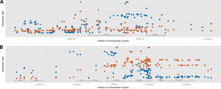
Clinical and neuropathological similarities between dementia with Lewy bodies (DLB), Parkinson's and Alzheimer's diseases (PD and AD, respectively) suggest that these disorders may share etiology. To test this hypothesis, we have performed an association study of 54 genomic regions, previously implicated in PD or AD, in a large cohort of DLB cases and controls. The cohort comprised 788 DLB cases and 2624 controls. To minimize the issue of potential misdiagnosis, we have also performed the analysis including only neuropathologically proven DLB cases (667 cases). The results show that the APOE is a strong genetic risk factor for DLB, confirming previous findings, and that the SNCA and SCARB2 loci are also associated after a study-wise Bonferroni correction, although these have a different association profile than the associations reported for the same loci in PD. We have previously shown that the p.N370S variant in GBA is associated with DLB, which, together with the findings at the SCARB2 locus, suggests a role for lysosomal dysfunction in this disease. These results indicate that DLB has a unique genetic risk profile when compared with the two most common neurodegenerative diseases and that the lysosome may play an important role in the etiology of this disorder. We make all these data available.
© The Author 2014. Published by Oxford University Press.
Figures
References
-
- Barber R., Panikkar A., McKeith I.G. Dementia with Lewy bodies: diagnosis and management. Int. J. Geriatr. Psychiatry. 2001;16(Suppl. 1):S12–S18. - PubMed
-
- Lippa C.F., Duda J.E., Grossman M., Hurtig H.I., Aarsland D., Boeve B.F., Brooks D.J., Dickson D.W., Dubois B., Emre M., et al. DLB and PDD boundary issues: diagnosis, treatment, molecular pathology, and biomarkers. Neurology. 2007;68:812–819. - PubMed
-
- McKeith I.G., Galasko D., Kosaka K., Perry E.K., Dickson D.W., Hansen L.A., Salmon D.P., Lowe J., Mirra S.S., Byrne E.J., et al. Consensus guidelines for the clinical and pathologic diagnosis of dementia with Lewy bodies (DLB): report of the consortium on DLB international workshop. Neurology. 1996;47:1113–1124. - PubMed
-
- McKeith I.G. Dementia with Lewy bodies. The British Journal of Psychiatry: the Journal of Mental Science. 2002;180:144–147. - PubMed
Publication types
MeSH terms
Substances
Grants and funding
- R01 NS078086/NS/NINDS NIH HHS/United States
- G-0907/PUK_/Parkinson's UK/United Kingdom
- G0701075/MRC_/Medical Research Council/United Kingdom
- CAPMC/ CIHR/Canada
- MC_G1000735/MRC_/Medical Research Council/United Kingdom
- R01 NS060113/NS/NINDS NIH HHS/United States
- P50 NS072187/NS/NINDS NIH HHS/United States
- UL1TR000040/TR/NCATS NIH HHS/United States
- MR/L010305/1/MRC_/Medical Research Council/United Kingdom
- 089701/WT_/Wellcome Trust/United Kingdom
- NS060113/NS/NINDS NIH HHS/United States
- P50AG008702/AG/NIA NIH HHS/United States
- 089703/WT_/Wellcome Trust/United Kingdom
- AG000951-12/AG/NIA NIH HHS/United States
- 089698/WT_/Wellcome Trust/United Kingdom
- Z01 AG000951/ImNIH/Intramural NIH HHS/United States
- MR/L016397/1/MRC_/Medical Research Council/United Kingdom
- 100140/WT_/Wellcome Trust/United Kingdom
- P50NS038370/NS/NINDS NIH HHS/United States
- 081864/WT_/Wellcome Trust/United Kingdom
- MR/L022656/1/MRC_/Medical Research Council/United Kingdom
- K-1204/PUK_/Parkinson's UK/United Kingdom
LinkOut - more resources
Full Text Sources
Other Literature Sources
Medical
Miscellaneous
