. 2014 Aug 1;193(3):1035-46.
doi: 10.4049/jimmunol.1302898. Epub 2014 Jun 27.

Dendritic cells and anergic type I NKT cells play a crucial role in sulfatide-mediated immune regulation in experimental autoimmune encephalomyelitis

Affiliations

Dendritic cells and anergic type I NKT cells play a crucial role in sulfatide-mediated immune regulation in experimental autoimmune encephalomyelitis

Igor Maricic et al. J Immunol. .

Abstract

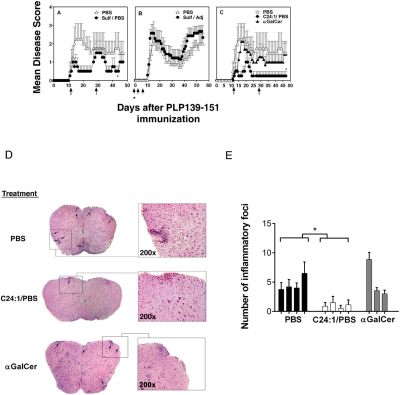
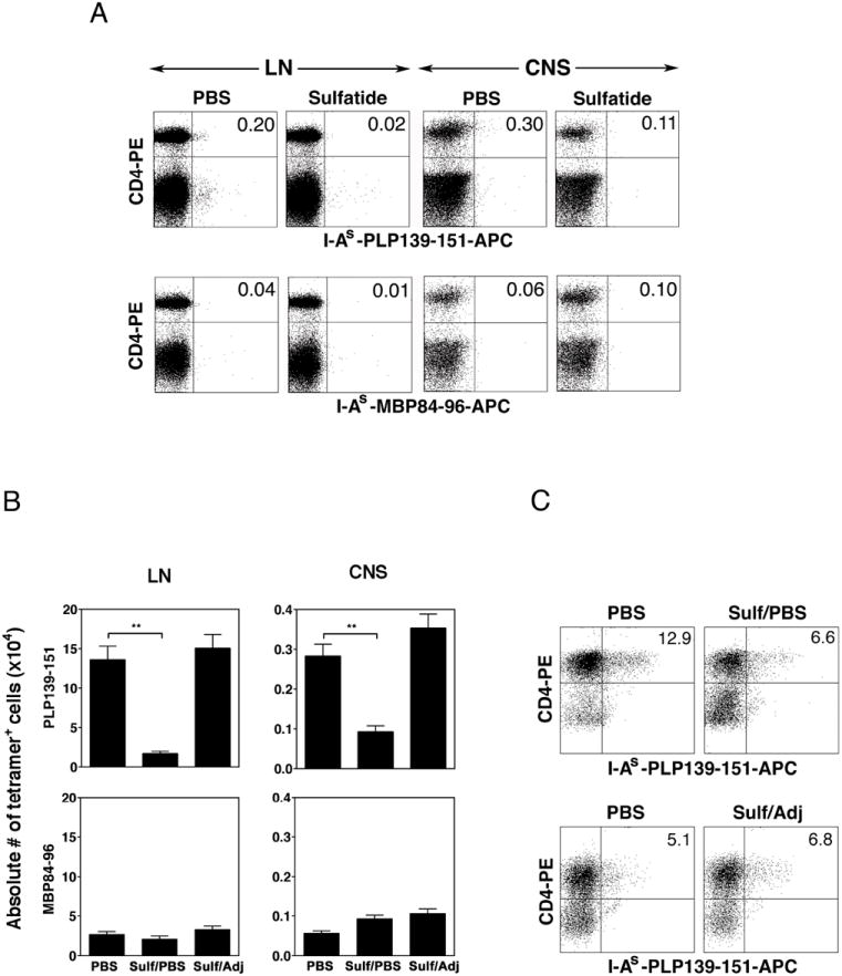
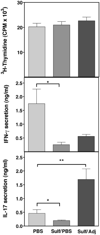
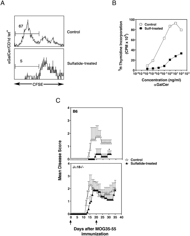
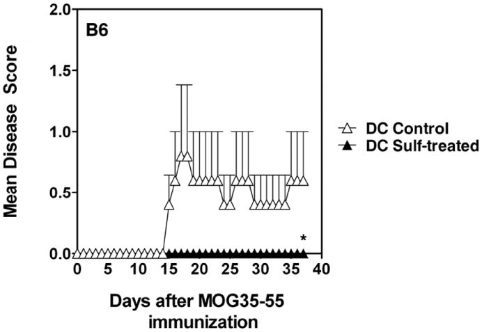
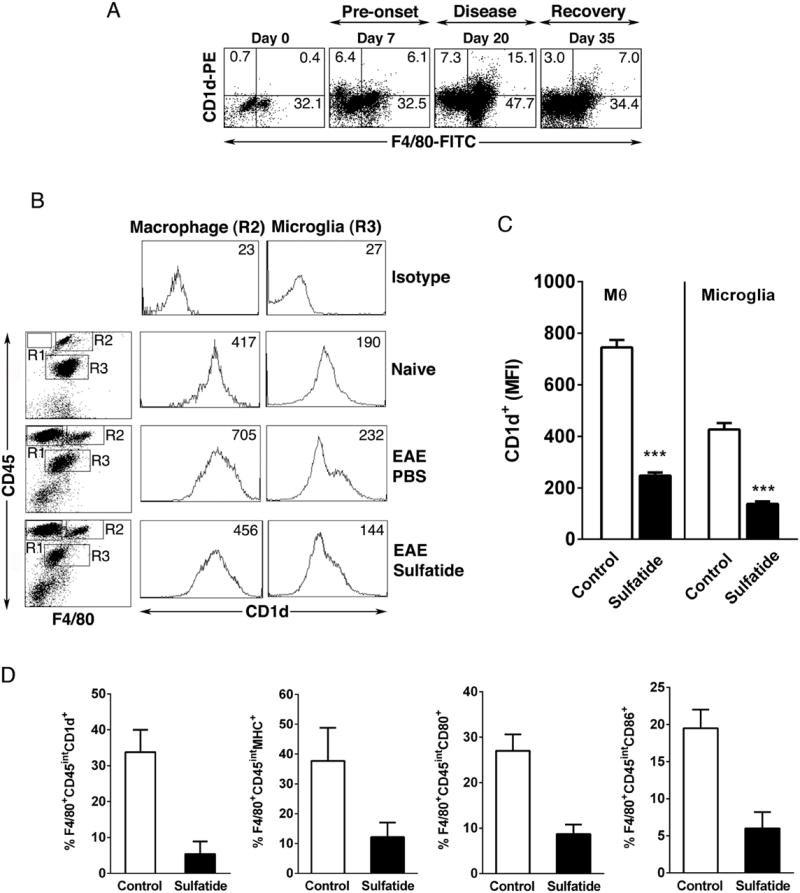
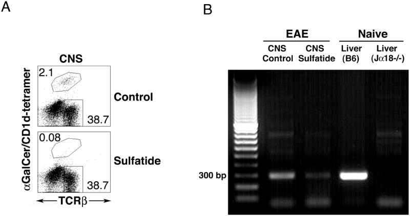
CD1d-restricted NKT cells can be divided into two groups: type I NKT cells use a semi-invariant TCR, whereas type II express a relatively diverse set of TCRs. A major subset of type II NKT cells recognizes myelin-derived sulfatides and is selectively enriched in the CNS tissue during experimental autoimmune encephalomyelitis (EAE). We have shown that activation of sulfatide-reactive type II NKT cells by sulfatide prevents induction of EAE. In this article, we have addressed the mechanism of regulation, as well as whether a single immunodominant form of synthetic sulfatide can treat ongoing chronic and relapsing EAE in SJL/J mice. We have shown that the activation of sulfatide-reactive type II NKT cells leads to a significant reduction in the frequency and effector function of myelin proteolipid proteins 139-151/I-A(s)-tetramer(+) cells in lymphoid and CNS tissues. In addition, type I NKT cells and dendritic cells (DCs) in the periphery, as well as CNS-resident microglia, are inactivated after sulfatide administration, and mice deficient in type I NKT cells are not protected from disease. Moreover, tolerized DCs from sulfatide-treated animals can adoptively transfer protection into naive mice. Treatment of SJL/J mice with a synthetic cis-tetracosenoyl sulfatide, but not α-galactosylceramide, reverses ongoing chronic and relapsing EAE. Our data highlight a novel immune-regulatory pathway involving NKT subset interactions leading to inactivation of type I NKT cells, DCs, and microglial cells in suppression of autoimmunity. Because CD1 molecules are nonpolymorphic, the sulfatide-mediated immune-regulatory pathway can be targeted for development of non-HLA-dependent therapeutic approaches to T cell-mediated autoimmune diseases.


Figures

Figure 1
Figure 1. Treatment of ongoing relapsing-remitting EAE in SJL/J mice following adjuvant-free administration of brain-derived sulfatide or its immunodominant synthetic form cis-tetracosenoyl sulfatide
Groups (4-8 mice in each) of female SJL/J mice were injected i.p. with 20 μg of bovine brain-derived sulfatide in PBS/vehicle (A) or emulsified in the complete Freund’s adjuvant (B) or with synthetic cis-teracosenoyl sulfatide (C24:1) or αGalCer in PBS/vehicle (C) or with PBS/vehicle alone following the onset of PLP139-151/CFA/PT-induced EAE. Sulfatides or αGalCer in PBS were administered on day 11 and 29 following PLP immunization. Sulfatide emulsified in adjuvant was administered at the time of immunization followed by day 4 and day 7. Sulfatide or αGalCer treatment data are representative of 4 and 2 experiments respectively. *P values <0.05 (D) Inhibition of histological severity of EAE in SJL mice following treatment with synthetic cis-tetracosenoyl sulfatide (C24:1) but not αGalCer. The spinal cords of SJL/J mice were analyzed 13 days after PLP/CFA/PT immunization. Histological sections following staining with H & E are shown. (E) Number of inflammatory foci in sections from groups (4 mice in each) of PBS/vehicle (PBS)- or C24:1 sulfatide/PBS- or αGalCer/PBS-treated mice are shown.
Figure 2
Figure 2. Decrease in numbers of pathogenic PLP reactive CD4+ T cells in the draining lymph nodes and in the CNS following treatment with sulfatide
(A) A representative flow cytometric profile of lymph node cells and CNS-infiltrating cells isolated ex vivo from SJL/J mice and stained with PLP139-151- I-As-tetramers or an irrelevant MBP84-96- I-As-tetramers and anti-CD4 mAb. The flow cytometric profile shown is derived from the gate that includes all of the mononuclear cells and excludes the dead cells and red blood cells. Groups of animals were treated with sulfatide in PBS/vehicle or sulfatide emulsified in CFA or with PBS/vehicle only, as in Figure 1. Single cells suspensions of draining lymph node cells or the CNS-infiltrating lymphocytes were stained with tetramers or other indicated antibodies and analyzed by flowcytometry. Lymph node cells and CNS-infiltrates were isolated 6 and 19 days, respectively following immunization with PLP139-151/CFA. (B) A summary of data from sulfatide-treated and control mice is shown. *P values <0.01. These data are representative of 3 independent experiments. (c) Flow cytometric profile of in vitro cultured lymph node cells stained with PLP139-151/I-As tetramers and anti-CD4 mAb. Groups of PLP139-151/CFA-immunized SJL/J mice were treated with PBS/vehicle (PBS), sulfatide/PBS (sulf/PBS) or sulfatide in adjuvant (sulf/adj). Lymph node cells were isolated 10 days following immunization and cultured with PLP139-151 peptide in vitro for 4-6 days and stained with indicated antibodies. Representative flow cytometry profiles are shown from each group.
Figure 3
Figure 3. Inhibition of pathogenic-PLP reactive, IFN-γ (Th1) or IL-17 (Th17) secreting CD4+ T cells is dependent upon the administration of sulfatide in PBS and not when sulfatide is administered emulsified with adjuvant
Groups of SJL/J mice were treated with sulfatide (20 μg/mouse) in PBS/vehicle or emulsified in CFA or with PBS/vehicle only. Subsequently all treated mice were subcutaneously immunized with PLP139-151/CFA. Six days later draining lymph node cells were prepared and used in in vitro proliferative recall assays with a graded concentration of PLP139-151 peptide. Supernatants from these cultures were examined for the secretion of IFN-γ and IL-17 using typical sandwich ELISA assays. *P values <0.05, **P values <0.01
Figure 4
Figure 4. Induction of anergy in type I NKT cells following adjuvant-free sulfatide administration in SJL/J mice
(A) CFSE dilution profiles of αGalCer/CD1d-tetramer+ cells in splenocytes from sulfatide- or PBS/vehicle-treated mice are shown. Splenocytes isolated 6 hr following sulfatide/PBS/vehicle injection were labeled with CFSE and stimulated in vitro with αGalCer, as described earlier (29). (B) In parallel incorporation of 3H-thymidine in triplicate cultures of splenocytes from sulfatide or PBS/vehicle-injected mice were measured in response to the in vitro challenge with αGalCer. Data are representative of 2 individual experiments. (C) Loss of sulfatide-mediated protection from EAE in mice deficient in type I NKT cells. Groups of C57BL/6 (B6) or type I NKT-deficient Jα18-/- (Jα18-/-) mice (4-5 mice in each group) were injected with sulfatide in PBS/vehicle or PBS/vehicle alone at day 1 and 20 in relation to MOG35-55/CFA/PT for the induction of EAE. Disease symptoms were monitored daily until day 32-35. Data are representative of 2 independent experiments. *P values <0.05
Figure 5
Figure 5. Adoptive transfer of hepatic DCs from sulfatide-treated but not from control mice prevents EAE in recipient animals
Groups of BL/6 mice were injected with sulfatide (20 μg, i.p.) or PBS/vehicle. One-day later, purified liver CD11c+ DCs (1 × 10⁶ cells/mouse) from both groups of donor mice were injected i.v. into recipients. Two days later, recipients were injected with MOG35-55/CFA/PT for the induction of EAE. Disease severity was monitored every day and until day 35. These data are representative of 2 independent experiments. *P values <0.05
Figure 6
Figure 6. Modulation of CD1d expression on macrophages and microglial populations during EAE and following treatment with sulfatide
(A) Up-regulation of CD1d expression in CNS during ongoing chronic-relapsing EAE in SJL/J mice. Total CNS cells were isolated from brain and spinal cord in the indicated time points from SJL/J mice (2-3 mice in each time points) immunized with PLP139-151/CFA for the induction of EAE. Two-color staining was performed using mAbs, e.g., CD1d-PE and F4/80-FITC and analyzed by flowcytometry. The percent positive cells are indicated in each quadrant. One of two representative experiments is shown. (B) Inhibition of CD1d expression on CNS-resident microglia and macrophage populations following sulfatide administration. (Top panels) Mononuclear cells specifically enriched in microglia (CD45int/F4/80+, gate R3) and macrophages (CD45hi/F4/80+, gate R2) were isolated on day 19 from the CNS of diseased SJL/J mice (n = 3) using percoll gradients. Cells were pooled from naïve unimmunized as well as PBS or sulfatide-treated PLP139-151/CFA-immunized mice and stained with the indicated CD45-PE, F4/80-FITC antibody and CD1d-APC and analyzed by flow cytometry. Numbers indicate mean fluorescence intensity (MFI). All of the flow cytometric profiles shown are derived from the gate that includes all of the mononuclear cells and excludes the dead cells and red blood cells. Sulfatide (20 μg) was injected on d14 and d17. EAE scores were lower in sulfatide-treated mice (2,1) in comparison to the control PBS/Vehicle-injected mice (4,3). One of 2 independent experiments is shown. (C) A summary from the data related to the CD1d expression (MFI) in macrophages and microglia from groups of mice in Fig. 1B treated with sulfatide/PBS (sulfatide) or with PBS as control are shown. *** P value <0.001 (D) Reduced expression of CD1d, MHC class II, CD80 and CD86 molecules on microglial populations following treatment with sulfatide. Mononuclear cells were isolated from the spinal chords of mice treated with sulfatide or PBS (control) day 19 following induction of EAE. The cells were gated on F4/80 and CD45intermediate expression and percentage of F4/80+CD45int cells expressing CD1d, MHC class II, CD80 and CD86 are shown in bar graph. Results are representative of 2 experiments with 4-5 mice per group. P value <0.05
Figure 7
Figure 7. Elimination of type I NKT cells in the CNS following sulfatide administration
(A) CNS-infiltrating cells were isolated from brain and spinal cord during chronic disease (day 19) in PLP139-151/CFA-immunized SJL/J mice (3-4 mice/groups) treated with PBS/vehicle (control) or sulfatide/PBS (Sulfatide) and two-color staining was performed using αGalCer/CD1d-tetramers and anti-TCRβ and analyzed by flowcytometry. The flow cytometric profile shown is derived from the gate that includes all of the mononuclear cells and excludes the dead cells and red blood cells. The numbers indicate % positive cells. Sulfatide was injected on day 14 and 17 in EAE mice. These data are representative of 2 independent experiments. (B) CNS-infiltrating cells as above in Figure 7a were isolated and 1.5 million cells from PBS/Vehicle- (control) or sulfatide/PBS-treated (sulfatide) mice and subjected to PCR analysis using Vα14- and Jα18-specific primers for the estimation of type I NKT cells. DNA samples from livers of BL/6 and Jα18-/- mice were included as positive and negative controls.

Similar articles

Cited by

References

    1. Bendelac A, Savage PB, Teyton L. The biology of NKT cells. Annu Rev Immunol. 2007;25:297–336. - PubMed
    1. Brigl M, Brenner MB. CD1: antigen presentation and T cell function. Annu Rev Immunol. 2004;22:817–890. - PubMed
    1. Arrenberg P, Halder R, Kumar V. Cross-regulation between distinct natural killer T cell subsets influences immune response to self and foreign antigens. J Cell Physiol. 2009;218:246–250. - PMC - PubMed
    1. Kronenberg M, Rudensky A. Regulation of immunity by self-reactive T cells. Nature. 2005;435:598–604. - PubMed
    1. Kumar V. NKT-cell subsets: Promoters and protectors in inflammatory liver disease. J Hepatol. 2013;59:618–620. - PMC - PubMed

Publication types

MeSH terms

Substances