. 2014 Sep:74:330-9.
doi: 10.1016/j.yjmcc.2014.06.010. Epub 2014 Jun 25.

Degradation of a connexin40 mutant linked to atrial fibrillation is accelerated

Affiliations

Degradation of a connexin40 mutant linked to atrial fibrillation is accelerated

Joanna Gemel et al. J Mol Cell Cardiol. 2014 Sep.

Abstract

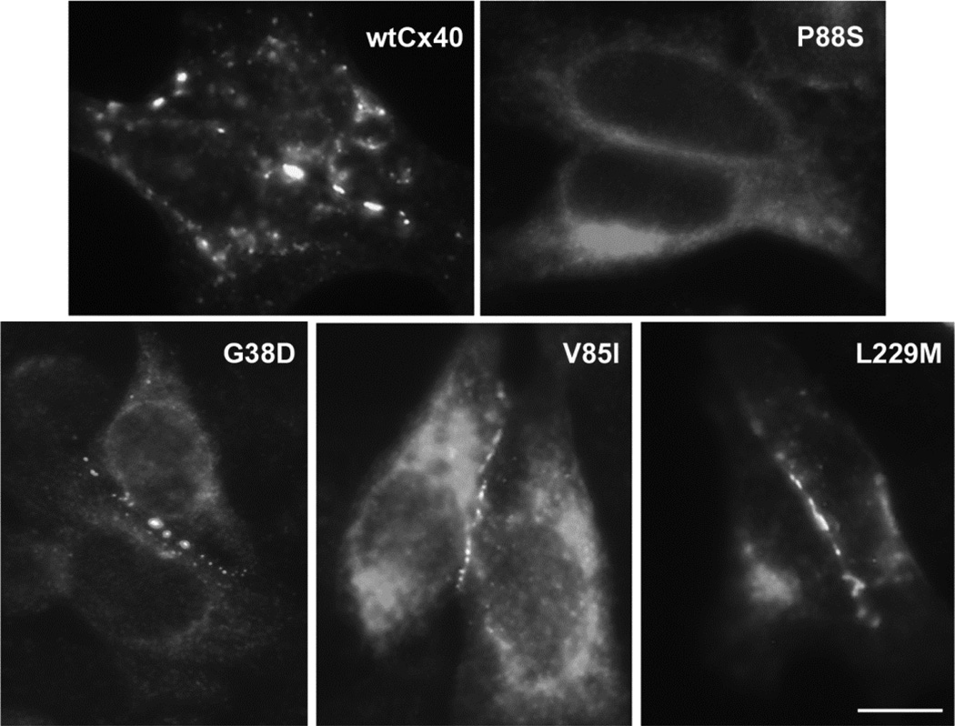
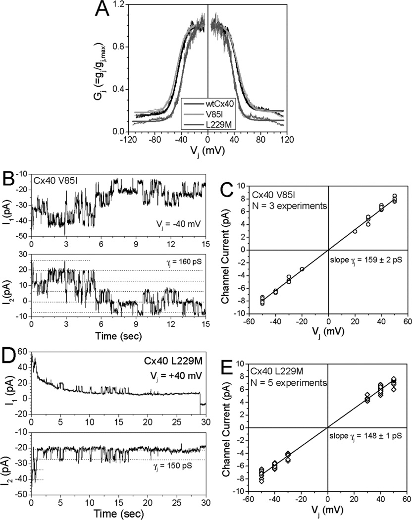
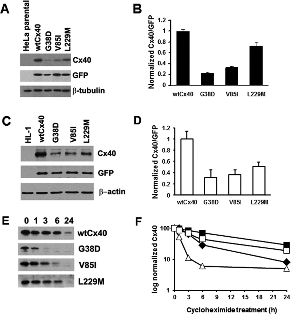
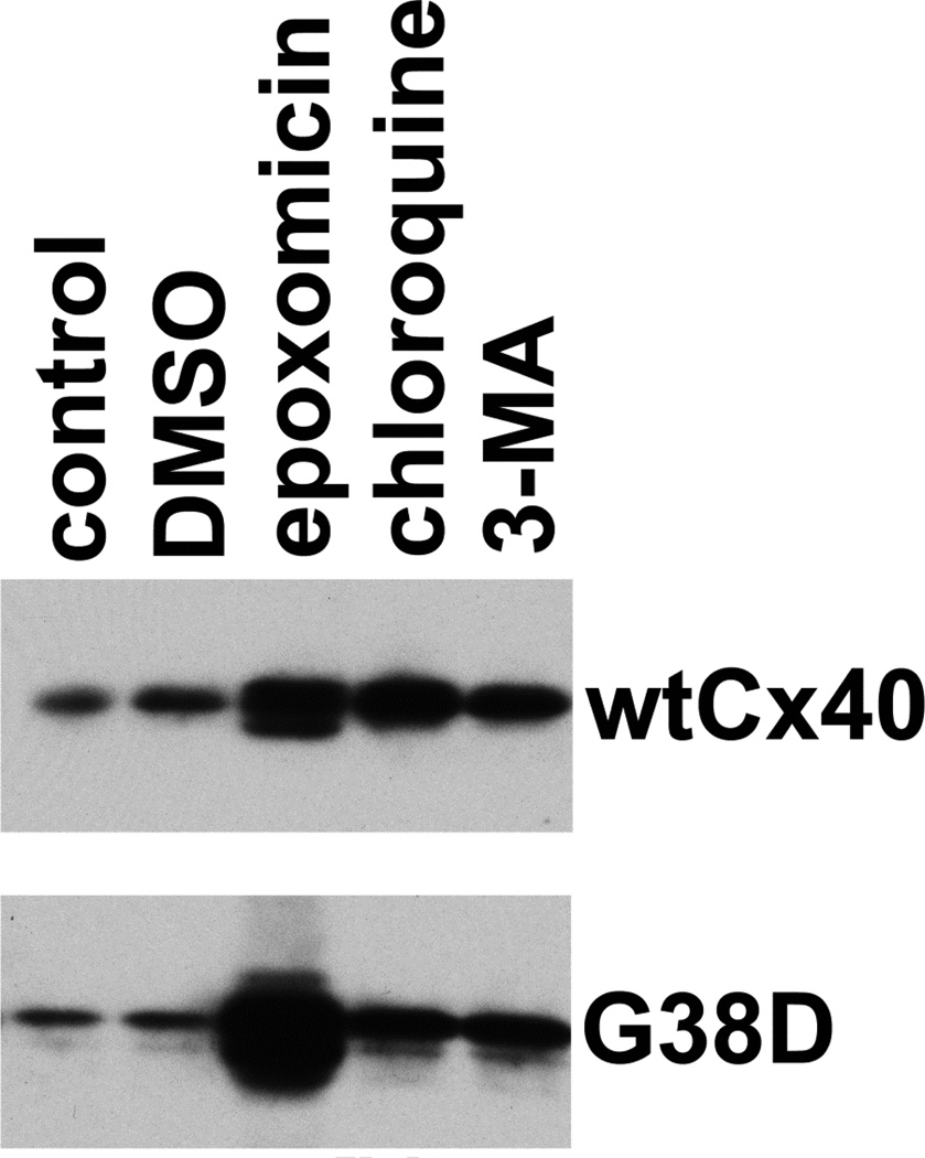
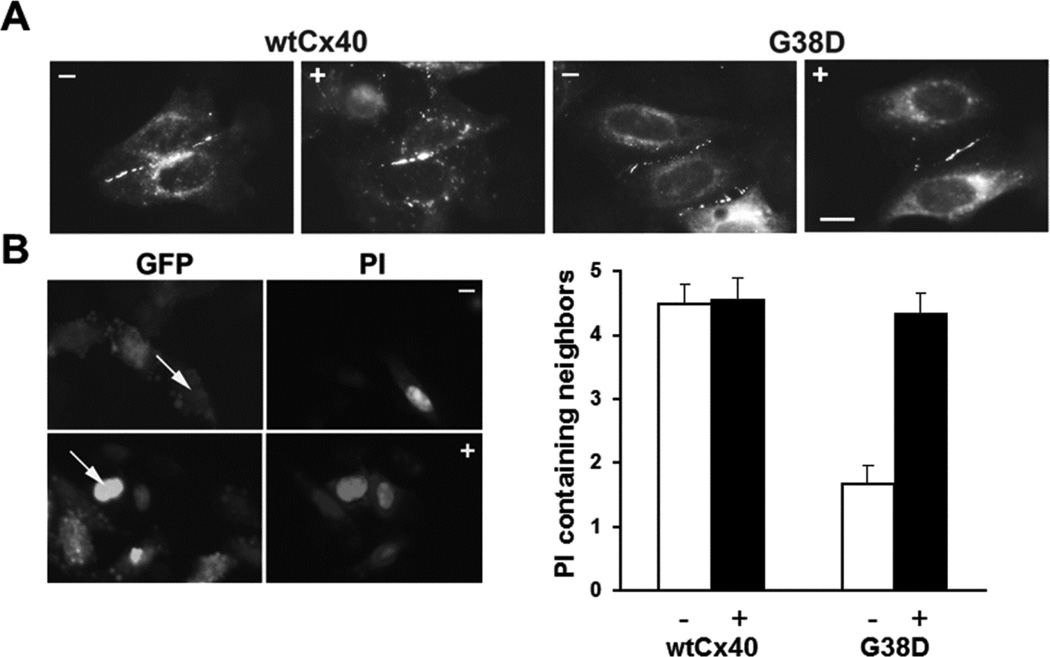
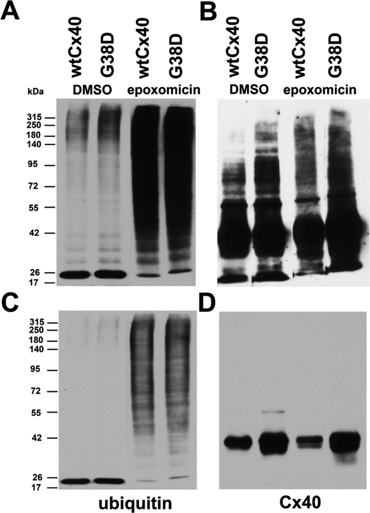
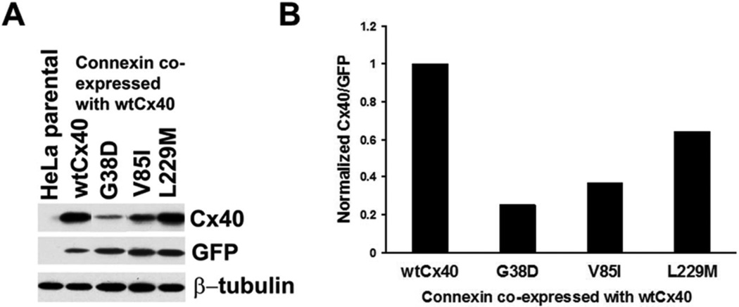
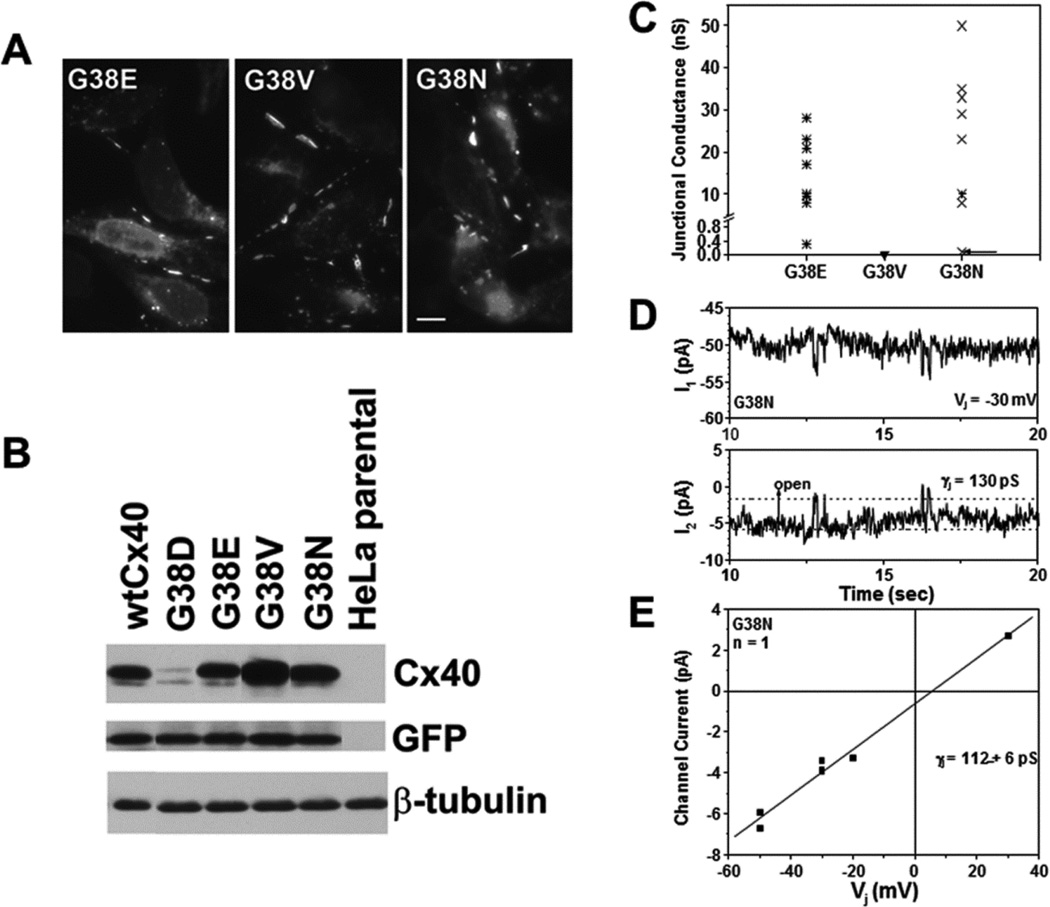
Several Cx40 mutants have been identified in patients with atrial fibrillation (AF). We have been working to identify physiological or cell biological abnormalities of several of these human mutants that might explain how they contribute to disease pathogenesis. Wild type (wt) Cx40 or four different mutants (P88S, G38D, V85I, and L229M) were expressed by the transfection of communication-deficient HeLa cells or HL-1 cardiomyocytes. Biophysical channel properties and the sub-cellular localization and protein levels of Cx40 were characterized. Wild type Cx40 and all mutants except P88S formed gap junction plaques and induced significant gap junctional conductances. The functional mutants showed only modest alterations of single channel conductances or gating by trans-junctional voltage as compared to wtCx40. However, immunoblotting indicated that the steady state levels of G38D, V85I, and L229M were reduced relative to wtCx40; most strikingly, G38D was only 20-31% of wild type levels. After the inhibition of protein synthesis with cycloheximide, G38D (and to a lesser extent the other mutants) disappeared much faster than wtCx40. Treatment with the proteasomal inhibitor, epoxomicin, greatly increased levels of G38D and restored the abundance of gap junctions and the extent of intercellular dye transfer. Thus, G38D, V85I, and L229M are functional mutants of Cx40 with small alterations of physiological properties, but accelerated degradation by the proteasome. These findings suggest a novel mechanism (protein instability) for the pathogenesis of AF due to a connexin mutation and a novel approach to therapy (protease inhibition).

Keywords: Atrial fibrillation; Connexin40; Gap junctions; Ion channels; Proteasome; Protein degradation.


Figures

Figure 1
Figure 1. Many, but not all, AF-associated Cx40 mutants make gap junctions when expressed in communication-deficient cells
Wild type Cx40 or AF-associated mutants (G38D, V85I, P88S, and L229M) were detected by immunofluorescent localization in transiently transfected HeLa cells. Wild type Cx40 and most of the mutants, including G38D, V85I, and L229M, were localized in a punctuate distribution along appositional membranes within the cytoplasm. P88S was retained intracellularly. Bar, 10 µm.
Figure 2
Figure 2. Gap junction channels formed by V85I or L229M had normal or mildly altered Vj-dependent gating and single channel conductances
(A) Plots show the relationships between normalized junctional conductance (Gj) and transjunctional voltage (Vj) for homomericwtCx40 (black), V85I (light gray) and L229M (dark gray) gap junctions. The Boltzmann parameters for the fitted curves are listed in Supplemental Table 1 (B) Whole cell currents (I1 and I2) were recorded simultaneously for 15 sec. from a pair of N2a cells expressing V85I during a 40 mV Vj step applied to cell 1. Current amplitudes are indicated by dashed lines in the I2 trace (Ij = −ΔI2). (C) Junctional current-voltage (Ij–Vj) relationship for V85I channels was generated from channel current amplitudes determined by Gaussian fits of the all points current histogram (not shown) for each 30 s Vj pulse. The mean slope conductance (γj) was 159 ± 2 pS. (D) Whole cell currents are shown for a 30 sec. recording of a L229M channel during a +40 mV Vj pulse. (E) The Ij−Vj relationship for L229M is shown. The mean slope γ j was 148 ± 2 pS.
Figure 3
Figure 3. Several mutants show reduced Cx40 levels and faster degradation than wild type Cx40
(A) Immunoblot of wtCx40 and Cx40 mutants expressed in HeLa cells. Lysates were prepared from (untransfected) HeLa cells and HeLa cells transiently transfected with Cx40 or AF mutants (in pTracer-CMV2) and subjected to immunoblotting using antibodies directed Cx40, GFP (to establish the efficiency of transfection) and β-tubulin (as a loading control). The identical blots were used for probing with each of the different antibodies after stripping. Representative lanes were cropped from a larger blot. (B) Graph shows levels of wtCx40 and mutants. The Cx40 and GFP bands were analyzed by densitometry for each construct. The ratio of Cx40:GFP was calculated for each construct (to correct for any differing efficiencies of transfection). Levels of expression were compared to the wtCx40. Values represent the mean +SEM, n=3. Levels of G38D, V85I, and L229M were all statistically different from wild type (Student’s t-test, p<0.05). (C) Immunoblot of wtCx40 and Cx40 mutants expressed in HL-1 cells. Lysates were prepared from (untransfected) HL-1 cells and HL-1 cells transiently transfected with Cx40 or AF mutants (in pTracer-CMV2) and subjected to immunoblotting using antibodies directed against Cx40, GFP (to establish the efficiency of transfection) and β-actin (as a loading control). The identical blots were used for probing with each of the different antibodies after stripping. (D) Graph shows levels of wtCx40 and mutants expressed in HL-1 cells. Densitometry was performed as described for panel B. Values represent the mean +SEM, n=3. Levels of G38D, V85I, and L229M were all statistically different from wild type (Student’s t-test, p<0.05). (E) Immunoblot analysis of HeLa cells transfected with wild type or mutant Cx40 and then treated with cycloheximide for 0, 1, 3, 6 or 24 h. (F) Graphs shows the levels of immunoreactiveCx40 (wild type or mutants) at different intervals of treatment with cycloheximide (based on quantification of the immunoblots). Values derive from analysis of the Cx40 bands by densitometry and normalization of the abundance at each time point to the abundance at time 0.
Figure 4
Figure 4. The proteasomal inhibitor, epoxomicin, blocks the accelerated degradation of G38D
HeLa cells were transiently transfected with wtCx40 or G38D, and 48 h later were treated for 18 hours with chloroquine (to inhibit the lysosome), 3-methyl adenine (3-MA; to inhibit autophagy) or epoxomicin (to block the proteasome). The cells were harvested and analyzed by immunoblotting to determine levels of immunoreactive Cx40. Although each of the drugs produced an increase in both wtCx40 and G38D, the relative increase for G38D was much greater following epoxomicin, implicating the proteasome in the accelerated degradation of this mutant.
Figure 5
Figure 5. The proteasomal inhibitor, epoxomicin also increases gap junction size/abundance and intercellular communication in HeLa cells expressing G38D
(A) Photomicrographs show immunofluorescent detection of the distributions of Cx40 in HeLa cells transiently transfected with wtCx40 or G38D. Cells were untreated (−) or exposed to 0.5µM epoxomicin (+) for 4 hours. Bar, 10 µm. (B) Photomicrographs show examples of the intercellular transfer of microinjected PI between HeLa cells transiently transfected with G38D. Cells were untreated (−) or exposed to 0.5µM epoxomicin (+) for 4 hours. GFP fluorescence (also expressed from the pTracer plasmid) allowed identification of the transfected cells. Arrows show injected cells. The bar graph shows the extent of dye transfer (PI containing neighbors) after injection of that tracer in cells transfected with wtCx40 or G38D. Cultures were untreated (−, white bars) or treated with 0.5µM epoxomicin (+, black bars).The extent of transfer was not statistically different between control wtCx40-expressing cells and those treated with epoxomicin. In HeLa cells transfected with G38D, transfer of PI was dramatically increased after epoxomicin treatment as compared to vehicle –treated control cultures (p<0.001). Epoxomicin treatment increased dye transfer in G38D–expresssing cells to levels comparable to those in cells expressing wtCx40. Values represent the mean + SEM, n=9−29.
Figure 6
Figure 6. Wild type Cx40 and G38D are ubiquitinated
HeLa cells stably transfected with wtCx40 or G38D were treated with DMSO (control) or with 0.5µM epoxomicin for 18h. Ubiquitinated proteins were isolated from cell lysates and analyzed by immunoblotting with antibodies directed against ubiquitin (A,C) or Cx40 (B,D). The same blot was probed sequentially with both antibodies with intervening stripping . Eluates equivalent to 10µg of total protein in the starting lysate were loaded in each lane. Top panels (A, B) show long exposures and bottom panels (C,D) show shorter exposures of the same blots. Epoxomicin increased the abundance of ubiquitinated proteins (A,C) in cells expressing either wtCx40 or G38D. Immunoreactive Cx40 was isolated with the ubiquitinated proteins from both cell lines (B,D), and the abundance of slower migrating forms (likely polyubiquitinated Cx40) was increased following epoxomicin treatment (B).
Figure 7
Figure 7. AF Cx40 mutants reduce total Cx40 levels when co-expressed with wild type Cx40
HeLa cells were transiently co-transfected with a plasmid encoding wild type Cx40 and an equal amount of plasmid encoding wild type or mutant Cx40. (A) Levels of total immunoreactive Cx40 were detected by immunoblotting. Blots were also probed with antibodies directed against GFP (transfection efficiency control) and β-tubulin (loading control). Although this experiment was performed in duplicate, some lanes were eliminated to include only representative examples. (B). Graph shows the total amounts of immunoreactive Cx40 (includes wild type and mutant) for cells transfected with wtCx40 plus wild type or mutant Cx40. Values were normalized according to the densities of the GFP bands and averaged from duplicates. Cx40 levels are lower in cells transfected with each of the mutants (and most severely reduced for G38D) suggesting that they also accelerate the degradation of the wild type protein.
Figure 8
Figure 8. While all site directed mutants at position G38 form gap junction plaques and differ in channel properties, only G38D has reduced protein levels as compared to wild type Cx40
(A) Immunofluorescent detection of the distributions of immunoreactiveCx40 in HeLa cells transiently transfected with G38E, G38V, or G38N. Each of these mutants localizes to appositional plasma membranes. Bar, 10 µm. (B) Immunoblot detection of Cx40 in untransfected HeLa cells or cells that were transiently transfected wtCx40, G38D, G38E, G38V, or G38N. Although this experiment was performed in triplicate, some lanes were eliminated to illustrate only representative examples. Blots were also probed with antibodies directed against GFP (transfection efficiency control) and β-tubulin (loading control). Only G38D shows reduced levels as compared to wtCx40. (C) Graph shows total junctional conductance in pairs of cells transiently transfected with G38E, G38V, or G38N. While both G38E and G38N formed large conductances in all cell pairs studied, gap junctional coupling was not detected in cell pairs expressing G38V. (D) Tracings show single channel events recorded from an N2a cell pair expressing G38N. Whole cell currents (I1 and I2) were recorded simultaneously for 10 sec. during a 30 mV Vj step applied to cell 1. (E) Junctional current-voltage (Ij –Vj) relationship for G38N channels was generated from channel current amplitudes determined by Gaussian fits of the all points current histogram. The mean slope conductance (γj) was 112 ± 6pS.

Similar articles

Cited by

References

    1. Allessie MA, Boyden PA, Camm AJ, Kleber AG, Lab MJ, Legato MJ, et al. Pathophysiology and prevention of atrial fibrillation. Circulation. 2001;103:769–777. - PubMed
    1. Iwasaki YK, Nishida K, Kato T, Nattel S. Atrial fibrillation pathophysiology: implications for management. Circulation. 2011;124:2264–2274. - PubMed
    1. Spach MS, Starmer CF. Altering the topology of gap junctions a major therapeutic target for atrial fibrillation. Cardiovasc Res. 1995;30:337–344. - PubMed
    1. Saffitz JE, Kanter HL, Green KG, Tolley TK, Beyer EC. Tissue-specific determinants of anisotropic conduction velocity in canine atrial and ventricular myocardium. Circ Res. 1994;74:1065–1070. - PubMed
    1. Lin X, Gemel J, Glass A, Zemlin CW, Beyer EC, Veenstra RD. Connexin40 and connexin43 determine gating properties of atrial gap junction channels. J Mol Cell Cardiol. 2010;48:238–245. - PMC - PubMed

Publication types

MeSH terms